Review
. 2017 Sep;22(9):1107-1116.
doi: 10.1634/theoncologist.2017-0081. Epub 2017 Jun 29.

Appendiceal Mucinous Neoplasms: Diagnosis and Management

Affiliations
Review

Appendiceal Mucinous Neoplasms: Diagnosis and Management

Walid L Shaib et al. Oncologist. 2017 Sep.

Erratum in

  • Appendiceal Mucinous Neoplasms: Diagnosis and Management.
    Shaib WL, Assi R, Shamseddine A, Alese OB, Staley C 3rd, Memis B, Adsay V, Bekaii-Saab T, El-Rayes BF. Shaib WL, et al. Oncologist. 2018 Jan;23(1):137. doi: 10.1634/theoncologist.2017-0081erratum. Oncologist. 2018. PMID: 29317549 Free PMC article. No abstract available.

Abstract

Objective: Appendiceal mucinous neoplasms (AMNs) are a rare and heterogeneous disease for which clinical management is challenging. We aim to review the literature regarding modalities of treatment to guide the management of AMNs.

Methods and review criteria: We conducted a PubMed search in February 2016 for English-language publications, using the terms "appendiceal," "appendix," "carcinoma," "cancer," "mucinous," "treatment," "genes," "target," "genomic," and terms listed in the articles' subheadings. Published reports and abstracts from the American Society of Clinical Oncology meetings were also searched.

Results: In this review, we summarize current data and controversies in AMN classification, clinical presentation, molecular alterations, treatment outcomes with regard to cytoreductive surgery, hyperthermic intraperitoneal chemotherapy (HIPEC), and the role of systemic chemotherapy.

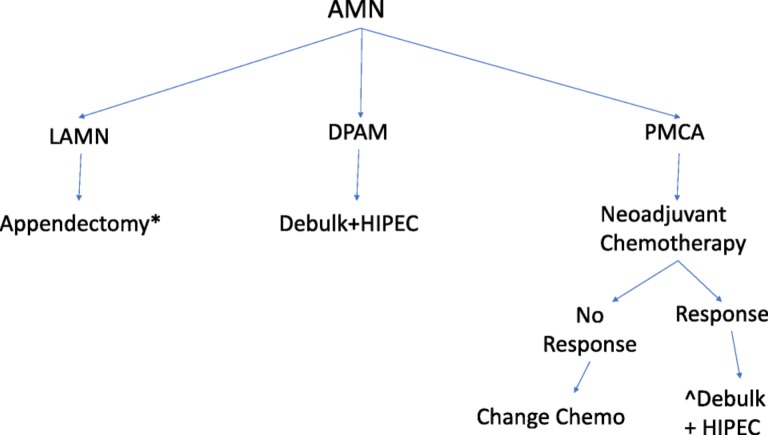
Conclusion: Appendiceal mucinous neoplasms are a heterogeneous group of tumors with a rising incidence. Treatment is based on stage and histology. Low-grade tumors are treated surgically with resection of the primary site in early stage disease, or peritoneal debulking and HIPEC in patients with advanced stage disease. Treatment of high-grade tumors requires further prospective trials, and options include debulking surgery and HIPEC with or without preoperative chemotherapy. Trials evaluating novel therapies based on the molecular profiling of AMN tumors are needed to evaluate therapeutic options in patients who are not surgical candidates.

Implications for practice: This review provides a reference to guide gastroenterologists, pathologists, surgeons, and oncologists in the management of appendiceal mucinous neoplasms (AMNs), a rare and heterogeneous disease with no consensus on histologic classification or guidelines for treatment algorithms. This review summarizes all AMN classifications and proposes a treatment algorithm based on stage and histology of disease.

Keywords: Abdominal surgery; Appendix carcinoma; Review.


Conflict of interest statement

Disclosures of potential conflicts of interest may be found at the end of this article.

Figures

Figure 1.
Figure 1.
Gross specimen. The appendix lumen is filled with gelatinous mucinous material characteristic of low‐grade appendiceal mucinous neoplasms LAMN (LAMN; so‐called “mucocele” formation). In this case, it shows protrusion into the mesoappendix in a diverticular fashion, which is not uncommon in LAMNs.
Figure 2.
Figure 2.
Low‐grade appendiceal mucinous neoplasms LAMN with invasive low‐grade mucinous adenocarcinoma component. The well‐formed and well‐preserved epithelium with papillary villous elements (upper middle) represents the mucosal adenomatous component. The mucin lakes, which are represented as pale nodules of white to slightly blue nodules with scant epithelial cells, represent invasive mucinous adenocarcinoma. Based on the degree of mucin and the relatively mild cytologic atypia, this qualifies as a “low‐ grade” mucinous adenocarcinoma.
Figure 3.
Figure 3.
Invasive intestinal type adenocarcinoma, not low‐grade appendiceal mucinous neoplasmsLAMN. Some invasive adenocarcinomas of the appendix are conventional intestinal (colonic) type, as illustrated in this example.
Figure 4.
Figure 4.
Low‐grade appendiceal mucinous neoplasms LAMN with paucicellullar mucinous spread to the peritoneal surfaces (low‐grade mucinous adenocarcinoma of the so‐called disseminated peritoneal adenomucinosis type). Some of the mucin nodules also contain detached signet‐ring like cells, but this does not qualify as signet ring (poorly cohesive) cell adenocarcinoma because there was no individual cell or cord‐like non‐mucinous infiltration into the stroma by the carcinoma cells.
Figure 5.
Figure 5.
Invasive mucinous adenocarcinoma of high grade involving the peritoneal surfaces (“peritoneal mucinous carcinomatosis”) which arose in a low‐grade appendiceal mucinous neoplasms LAMN (not shown here).
Figure 6.
Figure 6.
This is an example of an appendiceal crypt cell adenocarcinoma (so‐called adenocarcinoma ex‐goblet‐cell carcinoid), which can also show mucinous areas, as illustrated in the middle of the photograph and the upper left inset. The other components of this tumor, however, which are detailed in the insets in upper right and lower right, show features more characteristic of appendiceal crypt cell adenocarcinoma. In the upper right, there is a poorly cohesive (signet ring) cell pattern, and the lower right shows the classical goblet‐cell carcinoid pattern. Such cases should be distinguished from low‐grade appendiceal mucinous neoplasms LAMN or related appendiceal mucinous adenocarcinoma, as which they are commonly misclassified.
Figure 7.
Figure 7.
Treatment recommendations for appendiceal mucinous neoplasms. *Right hemicolectomy if tumor invades base of appendix, has high mitotic rate, size >2 cm, margin positive. ^Adjuvant chemotherapy if R1 or R2 surgery, or if lymph node positive. Abbreviations: AMN, appendiceal mucinous neoplasms; DPAM, disseminated peritoneal adenomucinosis; HIPEC, hyperthermic intraperitoneal chemotherapy; LAMN,: lLow‐grade appendiceal mucinous neoplasms; DPAM: disseminated peritoneal adenomucinosis, PMCA:, peritoneal mucinous carcinomatosis; PMCA I/D:, PMCAperiotoneal mucinous carcinomatosis of indeterminate or discordant features. The classification is dependent on the grade of differentiation rather than the anatomic invasion of the disease.

References

    1. McCusker ME, Cote TR, Clegg LX et al. Primary malignant neoplasms of the appendix: A population‐based study from the surveillance, epidemiology and end‐results program, 1973–1998. Cancer 2002;94:3307–3312. - PubMed
    1. Smeenk RM, van Velthuysen ML, Verwaal VJ et al. Appendiceal neoplasms and pseudomyxoma peritonei: A population based study. Eur J Surg Oncol 2008;34:196–201. - PubMed
    1. Rokitansky K, Swaine WE, Sir Sieveking EH, Moore CH, Day GE. A Manual of Pathological Anatomy. Vol. 2 Philadelphia, PA: Blanchard and Lea, 1855;24:100–118.
    1. Elting AW. IX. Primary carcinoma of the vermiform appendix, with a report of three cases. Ann Surg 1903;37:549–574. - PMC - PubMed
    1. Carr NJ, McCarthy WF, Sobin LH. Epithelial noncarcinoid tumors and tumor‐like lesions of the appendix. A clinicopathologic study of 184 patients with a multivariate analysis of prognostic factors. Cancer 1995;75:757–768. - PubMed

MeSH terms