. 2017 Mar:86:99-216.
doi: 10.1016/j.simyco.2017.04.002. Epub 2017 May 5.

Genera of phytopathogenic fungi: GOPHY 1

Affiliations

Genera of phytopathogenic fungi: GOPHY 1

Y Marin-Felix et al. Stud Mycol. 2017 Mar.

Abstract

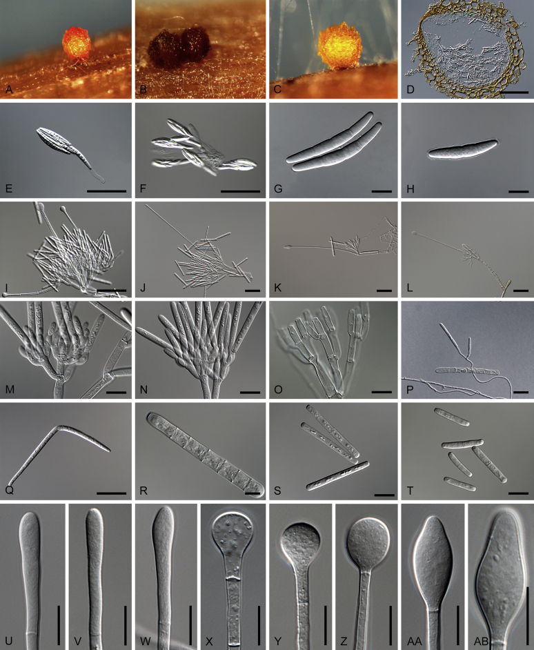
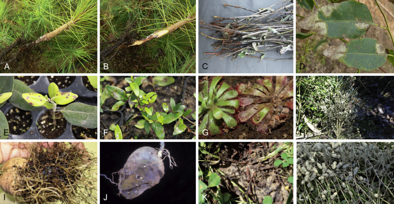
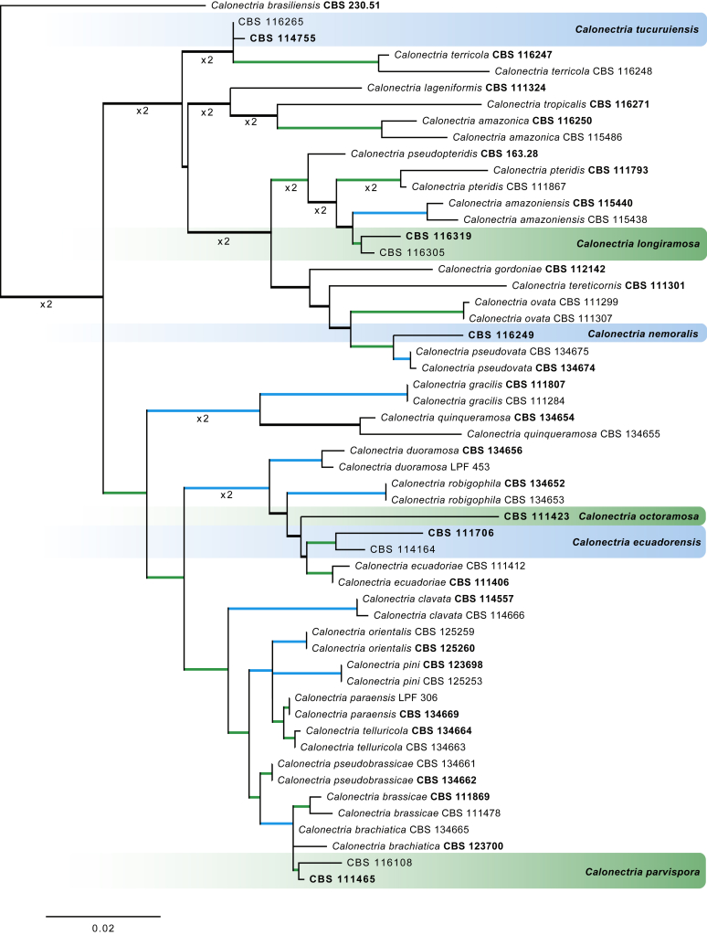
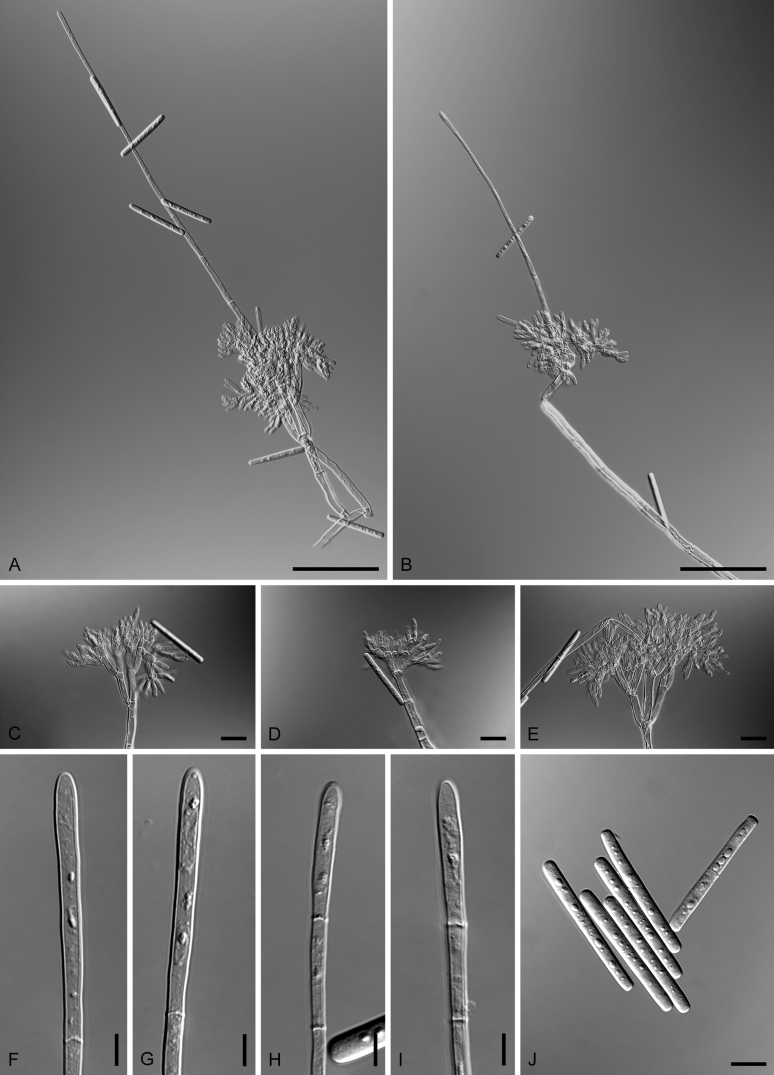
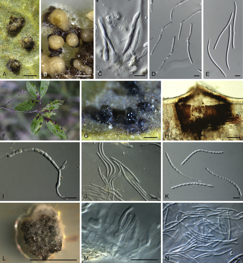
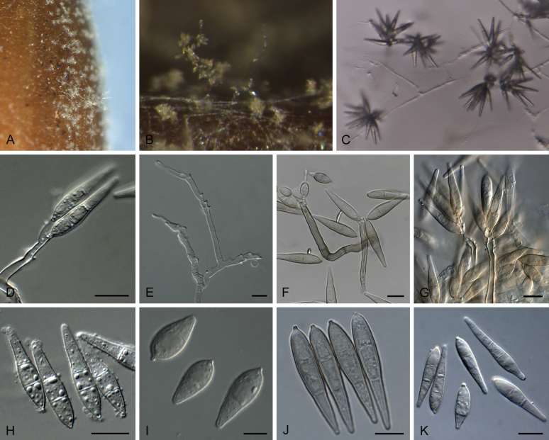
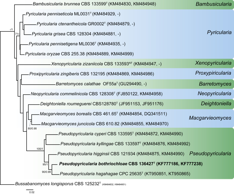
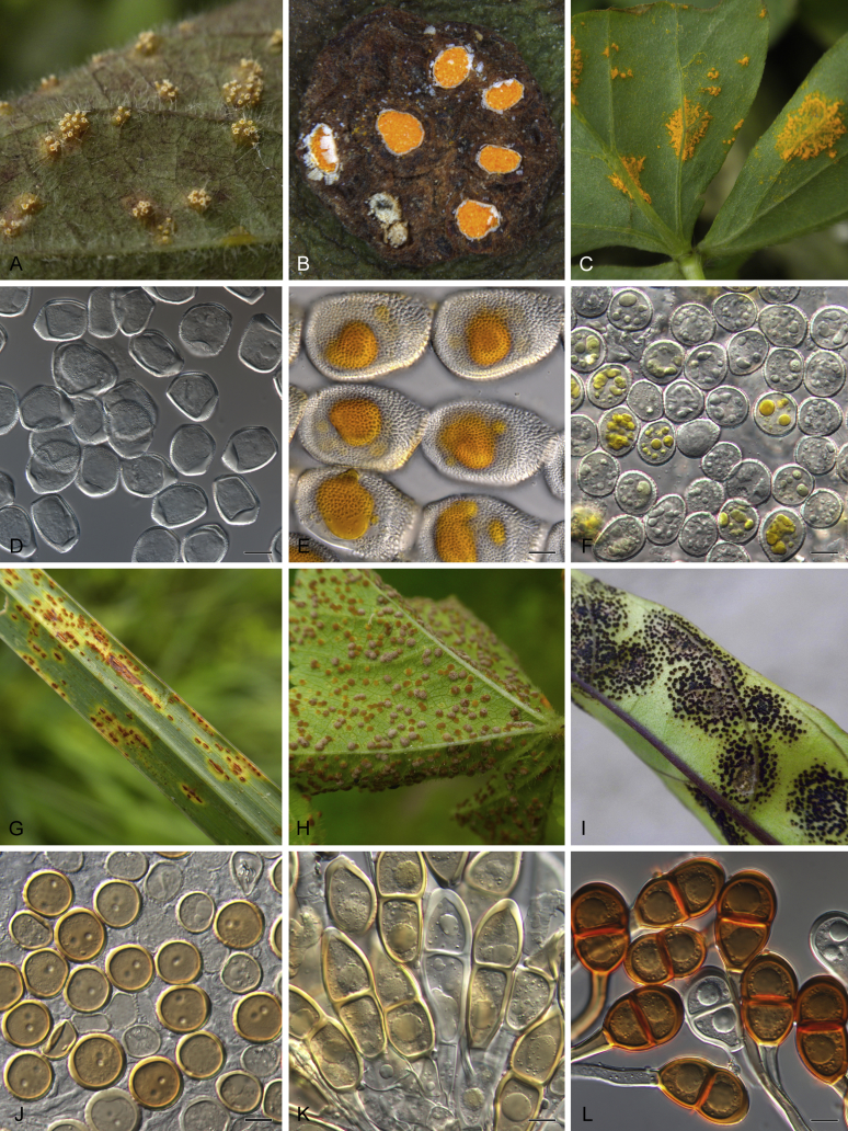
Genera of Phytopathogenic Fungi (GOPHY) is introduced as a new series of publications in order to provide a stable platform for the taxonomy of phytopathogenic fungi. This first paper focuses on 21 genera of phytopathogenic fungi: Bipolaris, Boeremia, Calonectria, Ceratocystis, Cladosporium, Colletotrichum, Coniella, Curvularia, Monilinia, Neofabraea, Neofusicoccum, Pilidium, Pleiochaeta, Plenodomus, Protostegia, Pseudopyricularia, Puccinia, Saccharata, Thyrostroma, Venturia and Wilsonomyces. For each genus, a morphological description and information about its pathology, distribution, hosts and disease symptoms are provided. In addition, this information is linked to primary and secondary DNA barcodes of the presently accepted species, and relevant literature. Moreover, several novelties are introduced, i.e. new genera, species and combinations, and neo-, lecto- and epitypes designated to provide a stable taxonomy. This first paper includes one new genus, 26 new species, ten new combinations, and four typifications of older names.

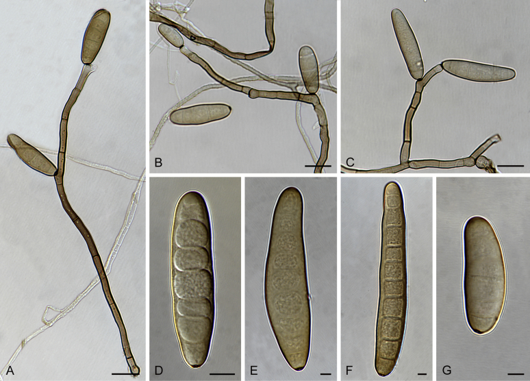
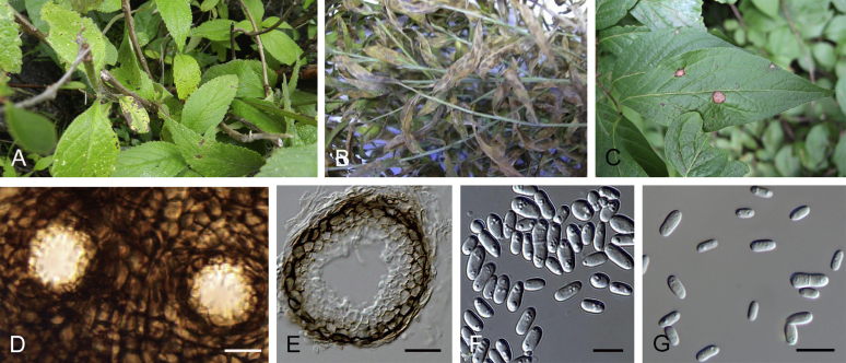
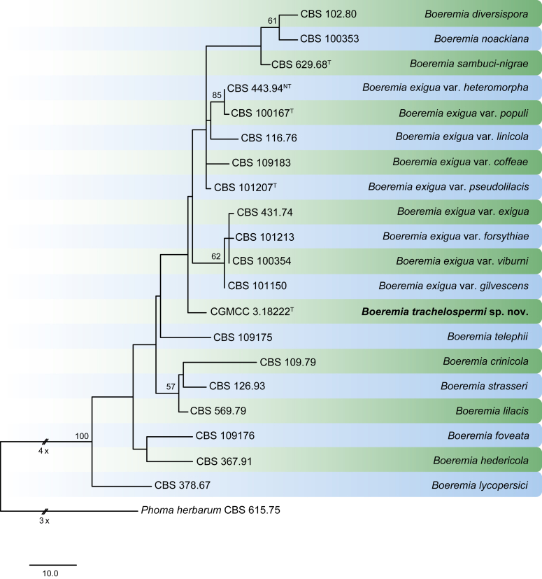
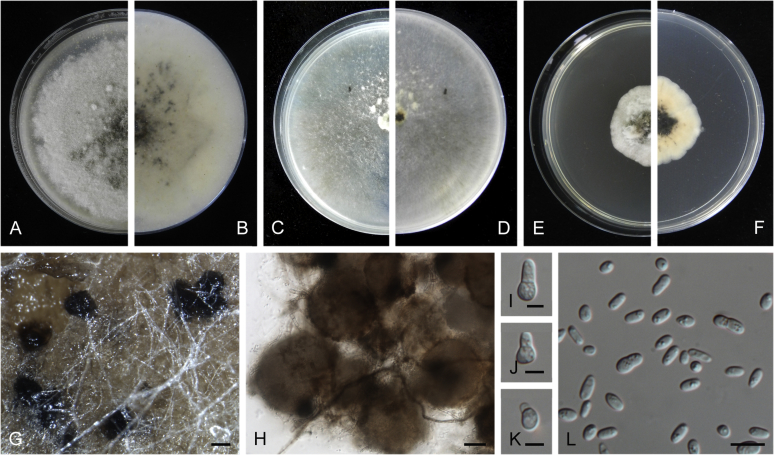
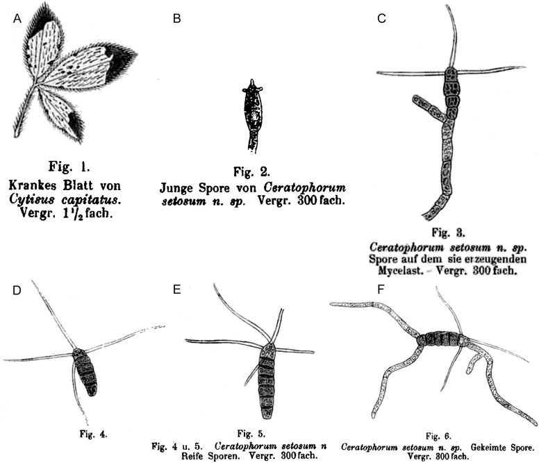
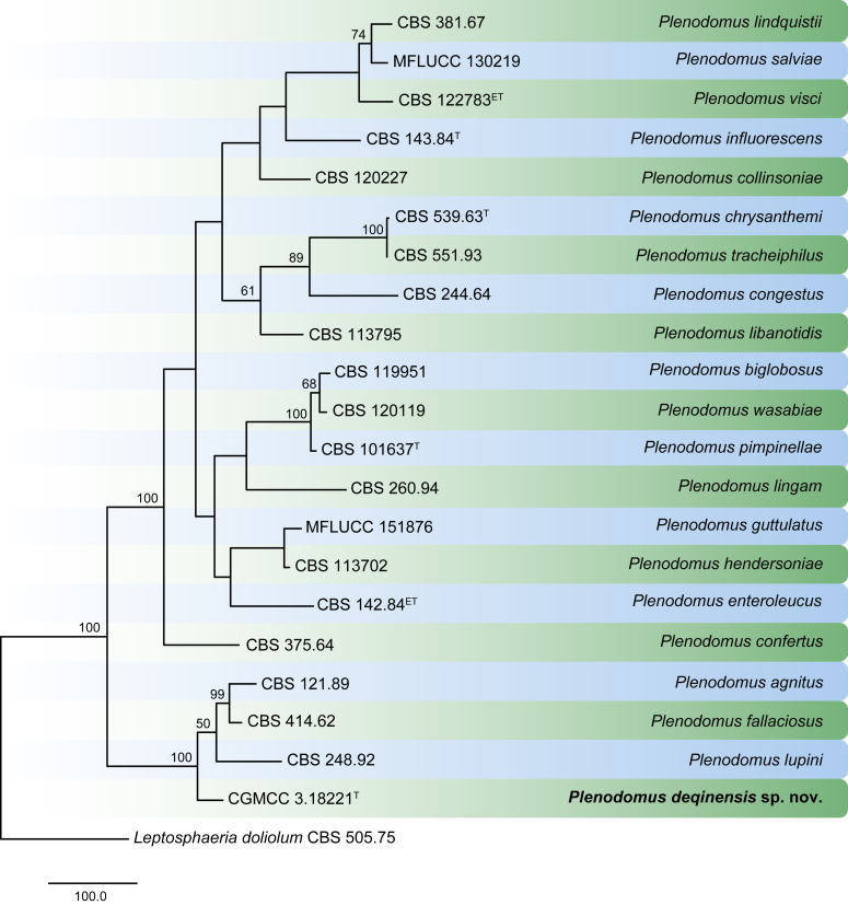
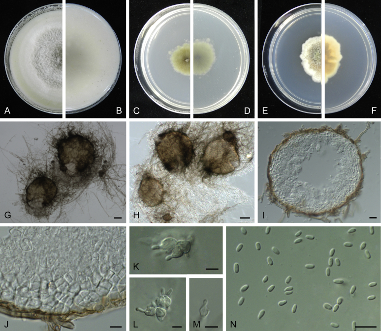
Keywords: Bi. variabilis Y. Marín, Y.P. Tan & Crous; Bipolaris saccharicola Y. Marín & Crous; Boeremia trachelospermi Q. Chen & L. Cai; Ca. longiramosa L. Lombard & Crous; Ca. nemoralis L. Lombard & Crous; Ca. octoramosa L. Lombard & Crous; Ca. parvispora L. Lombard & Crous; Ca. tucuruiensis L. Lombard & Crous; Calonectria ecuadorensis L. Lombard & Crous; Ceratophorum setosum Kirchn; Ceratophorum setosum Kirchn.; Cl. kenpeggii Bensch, U. Braun & Crous; Cl. welwitschiicola Bensch, U. Braun & Crous; Cladosporium chasmanthicola Bensch, U. Braun & Crous; Colletotrichum sydowii Damm; Coniella hibisci (B. Sutton) Crous; Coniella musaiaensis var. hibisci B. Sutton; Cu. soli Y. Marín & Crous; Curvularia pisi Y. Marín & Crous; DNA barcodes; Fungal systematics; Helminthosporium carpophilum Lév.; M. yunnanensis (M.J. Hu & C.X. Luo) Sandoval-Denis & Crous; Monilinia mumeicola (Y. Harada et al.) Sandoval-Denis & Crous; Neofusicoccum italicum Dissanayake & K.D. Hyde; Nm. pistaciicola Crous; Nm. pruni Crous; Phytopathogenic fungi; Pilidium septatum Giraldo & Crous; Plant pathology; Pleiochaeta carotae Hern.-Rest., van der Linde & Crous; Plenodomus deqinensis Q. Chen & L. Cai; Protostegia eucleicola Crous; Pseudopyricularia bothriochloae (Crous & Cheew.) Y. Marín & Crous; Pu. geitonoplesii (McAlpine) McTaggart & R.G. Shivas; Pu. merrilliana (Syd. & P. Syd.) McTaggart & R.G. Shivas; Pu. rhagodiae (Cooke & Massee) McTaggart & R.G. Shivas; Puccinia dianellae (Dietel) McTaggart & R.G. Shivas; S. protearum Crous; Saccharata leucospermi Crous; Taxonomy; Thyrostroma franseriae Crous; Typifications; Venturia martianoffiana (Thüm.) Y. Zhang ter & J.Q. Zhang; Venturia phaeosepta Y. Zhang ter & J.Q. Zhang; Verkleyomyces Y. Marín & Crous; Verkleyomyces illicii (X. Sun et al.) Y. Marín & Crous.


Figures

Fig. 1
Fig. 1
Bipolaris spp. A–F. Disease symptoms. A. Symptoms caused by Bipolaris eragrostiellae (ex-type IMI 155931). B. Symptoms caused by Bipolaris gossypina (IMI 123377). C. Symptoms caused by Bipolaris halepensis (ex-type BPI 1103129). D. Symptoms caused by Bipolaris microstegii. E. Symptoms caused by Bipolaris musae-sapientium (ex-type K (M) 181466). F. Symptoms caused by Bipolaris oryzae (ex-neotype MFLUCC 10-0715). G–L. Sexual morphs. G. Ascoma of Bipolaris luttrellii (IMI 345516). H–K. Asci. H.Bipolaris chloridis (ex-type IMI 213865). I.Bipolaris luttrellii (IMI 345516). J.Bipolaris maydis (CBS 241.92). K.Bipolaris microlaenae (IMI 338218). L. Ascospores of Bipolaris maydis (CBS 241.92). M–Z. Asexual morphs. M–R. Conidiophores and conidia. M.Bipolaris setariae (BPI 880305B). N.Bipolaris zeae (ex-type IMI 202085). O.Bipolaris bicolor (CBS 690.96). P.Bipolaris heveae (CBS 241.92). Q.Bipolaris sorokiniana (ex-type CBS 110.14). R.Bipolaris zeicola (ex-type BPI 626668). S–Z. Conidia. S.Bipolaris cookei (ex-type BPI 428852). T.Bipolaris costina (ex-type IMI 256417). U.Bipolaris crotonis (ex-type IMI 223682). V.Bipolaris gossypina (IMI 123377). W.Bipolaris obclavata (ex-type IMI 331725). X.Bipolaris oryzae (ex-neotype MFLUCC 10-0715). Y.Bipolaris salviniae (DAR 35056). Z.Bipolaris sorokiniana (ex-type CBS 110.14). Scale bars: A, N = 100 μm; B, E, F = 500 μm; C = 1 cm; G, H = 20 μm; I–L, O–Z = 10 μm; M = 50 μm. All pictures except for D taken from Manamgoda et al. (2014).
Fig. 2
Fig. 2
RAxML phylogram obtained from the combined ITS (478 bp), gapdh (472 bp) and tef1 (892 bp) sequences of all the accepted species of Bipolaris. The tree was rooted to Curvularia buchloës CBS 246.49 and Curvularia subpapendorfii CBS 656.74. The novel species described in this study are shown in bold. RAxML bootstrap support (BS) values above 70 % and Bayesian posterior probability scores ≥ 0.95 are shown at the nodes. GenBank accession numbers are indicated in Table 1. T, ET, IsoT, IsoLT, IsoPT, LT and NT indicate ex-type, ex-epitype, ex-isotype, ex-isolectotype, ex-isoparatype, ex-lectotype and ex-neotype strains, respectively. TreeBASE: S20877.
Fig. 3
Fig. 3
Bipolaris saccharicola (ex-type CBS 155.26). A–C. Conidiophores and conidia. D–H. Conidia. Scale bars: A–C = 20 μm; H applies to D–H = 10 μm.
Fig. 4
Fig. 4
Bipolaris variabilis (ex-type CBS 127716). A–C. Conidiophores and conidia. D–M. Conidia. Scale bars: A = 20 μm; B, C = 15 μm; H applies to D–H, M applies to I–M = 5 μm.
Fig. 5
Fig. 5
Bipolaris yamadae (CBS 127087). A–C. Conidiophores and conidia. D–G. Conidia. Scale bars: A–C = 20 μm; D–G = 10 μm.
Fig. 6
Fig. 6
Boeremia spp. A. Symptoms of Boeremia lilacis (LC 8116) on Ocimum sp. B. Symptoms of Boeremia exigua var. rhapontica (ex-type CBS 113651) on Rhaponticum repens. C. Symptoms of Boeremia lilacis (LC 5178) on Lonicera sp. D. Ostiole configuration of Boeremia exigua var. exigua (CBS 431.74). E. Section of young pycnidium of Boeremia exigua var. pseudolilacis (ex-type CBS 101207). F. Conidia of Boeremia exigua var. pseudolilacis (ex-type CBS 101207). G. Conidia of Boeremia exigua var. heteromorpha (ex-neotype CBS 443.94). Scale bars: D–E = 20 μm; F = 5 μm; G = 10 μm. Picture B taken from Berner et al. (2015); D–F from Aveskamp et al. (2010); G from Chen et al. (2015a).
Fig. 7
Fig. 7
Phylogenetic tree generated from a maximum parsimony analysis based on the combined LSU, ITS, tub2 and rpb2 sequences. Values above the branches represent parsimony bootstrap support values (> 50 %). The novel species are shown in bold. The tree is rooted with Phoma herbarum CBS 615.75. GenBank accession numbers are indicated in Table 2. T and NT indicate ex-type and ex-neotype strains, respectively. TreeBASE: S21048.
Fig. 8
Fig. 8
Boeremia trachelospermi (ex-type CGMCC 3.18222). A, B. Colony on OA (front and reverse). C, D. Colony on MEA (front and reverse). E, F. Colony on PDA (front and reverse). G. Pycnidia forming on OA. H. Pycnidia. I–K. Conidiogenous cells. L. Conidia. Scale bars: G = 200 μm; H = 50 μm; I–K = 5 μm; L = 10 μm.
Fig. 9
Fig. 9
Calonectria spp. A−H. Sexual morphs. A−D. Perithecia. A.Calonectria asiatica (ex-type CBS 114073). B.Calonectria braziliensis (ex-type CBS 230.51 × CBS 114257). b Calonectria fujianensis (ex-type CBS 127201). D. Section through perithecium of Calonectria asiatica (ex-type CBS 114073). E−F. Asci. E.Calonectria crousiana (ex-type CBS 127198). F.Calonectria asiatica (ex-type CBS 114073). G−H. Ascospores. G.Calonectria fujianensis (ex-type CBS 127201). H.Calonectria acicola (ex-type CBS 114813). I−AB. Asexual morphs. I−L. Macroconidiophores. I.Calonectria malesiana (ex-type CBS 112752). J.Calonectria macroconidialis (ex-type CBS 114880). K.Calonectria spathulata (ex-type CBS 555.92). L.Calonectria ovata (CBS 111307). M−O. Conidiogenous apparatus. M.Calonectria brachiatica (ex-type CBS 123700). N.Calonectria ecuadoriae (ex-type CBS 111406). O.Calonectria hurae (CBS 114551). P. Microconidiophore of Calonectria reteaudii (ex-type CBS 112144). Q. Megaconidia of Calonectria hurae (CBS 114551). R, S. Macroconidia. R.Calonectria angustata (ex-type CBS 109065). S.Calonectria chinensis (ex-type CBS 114827). T. Microconidia of Calonectria pteridis (ex-type CBS 111793). U−AB. Terminal vesicles of stipe extensions. U.Calonectria brassicae (ex-type CBS 111869). V.Calonectria rumohrae (CBS 109062). W.Calonectria cylindrospora (CBS 119670). X.Calonectria hongkongensis (ex-type CBS 114828). Y.Calonectria chinensis (ex-type CBS 114827). Z.Calonectria humicola (ex-type CBS 125251). AA.Calonectria mexicana (ex-type CBS 110918). AB.Calonectria spathulata (ex-type CBS 555.92). Scale bars: A−C = 500 μm; D−F = 100 μm; G, H, M−P, R−AB = 10 μm; I−L, Q = 20 μm.
Fig. 10
Fig. 10
Disease symptoms associated with Calonectria spp. A−B. Root and collar rot of Pinus spp. C. Cutting rot of Eucalyptus sp. D. Calonectria leaf blight of Eucalyptus sp. E. Calonectria leaf blight of Metrosideros thomasii. F. Calonectria leaf blight of Myrtus communis. G. Seedling blight of Drosera sp. H. Buxus blight. I. Root rot of Persea americana.J. Potato tuber rot. K−L. Calonectria black rot of Arachis hypogaea.
Fig. 11
Fig. 11
The Maximum Likelihood (ML) consensus tree inferred from the combined cmdA, tef1 and tub2 sequence alignments. Thickened lines indicate branches present in the ML, Maximum parsimony (MP) and Bayesian consensus trees. Branches with ML-bootstrap (BS) & MP-BS = 100 % and posterior probabilities (PP) = 1.00 are in blue. Branches with ML-BS & MP-BS ≥ 75 % and PP ≥ 0.95 are in green. The scale bar indicates 0.02 expected changes per site. The tree is rooted to Calonectria braziliensis (CBS 230.51). Ex-type strains are indicated in bold. GenBank accession numbers are indicated in Lombard et al., 2010a, Lombard et al., 2015, Lombard et al., 2016 and Alfenas et al. (2015). TreeBASE: S20877.
Fig. 12
Fig. 12
Calonectria ecuadorensis (ex-type CBS 111706). A, B. Macroconidiophores. C−E. Conidiogenous apparatus with conidiophore branches and doliiform to reniform phialides. F−I. Clavate vesicles. J. Macroconidia. Scale bars: A, B = 50 μm; C−J = 10 μm.
Fig. 13
Fig. 13
Calonectria longiramosa (ex-type CBS 116319). A−C. Macroconidiophores. D−F. Conidiogenous apparatus with conidiophore branches and elongate doliiform to allantoid phialides. G−J. Clavate vesicles. K. Macroconidia. Scale bars: A−C = 50 μm; D−K = 10 μm.
Fig. 14
Fig. 14
Calonectria nemoralis (ex-type CBS 116319). A, B. Macroconidiophores. C−D. Conidiogenous apparatus with conidiophore branches and doliiform to reniform phialides. E. Macroconidia. F−I. Fusiform to ovoid vesicles. Scale bars: A, B = 50 μm; C−I = 10 μm.
Fig. 15
Fig. 15
Calonectria octoramosa (ex-type CBS 111423). A−C. Macroconidiophores. D−F. Conidiogenous apparatus with conidiophore branches and doliiform to reniform phialides. G−J. Clavate vesicles. K. Macroconidia. Scale bars: A−C = 20 μm; D−K = 10 μm.
Fig. 16
Fig. 16
Calonectria parvispora (ex-type CBS 111465). A−C. Macroconidiophores. D−F. Conidiogenous apparatus with conidiophore branches and doliiform to reniform phialides. G−J. Clavate vesicles. K. Macroconidia. Scale bars: A−C = 20 μm; D−K = 10 μm.
Fig. 17
Fig. 17
Calonectria tucuruiensis (ex-type CBS 114755). A−C. Macroconidiophores. D−F. Conidiogenous apparatus with conidiophore branches and elongate doliiform to reniform phialides. G−J. Fusiform to ovoid to ellipsoid vesicles. K. Macroconidia. Scale bars: A−C = 50 μm; D−K = 10 μm.
Fig. 18
Fig. 18
A. Sweet potatoes (Ipomea batatas) infected with Ceratocystis fimbriata. B−O. Microscopic features of Ceratocystis fimbriata (CBS 114723 = CMW 14799) on 2 % MEA. B. Ascomata with yellowish droplets of ascospores at tips of necks, with asexual state (white background). C. Young ascoma. D. Mature ascoma. E. Ostiolar hyphae. F, G. Ascospores. H, I. Aleurioconidia. J. Conidiogenous cells producing aleurioconidia (black arrow) and cylindrical-shape conidia (white arrow). K–O. Conidia of various shapes in chains. Scale bars: B = 500 μm; C, D = 100 μm; E = 50 μm; F, G, K–O = 10 μm; H = 50 μm; I, J = 25 μm.
Fig. 19
Fig. 19
Cladosporium spp. A–H. Sexual morphs. A. Ascomata on host tissue (arrows) of Cladosporium silenes (holotype CBS H-19874). B. Ascoma and asci of Cladosporium herbarum (CPC 11600). C. Ostiole of Cladosporium macrocarpum (CBS 299.67). D, E. Asci of Cladosporium herbarum (CPC 11600). F, G. Ascospores of Cladosporium herbarum (CPC 11600). H. Ascospores (arrow denotes mucoid appendage) of Cladosporium silenes (holotype CBS H-19874). I–O. Asexual morphs. I. Conidiophores of Cladosporium halotolerans (ex-type CBS 119416). J. Fascicle of conidiophores of Cladosporium soldanellae (BPI 427476). K. Macronematous conidiophores and conidial chains of Cladosporium cladosporioides (ex-neotype CBS 112388). L. Conidial chains, septa of secondary ramoconidia distinctly darkened of Cladosporium paracladosporioides (ex-type CBS 171.54). M. CryoSEM of different types of conidia on aerial structures of Cladosporium exile (ex-type CBS 125987). Note a remarkable pattern of blastoconidium formation (backwards) (arrow). N. Secondary ramoconidia, conidia and scars of Cladosporium perangustum (ex-type CBS 125996). O. Whorls of secondary ramoconidia and conidia of Cladosporium scabrellum (ex-type CBS 126358). Scale bars: B, C, M, O = 5 μm; D–H, K, L = 10 μm; I = 100 μm; J = 50 μm; N = 2 μm. Pictures taken from Bensch et al. (2012).
Fig. 20
Fig. 20
The first of two equally most parsimonious trees obtained from a heuristic search of the combined ITS/tef1/actA alignment. The tree was rooted to Cladosporium allicinum CBS 121624 and the novel species described in this study are shown in bold. Bootstrap support values from 1000 replicates are shown at the nodes and the scale bar represents the number of changes. GenBank accession numbers are indicated in superscript (ITS/tef1/actA). TreeBASE: S20877.
Fig. 21
Fig. 21
Cladosporium chasmanthicola (ex-type CBS 142612). A–H. Conidiophores and conidial chains. Scale bars = 10 μm; C applies to C–G.
Fig. 22
Fig. 22
Cladosporium kenpeggii (ex-type CBS 142613). A. Part of the colony on SNA. B–H. Conidiophores and conidial chains. Note the microcyclic conidiogenesis in C, forming a secondary conidiophore at a still attached conidium with giving rise to secondary conidia and the germinating conidia in C and G. Scale bars = 10 μm; C applies to C, D; E applies to E–G.
Fig. 23
Fig. 23
Cladosporium welwitschiicola (ex-type CBS 142614). A–G. Conidiophores and conidial chains. H. Conidial chain. Scale bars = 10 μm; A applies to A–C; E applies to E, F.
Fig. 24
Fig. 24
Colletotrichum spp. A–AA. Asexual morphs. A–C. Conidiomata. A.Colletotrichum acutatum (ex-type CBS 112996). B.Colletotrichum destructivum (ex-type CBS 136228). C.Colletotrichum cymbidiicola (ex-type IMI 347923). D. Seta of Colletotrichum torulosum (ex-type CBS 128544). E. Tip of a seta of Colletotrichum gloeosporioides (ex-type CBS 112999). F. Basis of a seta of Colletotrichum gloeosporioides (ex-type CBS 112999). G–K. Conidiogenous cells. G. Colletotrichum brasiliense (ex-type CBS 128501). H.Colletotrichum scovillei (ex-type CBS 126529). I.Colletotrichum tofieldiae (CBS 495.85). J.Colletotrichum petchii (ex-type CBS 378.94). K.Colletotrichum gloeosporioides (ex-type CBS 112999). L–R. Appressoria. L.Colletotrichum americae-borealis (CBS 136855). M.Colletotrichum graminicola (ex-epitype CBS 130836). N.Colletotrichum gloeosporioides (ex-type CBS 112999). O.Colletotrichum laticiphilum (ex-type CBS 112989). P.Colletotrichum phormii (ex-type CBS 118194). Q.Colletotrichum liriopes (ex-type CBS 119444). R.Colletotrichum truncatum (ex-type CBS 151.35). S–AA. Conidia of the ex-type strains of the name-giving species of nine Colletotrichum species complexes. S.Colletotrichum dematium (ex-type CBS 125.25). T.Colletotrichum acutatum (ex-type CBS 112996). U.Colletotrichum truncatum (ex-type CBS 151.35). V.Colletotrichum gloeosporioides (ex-type CBS 112999). W.Colletotrichum graminicola (ex-epitype CBS 130836). X.Colletotrichum boninense (ex-type CBS 123755). Y.Colletotrichum destructivum (ex-type CBS 136228). Z.Colletotrichum orbiculare (ex-type CBS 570.97). AA.Colletotrichum gigasporum (ex-type CBS 133266). A–C, E–H, K. from Anthriscus stem. D, I, J, L–AA. from SNA. Scale bars: A = 200 μm; B applies to B, C = 100 μm; G applies to D–AA = 10 μm. A–AA Pictures taken by U. Damm; A, H, O–P, T from Damm et al. (2012b); B, L, Y from Damm et al. (2014); C, D, G, J, X from Damm et al. (2012a); I, Q–S, U from Damm et al. (2009); Z from Damm et al. (2013).
Fig. 25
Fig. 25
A–F. Disease symptoms caused by Colletotrichum spp. A. Anthracnose on fruit of Cucurbita maxima cv. Red Hokkaido caused by Colletotrichum coccodes. B. Leaf spot of red clover caused by Colletotrichum utrechtense. C. Anthracnose on bean hypocotyl caused by Colletotrichum lindemuthianum. D. Leaf spot of Paphiopedilum sp. caused by Colletotrichum arxii. E. Anthracnose on strawberry fruit caused by Colletotrichum nymphaeae. F. Leaf spot of Mahonia aquifolium caused by Colletotrichum godetiae. G–R. Sexual morphs of Colletotrichum spp. G, H. Ascomata. G. Colletotrichum petchii (ex-type CBS 378.94). H.Colletotrichum karstii (CBS 127597). I. Peridium in cross section of Colletotrichum karstii (CBS 127597). J. Outer surface of peridium of Colletotrichum constrictum (ex-type CBS 128504). K–N. Ascospores. K.Colletotrichum salicis (ex-type CBS 607.94). L. Colletotrichum constrictum (ex-type CBS 128504). M.Colletotrichum cymbidiicola (ex-type IMI 347923). N.Colletotrichum parsonsiae (ex-type CBS 128525). O–Q. Asci. O.Colletotrichum cymbidiicola (ex-type IMI 347923). P.Colletotrichum salicis (ex-type CBS 607.94). Q.Colletotrichum constrictum (ex-type CBS 128504). R. Paraphyses of Colletotrichum salicis (ex-type CBS 607.94). G, K, M, O–R. from Anthriscus stem. H–J, L, N. from SNA. Scale bars: G = 100 μm; H = 50 μm; I applies to I–R = 10 μm. A–R Pictures taken by U. Damm; E from Cannon et al. (2012); G–J, L–O, Q from Damm et al. (2012a); K, P, R from Damm et al. (2012b).
Fig. 26
Fig. 26
One of the 100 equally most parsimonious trees obtained from a heuristic search of the combined ITS, gapdh, chs-1, act and tub2 sequence data of the currently accepted species of Colletotrichum. Parsimony and likelihood bootstrap support values ≥ 50 % are indicated at the nodes and branches with Bayesian posterior probabilities above 0.80 given in bold. The tree is rooted with Monilochaetes infuscans CBS 869.96. GenBank accession numbers are listed in Table 6, Table 7. The ex-type strains are in bold. TreeBASE: S21045.
Fig. 26
Fig. 26
One of the 100 equally most parsimonious trees obtained from a heuristic search of the combined ITS, gapdh, chs-1, act and tub2 sequence data of the currently accepted species of Colletotrichum. Parsimony and likelihood bootstrap support values ≥ 50 % are indicated at the nodes and branches with Bayesian posterior probabilities above 0.80 given in bold. The tree is rooted with Monilochaetes infuscans CBS 869.96. GenBank accession numbers are listed in Table 6, Table 7. The ex-type strains are in bold. TreeBASE: S21045.
Fig. 27
Fig. 27
Colletotrichum sydowii (holotype CBS 135819). A–B. Conidiomata. C. Tip of seta. D. Base of seta. E–F. Conidiophores. G. Tip of seta. H. Base of seta. I–K. Conidiophores. L–P. Appressoria. Q–R. Conidia. A, C–F, Q. from Anthriscus stem. B, G–P, R. from SNA. A–B. DM; C–R. DIC. Scale bars: A applies to A, B = 100 μm; E applies to C–R = 10 μm.
Fig. 28
Fig. 28
Coniella spp. A–D. Disease symptoms. A, B.Coniella eucalyptorum on Eucalyptus sp. C.Coniella tibouchinae on Tibouchina granulosa. D.Coniella granati on Punica granatum (pictures taken by M. Mirabolfathy). E–I. Sexual morph of Coniella eucalyptigena (ex-type CBS 139893). E. Ascomata forming on OA. F. Ostiolar área. G, H. Asci. I. Ascospores. J–R. Asexual morphs. J. Conidiomata forming on OA of Coniella diplodiella (ex-epitype CBS 111858). K. Transverse section through a conidioma of Coniella eucalyptorum (ex-type CBS 112640). L, M. Conidiogenous cells giving rise to conidia. L.Coniella diplodiopsis (ex-type CBS 590.84). M.Coniella obovata (CBS 111025). N–R. Conidia. N.Coniella africana (ex-type CBS 114133). O.Coniella diplodiella (ex-epitype CBS 111858). P.Coniella fusiformis (ex-type CBS 141596). Q.Coniella limoniformis (ex-type CBS 111021). R.Coniella obovata (CBS 111025). Scale bars: E = 250 μm, others = 10 μm. Pictures taken from Alvarez et al. (2016).
Fig. 29
Fig. 29
Coniella duckerae (ex-type CBS 142045). A. Conidiomata forming on OA. B, C. Conidiogenous cells giving rise to conidia. D. Conidia. Scale bars: A = 350 μm, others = 10 μm.
Fig. 30
Fig. 30
Coniella hibisci (ex-epitype CBS 109757). A. Conidiomata forming on OA. B, C. Conidiogenous cells giving rise to conidia. D. Conidia. Scale bars: A = 350 μm, others = 10 μm.
Fig. 31
Fig. 31
Curvularia spp. A–F. Conidiophores and conidia. A.Curvularia geniculata. B.Curvularia neergaardii (CBS 277.91). C.Curvularia portulacea (ex-isotype BRIP 14541). D.Curvularia tropicalis (ex-isotype BRIP 14834). E.Curvularia hominis (ex-type CBS 136985). F.Curvularia muehlenbeckiae (ex-type CBS 144.63). G–I. Conidia. G.Curvularia crustacea (ex-epitype BRIP 13524). H.Curvularia nicotiae (ex-isotype BRIP 11983). I.Curvularia pseudolunata (ex-type CBS 136987). J. Germinating conidium of Curvularia neergaardii (CBS 277.91). K, L. Microconidiation. K.Curvularia americana (ex-type CBS 136983). L.Curvularia chlamydospora (ex-type CBS 136984). M. Chlamydospores of Curvularia pseudolunata (ex-type CBS 136987). Scale bars: A = 50 μm; the others = 10 μm. Picture A taken from Samson et al. (2010); C, D, G, H from Tan et al. (2014); E, F, I, K–M from Madrid et al. (2014).
Fig. 32
Fig. 32
RAxML phylogram obtained from the combined ITS (504 bp), gapdh (461 bp) and tef1 (893 bp), sequences of all the accepted species of Curvularia. The tree was rooted to Bipolaris panici-miliacei CBS 199.29 and Bipolaris peregianensis DAOM 221998. The novel species described in this study are shown in bold. RAxML bootstrap support (BS) values above 70 % and Bayesian posterior probability scores ≥ 0.95 are shown at the nodes. GenBank accession numbers are indicated in Table 9. ET, IsoT, IsoLT, PT,SynT and T indicate ex-epitype, ex-isotype, ex-isolectotype, ex-paratype, ex-syntype and ex-type strains, respectively. TreeBASE: S20877.
Fig. 33
Fig. 33
Curvularia pisi (ex-type CBS 190.48). A, B. Conidiophores and conidia. C–G. Conidia. Scale bars: A–C = 10 μm; D–G = 5 μm.
Fig. 34
Fig. 34
Curvularia soli (ex-type CBS 222.96). A–C. Conidiophores and conidia. D–G. Conidia. Scale bars: A = 10 μm; others = 5 μm.
Fig. 35
Fig. 35
Monilinia spp. A–C. Disease symptoms. A, B.Monilinia fructigena on Malus sp. (A, CBS 348.72) and on Sorbus aucuperiae mummified fruit (B, CBS H-14553). C.Monilinia laxa (CBS H-14556) leaf spot on Prunus padus. D. Sporodochia in vivo of Monilinia fructigena (CBS 348.72). E, F. Conidiophores. E.Monilinia fructigena (CBS 348.72). F.Monilinia fructicola (CBS 101512). G, H. Apothecia. G.Monilinia johnsonii (CBS H-005908) on Crategus sp. mummified fruit. H.Monilinia johnsonii (CBS H-005908) stipitate apothecia. I–K. Asci of Monilinia johnsonii (CBS H-14554). K. Tip of an ascus showing a blue reaction with Meltzer's solution. L. Ascospores of Monilinia johnsonii (CBS H-14554). M, N. Macroconidia. M.Monilinia fructicola (CBS 101512). N.Monilinia fructigena (CBS 348.72). O. Microconidia of Monilinia fructicola (CBS 101512). Scale bars: A–C, G, H = 1 mm; D = 100 μm; E, F, I = 20 μm; J–O = 10 μm.
Fig. 36
Fig. 36
RaxML phylogram obtained from the combined ITS (428 bp) and tef1 (420 bp) sequences of Monilinia spp. currently known by DNA data. Maximum parsimony and RaxML bootstrap support (BS) values above 70 % are shown at the nodes. Numbers between parentheses correspond to GenBank accession numbers for ITS and tef1 sequences, respectively. T indicates ex-type strain. TreeBASE: S20877.
Fig. 37
Fig. 37
Neofabraea malicorticis (ex-neotype CBS 122030). A. Colony on MEA. B. Colony on OA. C. Conidiomata on inoculated apple. D. Conidial mass on apple peel. E. Conidiogenous cells from sporodochium on OA. F. Conidiogenous cells giving rise to macroconidia. G. Microconidia on OA. H. Macroconidia from OA. I. Macroconidia from inoculated apple. J, K. Intermediate conidia between macro- and microconidia. Scale bars: 10 μm, I applies to H, I. Pictures taken from Chen et al. (2016).
Fig. 38
Fig. 38
RAxML phylogram obtained from the ITS (564 bp) sequences of Neofabraea spp. and related genera. Maximum parsimony and RAxML bootstrap support (BS) values above 70 % are shown in the nodes. The new genus introduced in this study is shown in bold. The tree was rooted to Sclerotinia sclerotiorum CBS 499.50. Numbers between parentheses correspond to GenBank accession numbers. T indicates ex-type strain. TreeBASE: S20877.
Fig. 39
Fig. 39
Neofusicoccum spp. A–D. Disease symptoms. A. Leaf blight on Protea sp. B. Canker on Vitis vinifera. C, D. Cankers on Eucalyptus sp. E–J. Sexual morphs. E, F. Ascomata. E.Neofusicoccum parvum (ex-type ATCC 58191). F.Neofusicoccum luteum (ex-type ATCC 58193). G, H. Asci. G.Neofusicoccum luteum (ex-type ATCC 58193). H.Neofusicoccum australe (ex-type CMW 6837). I. Detail of ascus apex of Neofusicoccum parvum (ex-type ATCC 58191). J. Ascospores of Neofusicoccum parvum (ex-type ATCC 58191). K–S. Asexual morph. K. Conidiomata on pine needles in culture of Neofusicoccum australe (CMW 6837). L, M. Conidiogenous cells. L.Neofusicoccum mediterraneum (ex-type CBS 121718). M.Neofusicoccum parvum (ex-type ATCC 58191). N–P. Conidia. N.Neofusicoccum arbuti (ex-type CBS 116131). O.Neofusicoccum australe (ex-type CMW 6837). P.Neofusicoccum vitifusiforme (ex-type CBS 110887). Q. Coloured, 1- and 2-septate conidia of Neofusicoccum parvum (ex-type ATCC 58191). R. Spermatogenous cells of Neofusicoccum mediterraneum (ex-type CBS 121718). S. Spermatia of Neofusicoccum mediterraneum (ex-type CBS 121718). Scale bar: E–G = 50 μm; H, J, L–P, R = 10 μm; I, Q, S = 5 μm; K = 1 mm. Pictures taken from Phillips et al. (2013).
Fig. 40
Fig. 40
RAxML phylogram obtained from the combined ITS (541 bp), tef1 (302 bp), rpb2 (594 bp) and tub2 (463 bp) sequences of Neofusicoccum spp. The tree was rooted to Botryosphaeria dothidea CBS 100564. The novel species described in this study are shown in bold. RAxML bootstrap support (BS) values above 70 % and Bayesian posterior probability scores ≥ 0.95 are shown at the nodes. GenBank accession numbers were listed in Berraf-Tebbal et al., 2014, Chen et al., 2015b, and Yang et al. (2017). T and NT indicate ex-type and ex-neotype strains, respectively. TreeBASE: S20877.
Fig. 41
Fig. 41
Neofusicoccum italicum (ex-type MFLUCC 15-0900). A. Conidiomata on host substrate. B, C. Cross section of conidiomata. D, E. Immature and mature conidia attached to conidiogenous cells. F. Mature conidia. Scale bars: B, C = 100 μm. D–F = 20 μm.
Fig. 42
Fig. 42
Neofusicoccum pistaciicola (ex-type CBS 113089). A. Conidiomata forming on PNA. B, C. Conidiomata cells giving rise to conidia. D. Conidia. Scale bars: 10 μm.
Fig. 43
Fig. 43
Neofusicoccum prunii (ex-type CBS 121112). A. Conidiomata forming on PNA. B, C. Conidiomata cells giving rise to conidia. D. Conidia. Scale bars: 10 μm.
Fig. 44
Fig. 44
Pilidium species. A, E, G.Pilidium eucalyptorum (CBS 140662). B, F, H.Pilidium pseudoconcavum (CBS 136433). C, D, I.Pilidium leucospermi (holotype PREM 59602). A, B. Conidiomata on OA and SNA, respectively. C. Vertical section of conidioma. D. Peridium. E, F. Conidiogenous cells. G–I. Conidia. Scale bars: C = 50 μm; D–I = 10 μm. Pictures C, D, I modified from Marincowitz et al. (2008a).
Fig. 45
Fig. 45
Maximum likelihood (ML) tree based on partial sequences of LSU (792 bp) and ITS (477 bp) regions from reference and ex-type strains of Pilidium species. Bootstrap support values and posterior probabilities above 70 % and 0.95, respectively are shown at the nodes. Chaetomella raphigera and Chaetomella oblonga (Chaetomellaceae, Helotiales) were used as outgroup taxa. Numbers within parentheses correspond to GenBank accession numbers of LSU and ITS sequences, respectively.T and ET indicate ex-type and ex-epitype strains, respectively. TreeBASE: S20877.
Fig. 46
Fig. 46
Pilidium septatum (ex-type BCC 79016). A. Conidiomata on OA. B, C. Longitudinal sections of pycnidia. D. Details of ostiolar region. E. Details of the outer and inner pycnidial walls. F, G. Conidiophores and conidiogenous cells. H. Conidia. Scale bars: B–H = 10 μm.
Fig. 47
Fig. 47
Pleiochaeta carotae (ex-type CBS 142644) A. Conidiophores with conidia. B. Conidiophores and conidiogenous cells. C–P. Conidia. Scale bars: 10 μm
Fig. 48
Fig. 48
Pleiochaeta setosa (ex-epitype CBS 496.63, CBS 502.80). A–D. Conidiophores with conidia (ex-epitype CBS 496.63). E, F. Conidiogenous cells (ex-epitype CBS 496.63). G–L. Conidia (ex-epitype CBS 496.63). M–O. Chlamydospores (CBS 502.80). Scale bars: 10 μm.
Fig. 49
Fig. 49
RAxML phylogram obtained from LSU (883 bp) sequences of Dothideomycetes. RAxML bootstrap support (BS) values above 70 % and Bayesian posterior probability scores ≥ 0.95 are shown at the nodes. The tree was rooted to Botryosphaeria dothidea CBS 115476, Neofusicoccum parvum CBS 124491 and Saccharata proteae CBS 115206. Numbers between parentheses correspond to GenBank accession numbers. T, ET, NT and PT indicate ex-type, ex-epitype, ex-neotype and ex-paratype, respectively. TreeBASE: S20877.
Fig. 50
Fig. 50
Phylogenetic tree resulting from a Bayesian analysis of the combined LSU and ITS sequences alignment of Pleiochaeta species. Bayesian posterior probabilities >0.95 are indicated at the nodes. The tree was rooted to Thyrostroma compactum and Thyrostroma cornicola. Numbers between parentheses correspond to GenBank accession numbers of ITS and LSU, respectively. T and ET indicate ex-type and ex-epitype strains, respectively. *, ITS and LSU sequences. TreeBASE: S20877.
Fig. 51
Fig. 51
Reproduction of the original drawings by Kirchner (1892) illustrating Ceratophorum setosum (original numbers are maintained to indicate the different structures). A. fig. 1. Symptoms in Cytisus capitatus. B. fig. 2. Young conidia. C. fig. 3. Conidiophores and conidia. D, E. figs 4, 5. Conidia. F. fig. 6. Germinating conidia.
Fig. 52
Fig. 52
Plenodomus spp. A. Symptoms of stem canker of Plenodomus biglobosa. B, C. Section through ascomata. B. Plenodomus guttulatus (holotype MFLU 15-1876). C.Plenodomus salviae (holotype MFLU 15-0515). D–G. Asci. D, E.Plenodomus guttulatus (holotype MFLU 15-1876); F, G.Plenodomus salviae (holotype MFLU 15-0515). H–K. Ascospores. H, I.Plenodomus guttulatus (holotype MFLU 15-1876). J, K.Plenodomus salviae (holotype MFLU 15-0515). Scale bars: B = 75 μm; C = 25 μm; D–G, J, K = 10 μm; H, I = 5 μm. Picture A taken from Fitt et al. (2008); B–K from Ariyawansa et al. (2015b).
Fig. 53
Fig. 53
Phylogenetic tree generated from a maximum parsimony analysis based on the combined LSU, ITS, tub2 and rpb2 sequences. Values above the branches represent parsimony bootstrap support values (> 50 %). Novel species are shown in bold. The tree is rooted with Leptosphaeria doliolum CBS 505.75. GenBank accession numbers are listed in Table 15. T and ET indicate ex-type and ex-epitype strains, respectively. TreeBASE: S21048.
Fig. 54
Fig. 54
Plenodomus deqinensis (ex-type CGMCC 3.18221). A, B. Colony on OA (front and reverse). C, D. Colony on MEA (front and reverse). E, F. Colony on PDA (front and reverse). G, H. Pycnidia. I. Section of pycnidium. J. Section of pycnidial wall. K–M. Conidiogenous cells. N. Conidia. Scale bars: G, H = 50 μm; I = 10 μm; J–M = 5 μm; N = 10 μm.
Fig. 55
Fig. 55
A–E.Protostegia eucleadicola (ex-type CBS 142615). A, B. Conidiomata on leaf and OA, respectively. C. Conidiogenous cells. D, E. Conidia. F–N.Protostegia eucleae (ex-epitype CBS 137232). F. Leaf symptoms. G. Close-up of conidiomata in vivo. H. Vertical section through conidioma. I–K. Conidia. L. Colony on MEA. M. Conidiogenous cells. N. Conidia in vitro. Scale bars: A, B, G = 250 μm, H = 60 μm, L = 5 mm, all others = 10 μm. Pictures G–N taken from Crous et al. (2015a).
Fig. 56
Fig. 56
Pseudopyricularia spp. A. Sporulation of Pseudopyricularia kyllingae (ex-type CBS 133597) on sterile barley seed on SNA. B. Sporulation of Pseudopyricularia bothriochloae (ex-type CBS 136427) on PNA. C–G. Conidiophores. C.Pseudopyricularia hagahagae (ex-type CPC 25635). D.Pseudopyricularia cyperi (ex-type CBS 133595). E.Pseudopyricularia bothriochloae (ex-type CBS 136427). F.Pseudopyricularia hagahagae (ex-type CPC 25635). G.Pseudopyricularia kyllingae (ex-type CBS 133597). H–K. Conidia. H.Pseudopyricularia cyperi (ex-type CBS 133595). I.Pseudopyricularia bothriochloae (ex-type CBS 136427). J.Pseudopyricularia hagahagae (ex-type CPC 25635). K.Pseudopyricularia kyllingae (ex-type CBS 133597). Scale bars = 10 μm. Pictures A, D, G, H, K taken from Klaubauf et al. (2014); B, E, I taken from Crous et al. (2013b), C, F, J taken from Crous et al. (2015e).
Fig. 57
Fig. 57
RAxML phylogram obtained from the combined ITS (546 bp) and LSU (750 bp) sequences of members of Pyriculariaceae. The tree was rooted to Bussabanomyces longisporus CBS 125232. The new combination proposed is indicated in bold. RAxML bootstrap support (BS) values above 70 % and Bayesian posterior probability scores ≥ 0.95 are shown at the nodes. Numbers between parentheses correspond to GenBank accession numbers of ITS and LSU sequences, respectively. T and NT indicate ex-type and ex-neotype strains, respectively. TreeBASE: S20877.
Fig 58
Fig 58
Puccinia spp. A, D. Aecia and aeciospores of Puccinia paederiae (BRIP 58338). B, E. Aecia and aeciospores of Puccinia loranthicola (BRIP 59685). C, F. Uredinia and urediniospores of Puccinia oxalidis (BRIP 58379). G, J. Uredinia and urediniospores of Puccinia philippinensis (BRIP 57418). H, K. Telia and teliospores of Puccinia malvacearum (BRIP 60128). I, L. Telia and teliospores of Puccinia thwaitesii (BRIP 58354). Scale bars = 10 μm.
Fig 59
Fig 59
Phylogram obtained from a maximum likelihood search of LSU and cytochrome c oxidase subunit 3 of mitochondrial DNA (co3), partitioned as two separate genes in RAxML. Bootstrap values (≥ 70 %) from 1 000 replicates in a maximum likelihood search shown above nodes. Posterior probabilities (≥0.95) summarised from 30 000 trees obtained by Bayesian inference in MrBayes are shown below nodes. General time-reversible (GTR) with GAMMA distribution was used as a model of evolution for both phylogenetic criteria. Major clades of Puccinia obtained in previous studies are shaded. New combinations made in the present study are in bold. Two species of Sphaerophragmiaceae were selected as outgroup to the Pucciniaceae. Numbers between parentheses correspond to GenBank accession numbers for LSU and co3 sequences, respectively. The locus co3 was used as a second, independent gene in the phylogenetic analyses; however it is not regarded as a molecular barcode for species of Puccinia as there are limited nucleotide differences between closely related species. TreeBASE: S21061.
Fig. 60
Fig. 60
Saccharata spp. A. On Banksia sp. B. Symptomatic leaves of Saccharata proteae (CBS 121406). C. Close-up of subepidermal conidiomata of Saccharata proteae.D–E. Sexual morph of Saccharata proteae (CBS 121406). D, E. Asci, paraphyses and ascospores. F–N. Asexual morphs. F. Colony sporulating on OA of Saccharata capensis (ex-type CBS 122693). G. Pycnidial conidioma of Saccharata capensis (ex-type CBS 122693). H–J. Conidiogenous cells and conidia. H. Saccharata proteae (CBS 121406). I, J.Saccharata capensis (ex-type CBS 122693). K–M. Conidia. K. Saccharata intermedia (ex-type CBS 125546). L.Saccharata kirstenboschensis (ex-type CBS 123537). M.Saccharata proteae (CBS 121406). N. Spermatia of Saccharata capensis (ex-type CBS 122693). Scale bars: G = 100 μm, others = 10 μm; I applies to I, J. Pictures B–K, J–N taken from Crous et al. (2013a); L from Crous et al. (2008).
Fig. 61
Fig. 61
Saccharata leucospermi (ex-type CBS 122694). A. Conidiomata forming on OA. B, C. Conidiogenous cells giving rise to conidia. D. Conidia. Scale bars: A = 300 μm, others = 10 μm.
Fig. 62
Fig. 62
Saccharata protearum (ex-type CBS 114569). A. Conidiomata forming on OA. B, C. Conidiogenous cells giving rise to conidia. D. Conidia. Scale bars: A = 400 μm, others = 10 μm.
Fig. 63
Fig. 63
A–H. Thyrostroma cornicola (ex-type CBS 141280). A, B. Symptomatic leaves of Cornus officinalis. C. Sporodochia on PNA. D. Sporulation on PNA. E–H. Conidiogenous cells giving rise to conidia. I–O.Thyrostroma franseriae (ex-type CBS 487.71). I. Sporulation on PNA. J–O. Conidiogenous cells giving rise to conidia. P. Conidia. Scale bars: 20 μm. Pictures B–F, H taken from Crous et al. (2016c).
Fig. 64
Fig. 64
RAxML phylogram obtained from the combined ITS (531 bp) and tef1 (389 bp) sequences of members of Dothidotthiaceae. The new species proposed is indicated in bold. RAxML bootstrap support (BS) values above 70 % and Bayesian posterior probability scores ≥ 0.95 are shown in the nodes. Numbers between parentheses correspond to GenBank accession numbers of ITS and tef1 sequences, respectively. T and ET indicate ex-type and ex-epitype strains, respectively. TreeBASE: S20877.
Fig. 65
Fig. 65
Venturia spp. A–C. Disease symptoms. A. Symptoms caused by Venturia martianoffiana (HMAS 247008). B. Symptoms caused by Venturia catenospora (HMAS 247006). C. Symptoms caused by Venturia fuliginosa (HMAS 247007). D–O. Sexual morphs. D–F. Ascomata on the host. D.Venturia chinensis (HMAS 246485). E.Venturia canadensis (NY 00914436). F.Venturia atriseda (K 189232). G–J. Asci. G.Venturia cephalariae (K 189236). H.Venturia chinensis (HMAS 246485). I.Venturia inaequalis (NY 00914442). J.Venturia asperata (PDD 31846). K–O. Ascospores. K.Venturia atriseda (K 189232). L.Venturia cephalariae (K 189236). M.Venturia carpophila (K 189234). N.Venturia inaequalis (NY 00914442). O.Venturia helvética (ZT 49111). P–T. Asexual morphs. P. Conidial chains of Venturia phaeosepta (ex-type CGMCC 3.18368). Q. Conidiophores sporulation of Venturia inaequalis (CGMCC 3.18372). R. Conidia of Venturia inaequalis (CGMCC 3.18372). S. Fasciculate conidiophores of Venturia pyrina (HMAS 03923). T. Conidium of Venturia pyrina (HMAS 03923). Scale bars: D = 300 μm; E, F = 0.2 mm; G, H, J–M, Q, T = 10 μm; I, N, O, S = 5 μm; P, R = 20 μm.
Fig. 66
Fig. 66
Maximum likelihood tree generated from a sequence analysis of the ITS rDNA dataset. The outgroup is Fusicladium africanum CPC 12829. Maximum likelihood bootstrap support values above 50 % are shown at the nodes and based on 1 000 replicates. Bayesian posterior probability values above 0.70 are shown at the nodes. The species from poplar are in bold. Numbers between parentheses correspond to GenBank accession numbers. T indicates ex-type strain. TreeBASE: S21068.
Fig. 67
Fig. 67
Venturia phaeosepta (ex-type CGMCC3.18368). A–F. On MEA. A. Colony on MEA. B. Conidial chains. C–D. Ramoconidia and conidia. E. Germinating conidium. F. Conidium. G–M. On leaves. G. Leaves infected by Venturia phaeosepta. H. Conidiophores and conidia. I. Conidiogenous cells giving rise conidia. J. Conidiogenous cell. K. Conidial chains. L. Conidia. M. Germinating conidium. Scale bars: B–D = 20 μm; E, F = 10 μm; G = 0.5 cm; H–M = 10 μm.
Fig. 68
Fig. 68
Wilsonomyces carpophilus (ex-epitype CBS 231.89). A. Conidiomata. B–G. Conidiogenous cells giving rise to conidia. E. Conidia. Scale bars: 10 μm.

References

    1. Adaskaveg J.E., Ogawa J.M., Butler E.E. Morphology and ontogeny of conidia in Wilsonomyces carpophilus, gen. nov. and comb. nov., causal pathogen of shot hole disease of Prunus species. Mycotaxon. 1990;37:275–290.
    1. Ahmadpour A., Ghosta Y., Javan-Nikkhah M. Study on morphology, pathogenicity and genetic diversity of Wilsonomyces carpophilus isolates, the causal agent of shot hole of stone fruit trees based on RAPD-PCR in Iran. Archives of Phytopathology and Plant Protection. 2012;45:1–11.
    1. Ahmadpour A., Heidarian Z., Donyadoost-Chelan M. A new species of Bipolaris from Iran. Mycotaxon. 2012;120:301–307.
    1. Aime M.C. Toward resolving family-level relationships in rust fungi (Uredinales) Mycoscience. 2006;47:112–122.
    1. Al Adawi A.O., Barnes I., Khan I.A. Ceratocystis manginecans associated with a serious wilt disease of two native legume trees in Oman and Pakistan. Australasian Plant Pathology. 2013;42:179–193.

LinkOut - more resources