

Surgical Treatment of a Dural Arteriovenous Fistula in the Sphenoid Wing with a Unique Drainage Pattern through the Basal Vein of Rosenthal
- PMID: 28663973
- PMCID: PMC5364890
- DOI: 10.2176/nmccrj.2014-0400
Surgical Treatment of a Dural Arteriovenous Fistula in the Sphenoid Wing with a Unique Drainage Pattern through the Basal Vein of Rosenthal
Abstract
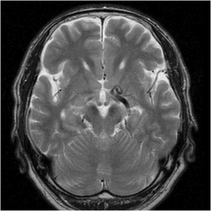
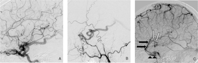
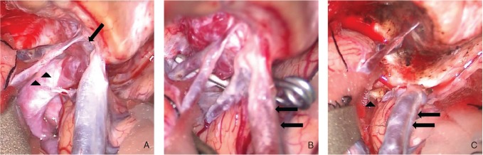
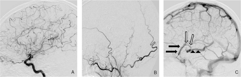
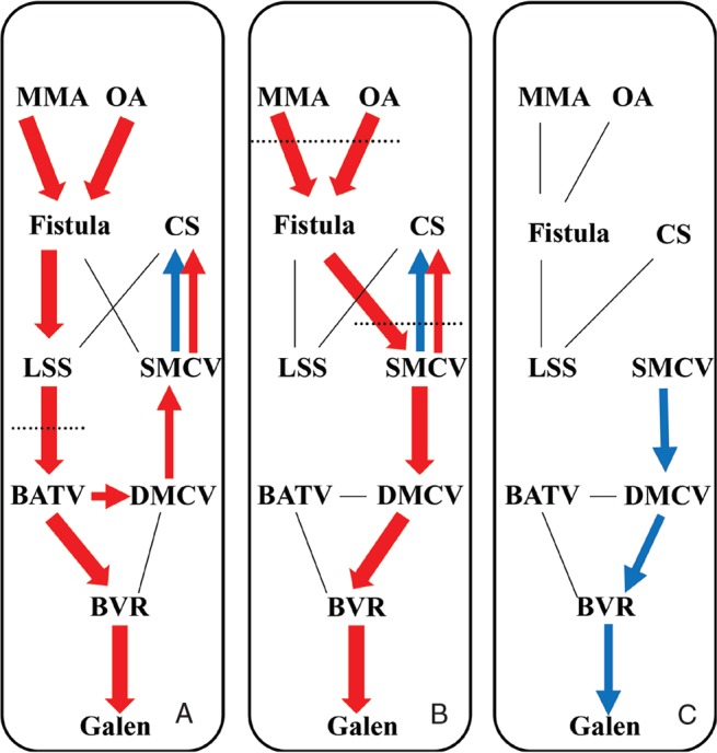
A 69-year-old man consulted our department regarding further examination because abnormal venous ectasia of the basal vein of Rosenthal (BVR) was accidentally found on magnetic resonance imaging. Angiography revealed a dural arteriovenous fistula (DAVF) in the left sphenoid wing; the fistula was supplied by the ophthalmic artery and the middle meningeal artery. Venous drainage from the fistula consisted of a large collector vein located in the temporal tip, which drained into the BVR. We selected open surgery because this lesion may be difficult to access and is associated with significant risks through an endovascular approach. After temporal clip ligation of the drainer, the superficial middle cerebral vein became the main drainage route directly in connection with the fistula. Therefore, both were ligated, after coagulation of feeding arterial networks on the dura around the sphenoid wing. The patient experienced no complications from the surgical procedure, and postoperative angiography demonstrated obliteration of the fistula. DAVF in the sphenoid wing with deep drainage is believed to carry a high risk of hemorrhage or venous infarction because of the presence of Galenic drainage, varix, and cortical venous reflux. Treatment is strongly recommended even if the symptoms are minimal. Open surgery appears to be safe and often the best therapeutic option.
Keywords: basal vein of Rosenthal; dural arteriovenous fistula; sphenoid wing; surgery.
Conflict of interest statement
Conflicts of Interest Disclosure The authors have no personal, financial, or institutional interest in any of the drugs, materials, or devices in the article. All authors who are members of the Japan Neurosurgical Society (JNS) have registered online Self-reported COI Disclosure Statement Forms through the website for JNS members.
Figures
Similar articles
-
Sphenoid Wing Dural Arteriovenous Fistulas.J Neuroendovasc Ther. 2025;19(1):2023-0034. doi: 10.5797/jnet.ra.2023-0034. Epub 2023 Dec 23. J Neuroendovasc Ther. 2025. PMID: 40018285 Free PMC article. Review.
-
Spontaneous obliteration of a greater sphenoid wing dural arteriovenous fistula involving the diploic venous system.Surg Neurol Int. 2025 Mar 21;16:99. doi: 10.25259/SNI_1113_2024. eCollection 2025. Surg Neurol Int. 2025. PMID: 40206771 Free PMC article.
-
Aberrant venous drainage pattern in a medial sphenoid wing dural arteriovenous fistula: a case report and review of the literature.World Neurosurg. 2013 Dec;80(6):e381-6. doi: 10.1016/j.wneu.2013.02.022. Epub 2013 Feb 9. World Neurosurg. 2013. PMID: 23403353
-
Dural Arteriovenous Fistula of the Sinus of the Lesser Sphenoid Wing Presenting with Pontine Hemorrhage.World Neurosurg. 2017 Feb;98:871.e17-871.e21. doi: 10.1016/j.wneu.2016.11.139. Epub 2016 Dec 6. World Neurosurg. 2017. PMID: 27931945
-
Spontaneous Resolution of Venous Aneurysms After Transarterial Embolization of a Variant Superior Sagittal Sinus Dural Arteriovenous Fistula: Case Report and Literature Review.Neurologist. 2017 Sep;22(5):186-195. doi: 10.1097/NRL.0000000000000137. Neurologist. 2017. PMID: 28859024 Review.
Cited by
-
Role of the ophthalmic artery in the endovascular treatment for intracranial vascular diseases.Acta Neurol Belg. 2021 Apr;121(2):321-330. doi: 10.1007/s13760-020-01576-z. Epub 2021 Jan 5. Acta Neurol Belg. 2021. PMID: 33400225 Review.
-
Sphenoid Wing Dural Arteriovenous Fistulas.J Neuroendovasc Ther. 2025;19(1):2023-0034. doi: 10.5797/jnet.ra.2023-0034. Epub 2023 Dec 23. J Neuroendovasc Ther. 2025. PMID: 40018285 Free PMC article. Review.
-
Spontaneous obliteration of a greater sphenoid wing dural arteriovenous fistula involving the diploic venous system.Surg Neurol Int. 2025 Mar 21;16:99. doi: 10.25259/SNI_1113_2024. eCollection 2025. Surg Neurol Int. 2025. PMID: 40206771 Free PMC article.
-
Trans-Galen Approach for Embolization of Sphenoparietal Sinus Dural Arteriovenous Fistulas.Neurointervention. 2025 Jul;20(2):114-118. doi: 10.5469/neuroint.2025.00094. Epub 2025 Mar 12. Neurointervention. 2025. PMID: 40068630 Free PMC article.
-
Sphenoid dural arteriovenous fistulas.Neurosurg Rev. 2021 Feb;44(1):77-96. doi: 10.1007/s10143-019-01209-x. Epub 2019 Dec 7. Neurosurg Rev. 2021. PMID: 31811518 Review.
References
-
- Osbun JW, Kim LJ, Spetzler RF, McDougall CG: Aberrant venous drainage pattern in a medial sphenoid wing dural arteriovenous fistula: a case report and review of the literature. World Neurosurg 80: e381– e386, 2013. - PubMed
-
- Fukuda H, Miyake K, Kunieda T, Murao K: Endovascular treatment of sphenoid wing dural arteriovenous fistula with pure cortical venous drainage. J Stroke Cerebrovasc Dis 23: 1730– 1735, 2014. - PubMed
-
- Kakarla UK, Deshmukh VR, Zabramski JM, Albuquerque FC, McDougall CG, Spetzler RF: Surgical treatment of high-risk intracranial dural arteriovenous fistulae: clinical outcomes and avoidance of complications. Neurosurgery 61: 447– 457; discussion 457–459, 2007. - PubMed
-
- Thompson BG, Doppman JL, Oldfield EH: Treatment of cranial dural arteriovenous fistulae by interruption of leptomeningeal venous drainage. J Neurosurg 80: 617– 623, 1994. - PubMed
Publication types
LinkOut - more resources
Full Text Sources