Neuroendoscopy via an Extremely Narrow Foramen of Monro: A Case Report
- PMID: 28664024
- PMCID: PMC5364906
- DOI: 10.2176/nmccrj.cr.2016-0157
Neuroendoscopy via an Extremely Narrow Foramen of Monro: A Case Report
Abstract
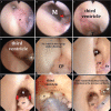
Herein, safe and reliable neuroendoscopic biopsy via an extremely narrow foramen of Monro (ENFM) for a non-hydrocephalic patient with hypothalamic and pineal region tumors was successfully applied. A 17-year-old boy presented with hypothalamic manifestations attributed to hypothalamic and pineal region tumors. Small ventricles were seen. Intraoperatively, to advance different diameter steerable fiberscopes via ENFM, the third ventricle was flushed to induce a moment increase in the intraventricular pressure with subsequent dilatation of FM. Postoperative course was uneventful. Histopathological studies revealed a yolk sac tumor. Adjuvant therapy was applied. Follow-up neuroimaging disclosed marvellous improvement of the condition. His symptoms gradually improved.
Keywords: endoscopic biopsy; fiberscope; foramen of Monro; pineal tumor; small ventricle.
Conflict of interest statement
Conflicts of Interest Disclosure The authors have no personal, financial, or institutional interest in any of the drugs, materials, or devices in the article. All authors who are members of the Japan Neurosurgical Society (JNS) have registered online Self-reporting Conflict Disclosure Statement Forms through the website for JNS members.
Figures


Similar articles
-
Endoscopic biopsy of foramen of Monro and third ventricle lesions guided by frameless neuronavigation: usefulness and limitations.Clin Neurol Neurosurg. 2009 Sep;111(7):579-82. doi: 10.1016/j.clineuro.2009.04.008. Epub 2009 May 26. Clin Neurol Neurosurg. 2009. PMID: 19473754
-
Intraventricular tumors.World Neurosurg. 2013 Feb;79(2 Suppl):S17.e15-9. doi: 10.1016/j.wneu.2012.02.023. Epub 2012 Feb 10. World Neurosurg. 2013. PMID: 22381840 Review.
-
An anatomical study of the foramen of Monro: implications in management of pineal tumors presenting with hydrocephalus.Acta Neurochir (Wien). 2019 May;161(5):975-983. doi: 10.1007/s00701-019-03887-4. Epub 2019 Apr 5. Acta Neurochir (Wien). 2019. PMID: 30953154
-
Endoscopic stent placement for treatment of secondary bilateral occlusion of the Monro foramina following endoscopic third ventriculostomy in a patient with aqueductal stenosis. Case report.J Neurosurg. 2007 Aug;107(2):416-20. doi: 10.3171/JNS-07/08/0416. J Neurosurg. 2007. PMID: 17695399
-
Neuroendoscopic management of intraventricular germinoma at the foramen of Monro: case report and review of the literature.Minim Invasive Neurosurg. 2011 Aug;54(4):191-5. doi: 10.1055/s-0031-1285887. Epub 2011 Sep 15. Minim Invasive Neurosurg. 2011. PMID: 21922450 Review.
Cited by
-
A preliminary study of the diagnostic efficacy and safety of the novel boring biopsy for brain lesions.Sci Rep. 2022 Mar 14;12(1):4387. doi: 10.1038/s41598-022-08366-y. Sci Rep. 2022. PMID: 35288608 Free PMC article.
-
Watertight Robust Osteoconductive Barrier for Complex Skull Base Reconstruction: An Expanded-endoscopic Endonasal Experimental Study.Neurol Med Chir (Tokyo). 2019 Mar 15;59(3):79-88. doi: 10.2176/nmc.oa.2018-0262. Epub 2019 Feb 21. Neurol Med Chir (Tokyo). 2019. PMID: 30787233 Free PMC article.
-
Multidisciplinary treatment of primary intracranial yolk sac tumor: A case report and literature review.Medicine (Baltimore). 2021 May 14;100(19):e25778. doi: 10.1097/MD.0000000000025778. Medicine (Baltimore). 2021. PMID: 34106610 Free PMC article. Review.
References
-
- Cappabianca P, Cinalli G, Gangemi M, Brunori A, Cavallo LM, de Divitiis E, Decq P, Delitala A, Di Rocco F, Frazee J, Godano U, Grotenhuis A, Longatti P, Mascari C, Nishihara T, Oi S, Rekate H, Schroeder HW, Souweidane MM, Spennato P, Tamburrini G, Teo C, Warf B, Zymberg ST: Application of neuroendoscopy to intraventricular lesions. Neurosurgery 62: 575– 597, 2008. - PubMed
-
- Song JH, Kong DS, Shin HJ: Feasibility of neuroendoscopic biopsy of pediatric brain tumors. Childs Nerv Syst 26: 1593– 1598, 2010. - PubMed
-
- Schroeder HW, Wagner W, Tschiltschke W, Gaab MR: Frameless neuronavigation in intracranial endoscopic neurosurgery. J Neurosurg 94: 72– 79, 2001. - PubMed
-
- Ogiwara T, Goto T, Aoyama T, Nagm A, Yamamoto Y, Hongo K: Bony surface registration of navigation system in the lateral or prone position: technical note. Acta Neurochir (Wien) 157: 2017– 2022, 2015. - PubMed
-
- Hayashi N, Murai H, Ishihara S, Kitamura T, Miki T, Miwa T, Miyajima M, Nishiyama K, Ohira T, Ono S, Suzuki T, Takano S, Date I, Saeki N, Endo S: Nationwide investigation of the current status of therapeutic neuroendoscopy for ventricular and paraventriculartumors in Japan. J Neurosurg 115: 1147– 1157, 2011. - PubMed
Publication types
LinkOut - more resources
Full Text Sources
Other Literature Sources
