. 2017 Jun 13:15:633-643.
doi: 10.1016/j.nicl.2017.06.016. eCollection 2017.

Fully automatic acute ischemic lesion segmentation in DWI using convolutional neural networks

Affiliations

Fully automatic acute ischemic lesion segmentation in DWI using convolutional neural networks

Liang Chen et al. Neuroimage Clin. .

Abstract

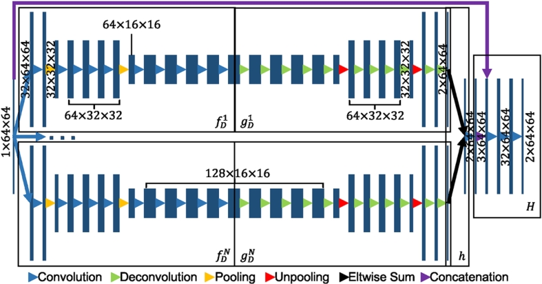
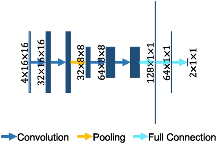
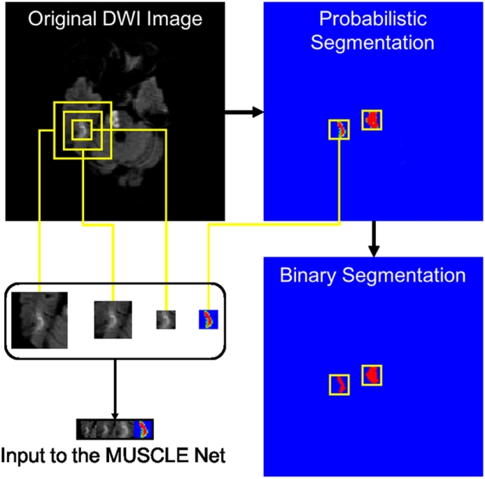
Stroke is an acute cerebral vascular disease, which is likely to cause long-term disabilities and death. Acute ischemic lesions occur in most stroke patients. These lesions are treatable under accurate diagnosis and treatments. Although diffusion-weighted MR imaging (DWI) is sensitive to these lesions, localizing and quantifying them manually is costly and challenging for clinicians. In this paper, we propose a novel framework to automatically segment stroke lesions in DWI. Our framework consists of two convolutional neural networks (CNNs): one is an ensemble of two DeconvNets (Noh et al., 2015), which is the EDD Net; the second CNN is the multi-scale convolutional label evaluation net (MUSCLE Net), which aims to evaluate the lesions detected by the EDD Net in order to remove potential false positives. To the best of our knowledge, it is the first attempt to solve this problem and using both CNNs achieves very good results. Furthermore, we study the network architectures and key configurations in detail to ensure the best performance. It is validated on a large dataset comprising clinical acquired DW images from 741 subjects. A mean accuracy of Dice coefficient obtained is 0.67 in total. The mean Dice scores based on subjects with only small and large lesions are 0.61 and 0.83, respectively. The lesion detection rate achieved is 0.94.

Keywords: Acute ischemic lesion segmentation; Convolutional neural networks; DWI; Deep learning.


Figures

Fig. 1
Fig. 1
Examples of acute ischemic lesions in DWI. The red circles indicate the acute ischemic lesions and the yellow ones show the artefacts. (For interpretation of the references to color in this figure legend, the reader is referred to the web version of this article.)
Fig. 2
Fig. 2
The overview of the proposed CNN based system to segment the acute ischemic lesions in DWI. It comprises the EDD Net and the MUSCLE Net. The EDD Net conducts the semantic segmentation on the input DWI. Based on the output of the EDD Net, patches containing small lesions are extracted and they are evaluated by the MUSCLE Net so that many false positives are removed. The refined segmentation is therefore obtained.
Fig. 3
Fig. 3
The architecture of the proposed EDD Net. The rectangles in different sizes indicate data blobs in different sizes. The height shows the size of each piece of data, e.g. 64 × 64. The width shows the number of data pieces in each blob, e.g. 1, 32. Arrows in difference colors stand for different operations. (For interpretation of the references to color in this figure legend, the reader is referred to the web version of this article.)
Fig. 4
Fig. 4
The max pooling and unpooling strategy demonstrated in the DeconvNet approach (Noh et al., 2015). In the pooling stage, the position of the maximum activation is recorded within each filter window by a mask. In the unpooling stage, the entries are placed in the unpooled map according to the mask.
Fig. 5
Fig. 5
The architecture of the MUSCLE Net. The rectangles stand for the data blobs. Their heights represent the sizes of data pieces, e.g. 16 × 16. Their widths show the number of data pieces in the blobs, e.g. 4, 32. In the fully connected layers, the lengths of strings demonstrate the number of elements in the layers. Arrows in different colors show different operations. (For interpretation of the references to color in this figure legend, the reader is referred to the web version of this article.)
Fig. 6
Fig. 6
The derivation of the input to the MUSCLE Net. The probabilistic segmentation is obtained from the EDD Net. The binary segmentation is obtained by thresholding the probabilistic segmentation. Candidate small blobs are detected in the binary segmentation. The corresponding patches are extracted in the original DWI in multiple scales and the probabilistic segmentation map. They are then resized and concatenated resulting in the input to the MUSCLE Net.
Fig. 7
Fig. 7
The statistics of the false positives on the validation dataset provided by the EDD Net.
Fig. 8
Fig. 8
The results of the proposed method. The first column shows the original DWI. The second column displays the manual annotations of the acute ischemic lesions. The third column demonstrates the results given by the EDD Net. The last column illustrates the lesion segmentations refined by the MUSCLE Net.

References

    1. Avendi M., Kheradvar A., Jafarkhani H. A combined deep-learning and deformable-model approach to fully automatic segmentation of the left ventricle in cardiac MRI. Med. Image Anal. 2016;30:108–119. - PubMed
    1. Bengio Y., Courville A., Vincent P. Representation learning: a review and new perspectives. IEEE Trans. Pattern Anal. Mach. Intell. 2013;35(8):1798–1828. - PubMed
    1. Chang P.D. Proceedings of MICCAI-BRATS 2016 Multimodal Brain Tumor Image Segmentation Benchmark: “Change Detection”. 2016. Fully convolutional neural networks with hyperlocal features for brain tumor segmentation; pp. 4–9.
    1. Charoensuk W., Covavisaruch N., Lerdlum S., Likitjaroen Y. Acute stroke brain infarct segmentation in DWI images. Int. J. Pharm. Med. Biol. Sci. 2015;4:115.
    1. Chen H., Qi X., Yu L., Heng P.A. 2016. DCAN: Deep Contour-Aware Networks for Accurate Gland Segmentation. (arXiv preprint arXiv:1604.02677) - PubMed