

Evaluation of cytotoxic activity of platinum nanoparticles against normal and cancer cells and its anticancer potential through induction of apoptosis
- PMID: 28664137
- PMCID: PMC5478255
- DOI: 10.1016/j.imr.2017.01.006
Evaluation of cytotoxic activity of platinum nanoparticles against normal and cancer cells and its anticancer potential through induction of apoptosis
Abstract
Background: Plant mediated green synthesis of nanoparticles is an eco-friendly and efficacious approach which finds immense application in the field of medicine. This study aimed to evaluate the cytotoxicity of platinum nanoparticles (ptNPs) synthesized through green technology against normal and different cancer cell lines.
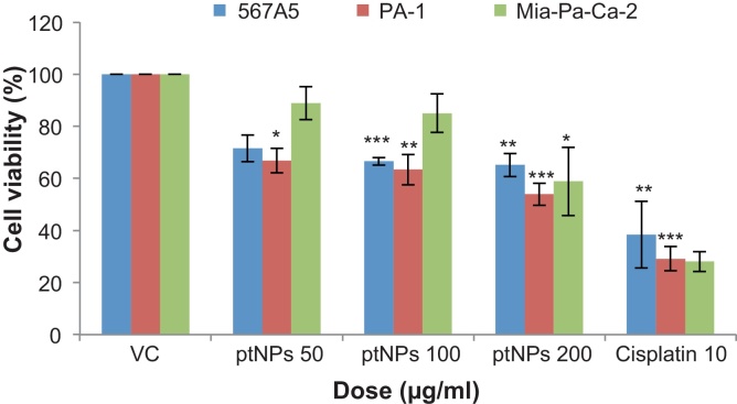
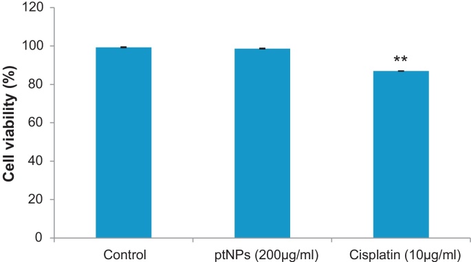
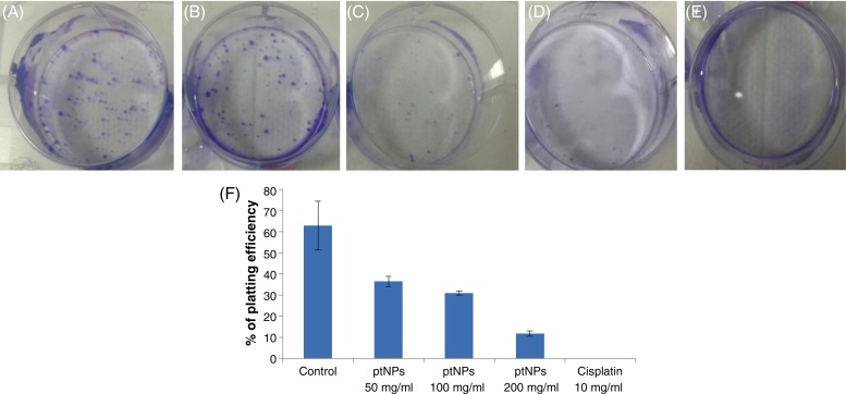
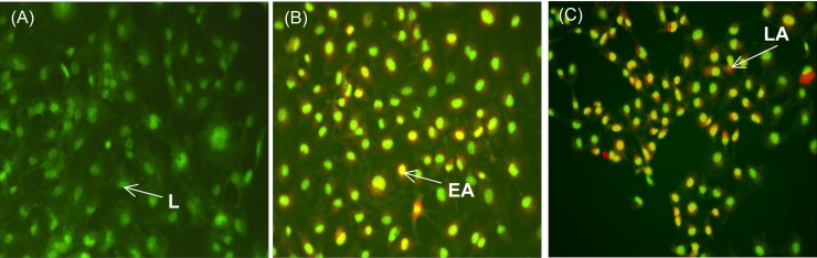
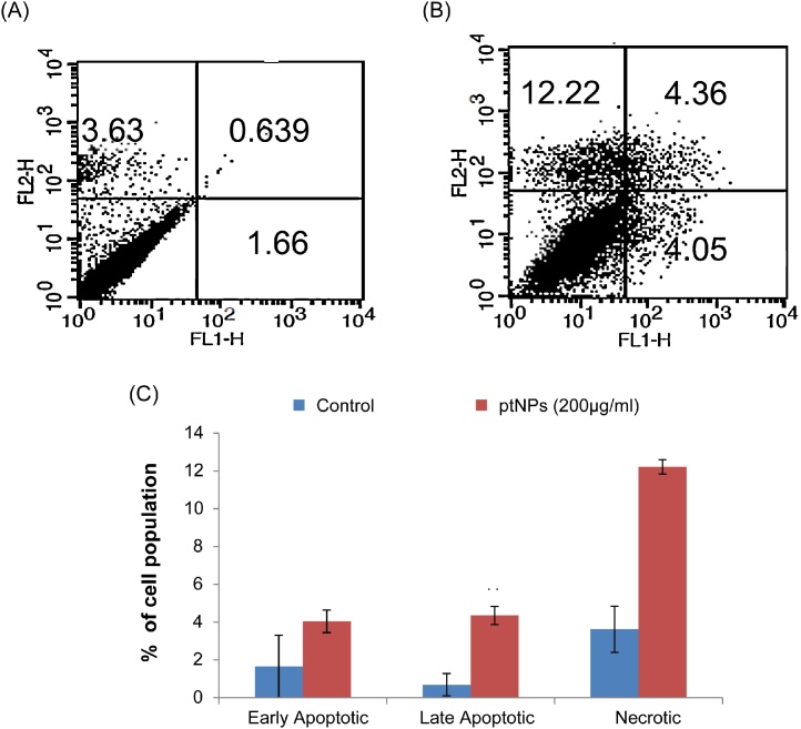
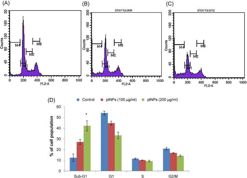
Methods: Platinum nanoparticles were synthesized by green technology and characterized earlier. In this study we examined the cytotoxic effect of platinum nanoparticles (ptNPs) on human lung adenocarcinoma (A549), ovarian teratocarcinoma (PA-1), pancreatic cancer (Mia-Pa-Ca-2) cells and normal peripheral blood mononucleocyte (PBMC) cells and evaluate anticancer potential through induction of apoptosis on PA-1 cells if any. Cytotoxicity was evaluated using MTT assay, trypan blue dye exclusion assay and anticancer potential assessed through clonogenic assay, apoptosis assay, cell cycle analysis.
Results: We found that ptNPs exerted cytotoxic effect on cancer cell lines, whereas no cytotoxic effect was observed at highest dose on normal cells. The results showed that ptNPs had potent anticancer activities against PA-1 cell line via induction of apoptosis and cell cycle arrest.
Conclusion: Overall, these findings have proved that biosynthesized ptNPs could be potent anti-ovarian cancer drugs. Further studies are required to elucidate the molecular mechanism of ptNPs induced anti-tumor effect in vivo.
Keywords: Anti-cancer potential; Apoptosis; Cytotoxicity; In vitro; Platinum nanoparticles.
Figures
References
-
- Henglein A. Small-particle research: physicochemical properties of extremely small colloidal metal and semiconductor particles. Chem Rev. 1989;89:1861.
-
- Oggawa S., Hayashi Y., Kobayashi N., Tokizakiand T., Nakamura A. Novel preparation method of metal particles dispersed in polymer films and their third-order optical nonlinearities. Jpn J Appl Phys. 1994;33:331.
-
- Chen X., Schluesener H.J. Nanosilver: a nanoproduct in medical application. Toxicol Lett. 2008;4:176. - PubMed
-
- Stephens I.E.L., Bondarenko A.S., Grønbjerg U., Rossmeisl J., Chorkendorff I. Understanding the electrocatalysis of oxygen reduction on platinum and its alloys. Energy Environ Sci. 2012;5:6744–6762.
-
- Kostova I. Platinum complexes as anticancer agents. Recent Pat Anti-Cancer Drug Discov. 2006;1:1–22. - PubMed
LinkOut - more resources
Full Text Sources
Other Literature Sources
