Review
. 2017 Sep;174(18):2967-2983.
doi: 10.1111/bph.13936. Epub 2017 Aug 10.

The macrolide antibiotic renaissance

Affiliations
Review

The macrolide antibiotic renaissance

George P Dinos. Br J Pharmacol. 2017 Sep.

Abstract

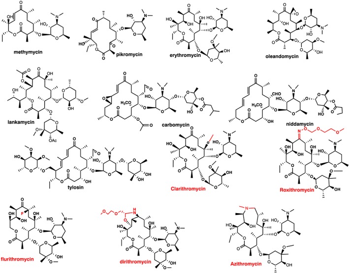
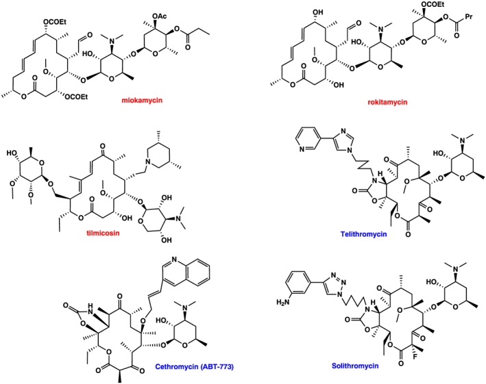
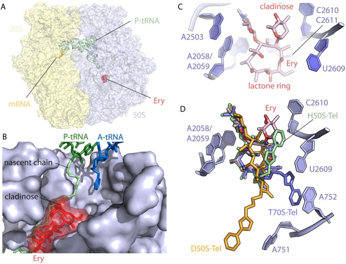
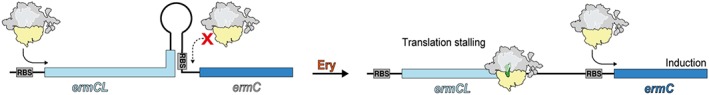
Macrolides represent a large family of protein synthesis inhibitors of great clinical interest due to their applicability to human medicine. Macrolides are composed of a macrocyclic lactone of different ring sizes, to which one or more deoxy-sugar or amino sugar residues are attached. Macrolides act as antibiotics by binding to bacterial 50S ribosomal subunit and interfering with protein synthesis. The high affinity of macrolides for bacterial ribosomes, together with the highly conserved structure of ribosomes across virtually all of the bacterial species, is consistent with their broad-spectrum activity. Since the discovery of the progenitor macrolide, erythromycin, in 1950, many derivatives have been synthesised, leading to compounds with better bioavailability and acid stability and improved pharmacokinetics. These efforts led to the second generation of macrolides, including well-known members such as azithromycin and clarithromycin. Subsequently, in order to address increasing antibiotic resistance, a third generation of macrolides displaying improved activity against many macrolide resistant strains was developed. However, these improvements were accompanied with serious side effects, leading to disappointment and causing many researchers to stop working on macrolide derivatives, assuming that this procedure had reached the end. In contrast, a recent published breakthrough introduced a new chemical platform for synthesis and discovery of a wide range of diverse macrolide antibiotics. This chemical synthesis revolution, in combination with reduction in the side effects, namely, 'Ketek effects', has led to a macrolide renaissance, increasing the hope for novel and safe therapeutic agents to combat serious human infectious diseases.


Figures

Figure 1
Figure 1
Macrolide structures. First generation: 12‐membered (methymycin), 14‐membered (pikromycin, erythromycin, oleandomycin and lankamycin) and 16‐membered (carbomycin, niddamycin and tylosin), all natural products. Second generation: 14‐membered (clarithromycin, roxithromycin, flurithromycin dirithromycin) and 15‐membered (azithromycin). Red colour indicates modifications inserted in the erythromycin molecule to generate the second generation of 14‐ and 15‐membered macrolides.
Figure 2
Figure 2
Macrolides structure. Second generation: 16‐membered (miokamycin, rokitamycin and tilmicosin). Third generation: ketolides: Telithromycin, cethromycin and solithromycin. Red and blue colour indicates second‐ and third‐generation macrolides respectively.
Figure 3
Figure 3
Interaction of macrolides with the ribosome: (A) Overview of the antibiotic erythromycin (Ery, red) bound to the 70S E. coli ribosome (PDB entry 4V7U; Dunkle et al., 2010). (B) Close‐up view of the erythromycin binding site in the ribosomal exit tunnel in the presence of the P‐site peptidyl‐tRNA (green) and the A‐site tRNA (blue) (prepared with modifications from Wilson 2014). (C) Erythromycin binding pocket within the E. coli 70S ribosome is located adjacent to bases A2058, A2059, A2503 and U2609. The desosamine amino sugar of Ery at position 5 of the lactone ring contains a dimethyl amine that makes pivotal contact with the base A2058 (PDB entry 4V7U; Dunkle et al., 2010). (D) Superposition of telithromycin (Tel) bound to the ribosomes from different species. All structures of ribosome‐bound Tel were aligned based on domain V of the 23S rRNA. Note that although the lactone rings almost perfectly match in all cases, the position of the alkyl–aryl groups varies significantly depending on the species. Shown are Haloarcula marismortui (green, PDB entry 1YIJ; Tu et al., 2005), D. radiodurans (orange, PDB entry 1P9X; Berisio et al., 2003) and T. thermophilus (blue, PDB entry 4V7Z; Bulkley et al., 2010). All figures were prepared using PyMol software.
Figure 4
Figure 4
Regulation of gene expression by ribosomal stalling. In the absence of erythromycin, there is no stalling and no translation of ermC. In the presence of erythromycin, there is stalling during translation of the leader peptide ermCL, which causes the ribosome to block stem‐loop formation and exposes the ribosome binding site (RBS) of the downstream cistrons, allowing its expression. (Figure was prepared with modifications from Wilson et al., 2016).

Similar articles

Cited by

References

    1. Akshay S, Bertea M, Hobbie SN, Oettinghaus B, Shcherbakov D, Böttger EC et al. (2011). Phylogenetic sequence variations in bacterial rRNA affect species‐specific susceptibility to drugs targeting protein synthesis. Antimicrob Agents Chemother 55: 4096–4102. - PMC - PubMed
    1. Alekshun MN, Levy SB (2007). Molecular mechanisms of antibacterial multidrug resistance. Cell 128: 1037–1050. - PubMed
    1. Alexander SPH, Fabbro D, Kelly E, Marrion N, Peters JA, Benson HE et al. (2015a). The Concise Guide to PHARMACOLOGY 2015/16: Enzymes. Br J Pharmacol 172: 6024–6109. - PMC - PubMed
    1. Alexander SPH, Peters JA, Kelly E, Marrion N, Benson HE, Faccenda E et al. (2015b). The Concise Guide to PHARMACOLOGY 2015/16: Ligand‐gated ion channels. Br J Pharmacol 172: 5870–5903. - PMC - PubMed
    1. Almutairi MM, Park SR, Rose S, Hansen DA, Vazquez‐Laslop N, Douthwaite S et al. (2015). Resistance to ketolide antibiotics by coordinated expression of rRNA methyltransferases in a bacterial producer of natural ketolides. Proc Natl Acad Sci U S A 112: 12956–12961. - PMC - PubMed

MeSH terms