. 2017 Jun;98(3):134-146.
doi: 10.1111/iep.12232. Epub 2017 Jun 29.

Corticosterone influences gerbil (Meriones unguiculatus) prostatic morphophysiology and alters its proliferation and apoptosis rates

Affiliations

Corticosterone influences gerbil (Meriones unguiculatus) prostatic morphophysiology and alters its proliferation and apoptosis rates

Julia Quilles Antoniassi et al. Int J Exp Pathol. 2017 Jun.

Abstract

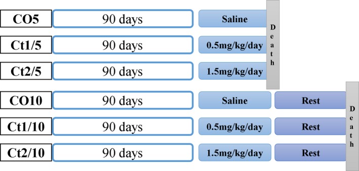
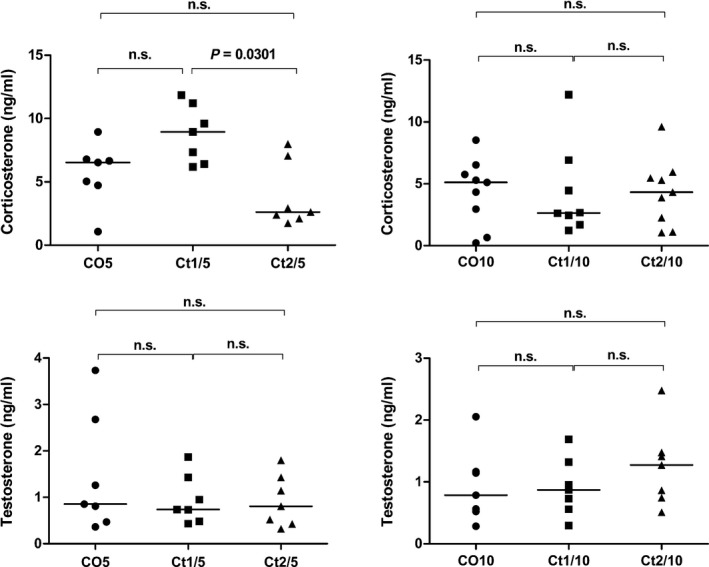
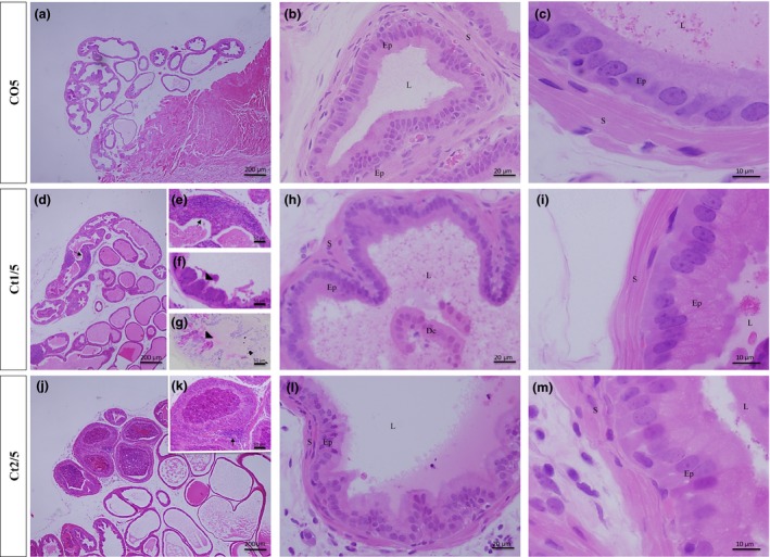
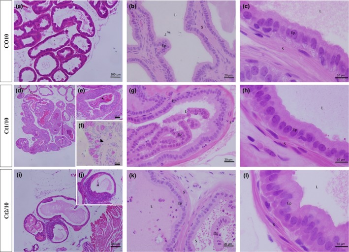
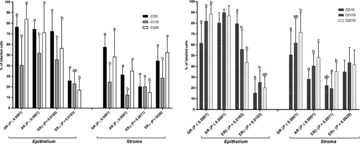
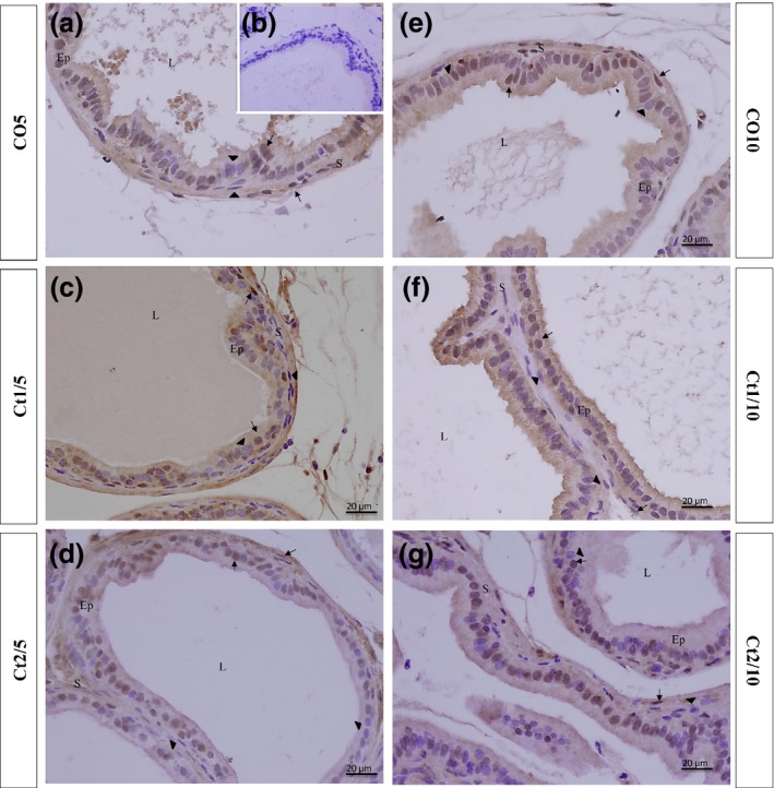
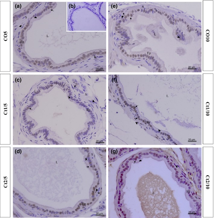
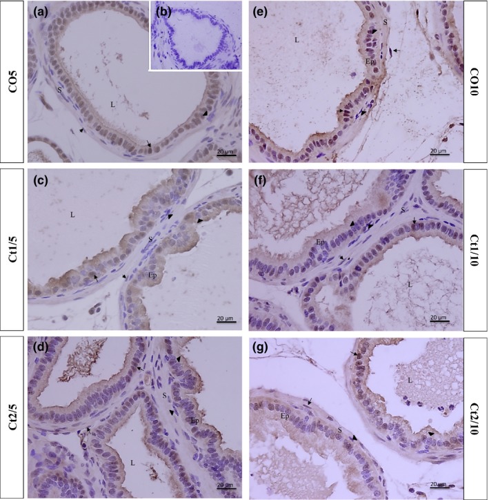
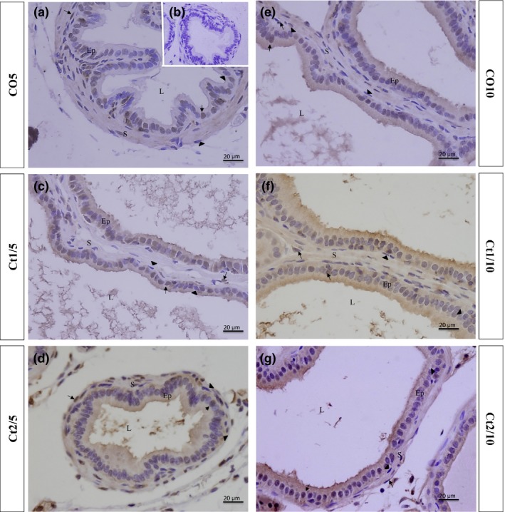
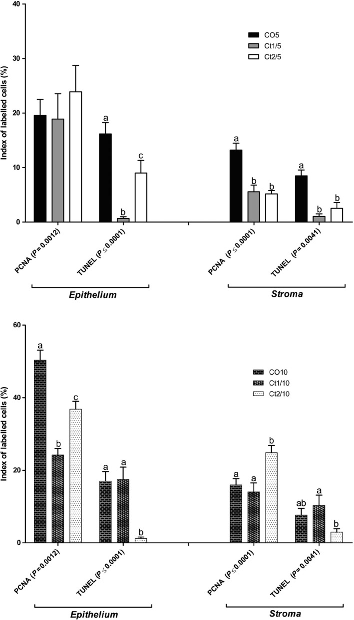
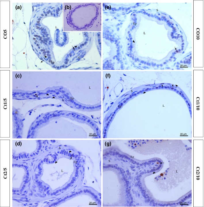
Glucocorticoids (GCs) are hormones that are widely used in medicine; but although side effects are generally recognised, little is known about the precise mechanisms that is implicated in many of these side effects. Furthermore, GCs are highly correlated with stress and behaviour disorders. This study evaluated the effects of the glucocorticoid corticosterone on the ventral prostate of the Mongolian gerbil. Male gerbils (Meriones unguiculatus) (n = 5) received intraperitoneal injections of saline or corticosterone in doses of 0.5 mg/kg/day and 1.5 mg/kg/day for 5 days; while some of the animals were killed immediately after the treatment, the others were killed 5 days after the treatment period. The data show that corticosterone influences the structure and functionality of this organ. This hormone has anti-proliferative and anti-apoptotic properties in the prostate. In addition, the frequencies of the androgen (AR), oestrogen (ERα, ERβ) and glucocorticoid (GR) receptors changed. The frequencies of AR, GR and ERβ decreased in the Ct1/5 group; in the groups with rest period, the frequencies of GR increased and ERβ decreased in the epithelium. Changes in the proliferative index, apoptotic index and receptor activity may have contributed to the emergence of prostatic morphological alterations, such as the presence of cellular debris and inflammatory cells. Different doses of corticosterone had variable effects on the prostate, with a higher dose showing subtler effects and a lower dose showing more striking effects. The corticosterone effects on nuclear receptors were reverted or attenuated after a rest period, which was not observed for proliferation and apoptosis. In summary, we have demonstrated that corticosterone might influence the prostatic morphophysiology and that these changes may be linked in some way to the altered receptor distribution.

Keywords: corticosterone; gerbil; glucocorticoids; ventral prostate.


Figures

Figure 1
Figure 1
Schematic representation of treatments and experimental groups.
Figure 2
Figure 2
Serum testosterone (ng/ml) and corticosterone (ng/ml) levels. Different letters indicate significant differences between groups. Two‐way anova showed a significant dose vs. rest period interaction (= 0.0108) for corticosterone levels (Bonferroni post‐tests,  0.05).
Figure 3
Figure 3
Histologic sections of prostate of gerbils, stained with haematoxylin and eosin. Morphological alterations in the treated groups can be observed, such as cell debris. Note an increase in epithelial compartment height in the Ct1/5 (d–i), Ct2/5 (j–m) groups and a decrease in stromal compartment height in the Ct1/5 (d–i) and Ct2/5 (j–m) groups. Legend: Ep – epithelium; S – stroma; L – lumen; Dc – cellular debris; arrow – inflammatory cells; arrowhead – atypical cells.
Figure 4
Figure 4
Histologic sections of prostate of gerbils, stained with haematoxylin and eosin. Epithelial compartment height increased in the Ct2/10 group (i–l) and the stromal compartment increased in the Ct2/10 group (i–l). Legend: Ep – epithelium; S – stroma; L – lumen; Dc – cellular debris; arrow – inflammatory cells; arrowhead – atypical cells.
Figure 5
Figure 5
Quantification of cells immunostained for GR, AR, ERβ and ERα of the different experimental groups (= 5). The values shown are the average and standard deviation. Two‐way anova showed a significant dose vs. rest period interaction for all receptors analysed in the epithelial and stromal compartments (P value presented on figure). The letters a and b represent the significant differences between the groups (Bonferroni post‐tests,  0.05).
Figure 6
Figure 6
Histologic sections of the prostate of gerbils subjected to immunostaining for anti‐GR. Counterstaining: Harris haematoxylin. B: negative control. EP: alveolar epithelium; L: alveolar lumen; S: prostatic stroma. Arrow: GR‐positive cell; arrowhead: GR‐negative cell.
Figure 7
Figure 7
Histologic sections of the prostate of gerbils subjected to immunostaining for anti‐AR. Counterstaining: Harris haematoxylin. B: negative control. EP: alveolar epithelium; L: alveolar lumen; S: prostatic stroma. Arrow: AR‐positive cell; arrowhead: AR‐negative cell.
Figure 8
Figure 8
Histologic sections of the prostate of gerbils subjected to immunostaining for anti‐ERβ. Counterstaining: Harris haematoxylin. B: negative control. EP: alveolar epithelium; L: alveolar lumen; S: prostatic stroma. Arrow: ERβ‐positive cell; arrowhead: ERβ‐negative cell.
Figure 9
Figure 9
Histologic sections of the prostate of gerbils subjected to immunostaining for anti‐ERα. Counterstaining: Harris haematoxylin. B: negative control. EP: alveolar epithelium; L: alveolar lumen; S: prostatic stroma. Arrow: ERα‐positive cell; arrowhead: ERα‐negative cell.
Figure 10
Figure 10
Quantification of cells immunostained for proliferation and apoptosis of the different experimental groups (= 5). The values shown are the average and standard error. Two‐way anova showed a significant dose vs. rest period interaction (P value presented on figure) for proliferation and apoptosis. The letters a, b and c represent the significant differences between the groups (Bonferroni post‐tests,  0.05).
Figure 11
Figure 11
Histologic sections of the prostate of gerbils subjected to immunostaining for proliferating cells (PCNA). B: negative control. EP: alveolar epithelium; L: alveolar lumen; S: prostatic stroma. Arrow: immunostained cell; arrowhead: unstained cell.
Figure 12
Figure 12
Histologic sections of the prostate of gerbils subjected to immunostaining for apoptotic cells (TUNEL). Counterstaining: Harris haematoxylin. B: negative control. EP: alveolar epithelium; L: alveolar lumen; S: prostatic stroma. Arrow: immunostained cell; arrowhead: unstained cell.

Similar articles

Cited by

References

    1. Andersen ML, Ribeiro DA, Bergamaschi CT et al (2009) Distinct effects of acute and chronic sleep loss on DNA damage in rats. Prog.Neuropsychopharmacol. Biol. Psychiatry 33, 562–567. - PubMed
    1. Behmer A.O., Tolosa E.M.C. & Neto A.G.F. (1976) Manual de praticas para histologia normal e patologica. São Paulo, Brazil: Edart‐Edusp.
    1. Biddie S.C., Conway‐Campbell B.L. & Lightman S.L. (2011) Dynamic regulation of glucocorticoid signalling in health and disease. Rheumatology 51. - PMC - PubMed
    1. Bland R., Worker C.A., Noble B.S. et al (1999) Characterization of 11b‐hydroxysteroid dehydrogenase activity and corticosteroid receptor expression in human osteosarcoma cell lines. J. Endocrinol. 161, 455–464. - PubMed
    1. Bruni‐Cardoso A., Vilamaior P.S., Taboga S.R. & Carvalho H.F. (2008) Localized matrix metalloproteinase (MMP)‐2 and MMP‐9 activity in the rat ventral prostate during the first week of postnatal development. Histochem. Cell Biol. 129(6), 805–815. - PubMed

Publication types

LinkOut - more resources