. 2017 Jun 30;7(7):162.
doi: 10.3390/nano7070162.

Biological Fate of Fe₃O₄ Core-Shell Mesoporous Silica Nanoparticles Depending on Particle Surface Chemistry

Affiliations

Biological Fate of Fe₃O₄ Core-Shell Mesoporous Silica Nanoparticles Depending on Particle Surface Chemistry

Estelle Rascol et al. Nanomaterials (Basel). .

Abstract

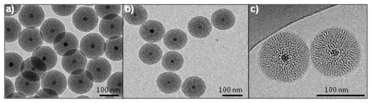
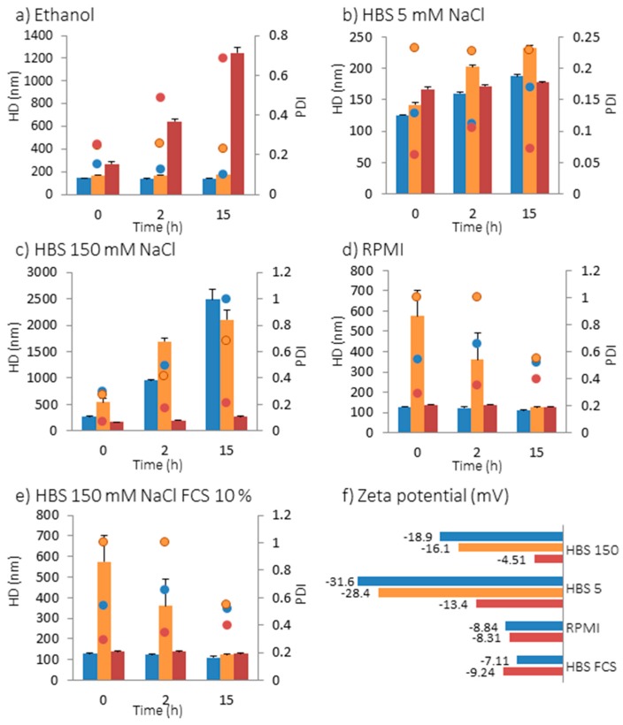
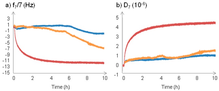
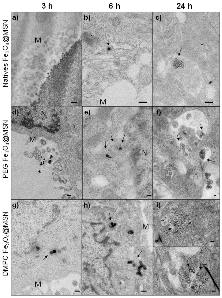
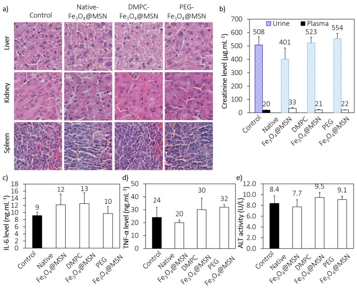
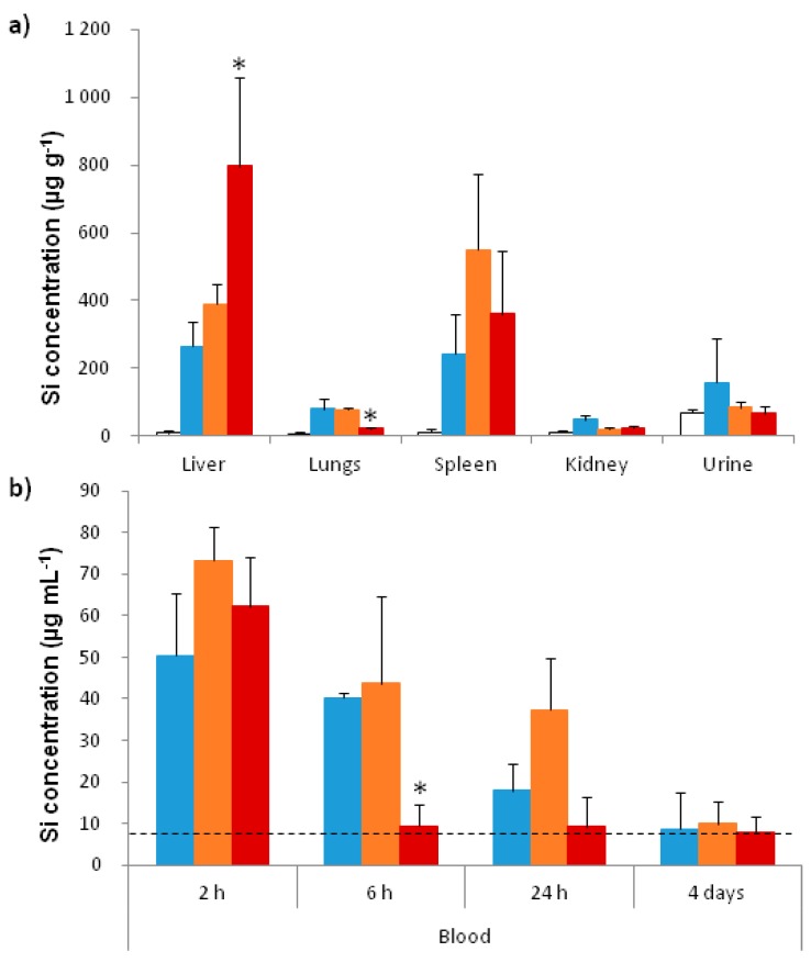
The biological fate of nanoparticles (NPs) for biomedical applications is highly dependent of their size and charge, their aggregation state and their surface chemistry. The chemical composition of the NPs surface influences their stability in biological fluids, their interaction with proteins, and their attraction to the cell membranes. In this work, core-shell magnetic mesoporous silica nanoparticles (Fe₃O₄@MSN), that are considered as potential theranostic candidates, are coated with polyethylene glycol (PEG) or 1,2-dimyristoyl-sn-glycero-3-phosphocholine (DMPC) lipid bilayer. Their biological fate is studied in comparison to the native NPs. The physicochemical properties of these three types of NPs and their suspension behavior in different media are investigated. The attraction to a membrane model is also evaluated using a supported lipid bilayer. The surface composition of NPs strongly influences their dispersion in biological fluids mimics, protein binding and their interaction with cell membrane. While none of these types of NPs is found to be toxic on mice four days after intravenous injection of a dose of 40 mg kg-1 of NPs, their surface coating nature influences the in vivo biodistribution. Importantly, NP coated with DMPC exhibit a strong accumulation in liver and a very low accumulation in lung in comparison with nude or PEG ones.

Keywords: biodistribution; cell-membrane interactions; nanoparticles; safety; surface coating.


Conflict of interest statement

The authors declare no conflict of interest.

Figures

Figure 1
Figure 1
TEM and cryoTEM images of the nanoparticles (NPs); (a) Native magnetic mesoporous silica core-shell nanoparticles (Fe3O4@MSN); (b) polyethylene glycol (PEG) Fe3O4@MSN; (c) 1,2-dimyristoyl-sn-glycero-3phosphocholine (DMPC) Fe3O4@MSN observed by cryoTEM.
Figure 2
Figure 2
Hydrodynamic diameter (HD) represented by bars and polydispersity index (PDI) represented as dots of native (blue), PEG (orange) and DMPC (red) Fe3O4@MSN in different media: (a) in ethanol; (b) in HBS 5 mM NaCl, pH 7.4; (c) in HBS 150 mM NaCl, pH 7.4; (d) in RPMI cell culture medium (10% fetal calf serum (FCS), pH 7.4) and (e) in HBS 150 mM NaCl (pH 7.4) containing 10% FCS; (f) Zeta potential in HBS 150, HBS 5, RPMI and HBS 150 mM NaCl containing 10% FCS.
Figure 3
Figure 3
Quartz crystal microbalance with dissipation (QCM-D) sensorgrams following the interaction between NPs and egg phosphatidyl choline (EPC) supported lipid bilayer (SLB). Native (blue), PEG (orange) and DMPC (red) Fe3O4@MSN were flowed into HBS 150 mM NaCl 10% SCF medium on the top of EPC SLB, at a concentration of 0.25 mg mL−1 of NPs. After Fe3O4@MSN addition in the medium on the top of the EPC SLB during 15 min, the flow was stopped for 10 h. The results on the variations of frequency (a) and of the dissipation (b) are presented after offset of the lipid bilayer formation.
Figure 4
Figure 4
TEM imaging of Hep-G2 cells exposed for 3, 6, and 24 h at 50 µg mL−1 for native (ac), PEG (df) or DMPC Fe3O4@MSN (gi). The NPs are localized by arrows, near the cell membrane (M) or the nucleus (N).
Figure 5
Figure 5
Preliminary toxicological assessment. (a) Hematoxylin-eosin sections from paraffin-embedded tissues (kidney, liver and spleen) of control and treated mice; (b) Plasma and urine levels of renal biomarker (creatinine); (c,d) Plasma levels of systemic inflammation biomarkers (IL-6 and TNF-α); (e) Plasma level of liver biomarker (ALT).
Figure 6
Figure 6
Biodistribution of Fe3O4@MSN in mice. (a) Quantification of silicium in different organs 4 days after injection. Inductively coupled plasma-mass spectrometry (ICPMS) was used after acid digestion to quantify the silicium in the liver, the lungs, the spleen, the kidneys, and urine 4 days after intravenous injection of native (blue), PEG (orange) and DMPC (red) Fe3O4@MSN at a concentration of 40 mg kg−1 in comparison to control mice (white); (b) NPs level in blood. The silicium was quantified in the blood 2 h, 6 h, 24 h, and 4 days after intravenous injection of native (blue), PEG (orange) and DMPC (red) Fe3O4@MSN at a concentration of 40 mg kg−1. The dashed line indicates the silicium level found in the blood of control mice. For this experiment 20 mice were divided in 4 groups of 5 animals. Values of histograms represent mean ± SD of values of each animal of a group. * p < 0.05 statistically different from all other groups treated with NPs.

References

    1. Tarn D., Ashley C.E., Xue M., Carnes E.C., Zink J.I., Brinker C.J. Mesoporous silica nanoparticle nanocarriers: Biofunctionality and biocompatibility. Acc. Chem. Res. 2012;46:792–801. doi: 10.1021/ar3000986. - DOI - PMC - PubMed
    1. Lazarovits J., Chen Y.Y., Sykes E.A., Chan W.C. Nanoparticle-blood interactions: The implications on solid tumour targeting. Chem. Commun. 2015;51:2756–2767. doi: 10.1039/C4CC07644C. - DOI - PubMed
    1. Monopoli M.P., Walczyk D., Campbell A., Elia G., Lynch I., Bombelli F.B., Dawson K.A. Physical-chemical aspects of protein corona: Relevance to in vitro and in vivo biological impacts of nanoparticles. J. Am. Chem. Soc. 2011;133:2525–2534. doi: 10.1021/ja107583h. - DOI - PubMed
    1. Westmeier D., Stauber R.H., Docter D. The concept of bio-corona in modulating the toxicity of engineered nanomaterials (ENM) Toxicol. Appl. Pharmacol. 2015;299:53–57. doi: 10.1016/j.taap.2015.11.008. - DOI - PubMed
    1. Tada D.B., Suraniti E., Rossi L.M., Leite C.A., Oliveira C.S., Tumolo T.C., Calemczuk R., Livache T., Baptista M.S. Effect of lipid coating on the interaction between silica nanoparticles and membranes. J. Biomed. Nanotechnol. 2014;10:519–528. doi: 10.1166/jbn.2014.1723. - DOI - PubMed

LinkOut - more resources