

Signaling Pathways Critical for Tooth Root Formation
- PMID: 28665752
- PMCID: PMC5613878
- DOI: 10.1177/0022034517717478
Signaling Pathways Critical for Tooth Root Formation
Abstract
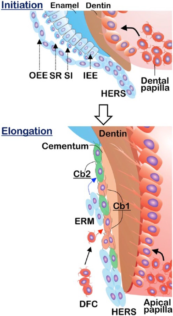
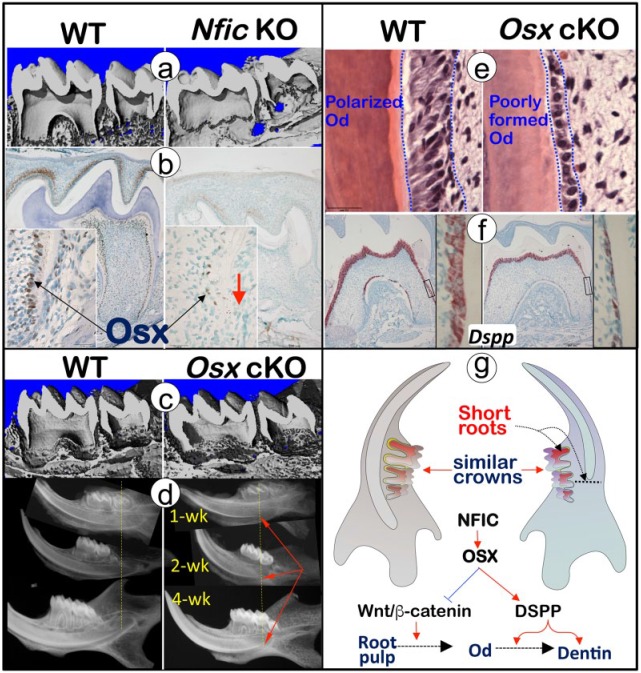
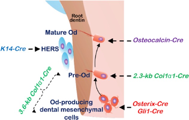
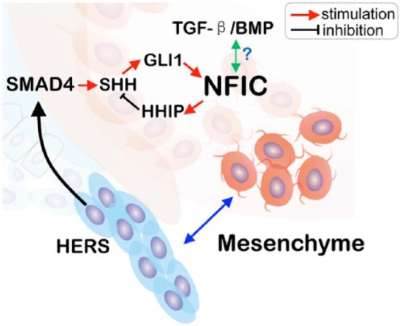
Tooth is made of an enamel-covered crown and a cementum-covered root. Studies on crown dentin formation have been a major focus in tooth development for several decades. Interestingly, the population prevalence for genetic short root anomaly (SRA) with no apparent defects in crown is close to 1.3%. Furthermore, people with SRA itself are predisposed to root resorption during orthodontic treatment. The discovery of the unique role of Nfic (nuclear factor I C; a transcriptional factor) in controlling root but not crown dentin formation points to a new concept: tooth crown and root have different control mechanisms. Further genetic mechanism studies have identified more key molecules (including Osterix, β-catenin, and sonic hedgehog) that play a critical role in root formation. Extensive studies have also revealed the critical role of Hertwig's epithelial root sheath in tooth root formation. In addition, Wnt10a has recently been found to be linked to multirooted tooth furcation formation. These exciting findings not only fill the critical gaps in our understanding about tooth root formation but will aid future research regarding the identifying factors controlling tooth root size and the generation of a whole "bio-tooth" for therapeutic purposes. This review starts with human SRA and mainly focuses on recent progress on the roles of NFIC-dependent and NFIC-independent signaling pathways in tooth root formation. Finally, this review includes a list of the various Cre transgenic mouse lines used to achieve tooth root formation-related gene deletion or overexpression, as well as strengths and limitations of each line.
Keywords: NFIC; cell signaling; dentin; odontogenesis; osterix; tooth regeneration.
Conflict of interest statement
The authors declare no potential conflicts of interest with respect to the authorship and/or publication of this article.
Figures
References
-
- Ahn S, Joyner AL. 2004. Dynamic changes in the response of cells to positive hedgehog signaling during mouse limb patterning. Cell. 118(4):505–516. - PubMed
-
- Ando S, Aizawa K, Oshima S, Nakamura Y, Sato A, Suzuki Y, Arita I, Kaida Y, Nagayama R. 1976. Studies on the consecutive survey of succedaneous and permanent dentition in the Japanese children: part 11. On the difference between variations in the eruptive time of mandibular first premolars and their root formation. J Nihon Univ Sch Dent. 18(2):25–28. - PubMed
-
- Baccetti T. 1998. A controlled study of associated dental anomalies. Angle Orthod. 68(3):267–274. - PubMed
-
- Bae CH, Lee JY, Kim TH, Baek JA, Lee JC, Yang X, Taketo MM, Jiang R, Cho ES. 2013. Excessive Wnt/beta-catenin signaling disturbs tooth-root formation. J Periodontal Res. 48(4):405–410. - PubMed
Publication types
MeSH terms
Substances
Grants and funding
LinkOut - more resources
Full Text Sources
Other Literature Sources
