. 2017 Jun 30;12(6):e0180242.
doi: 10.1371/journal.pone.0180242. eCollection 2017.

Biochemical characterization of functional domains of the chaperone Cosmc

Affiliations

Biochemical characterization of functional domains of the chaperone Cosmc

Melinda S Hanes et al. PLoS One. .

Abstract

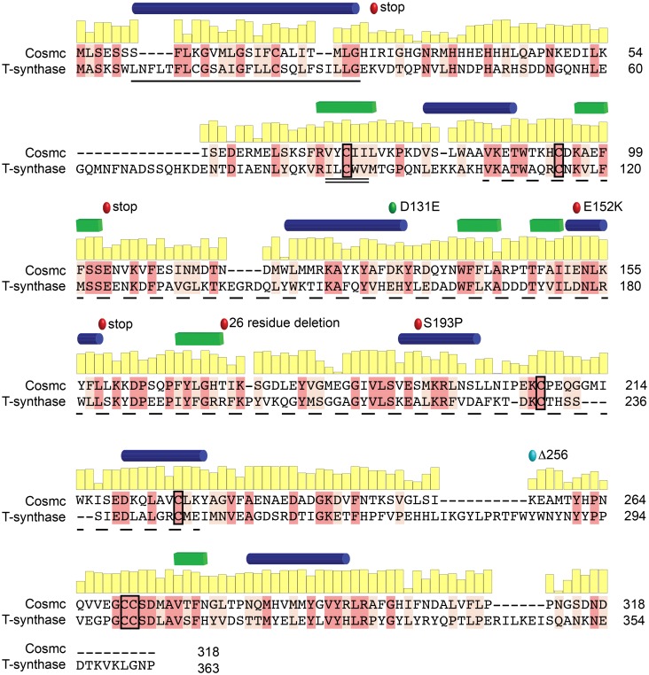
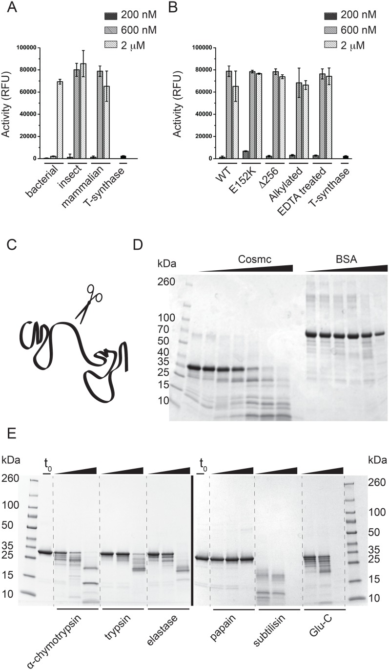
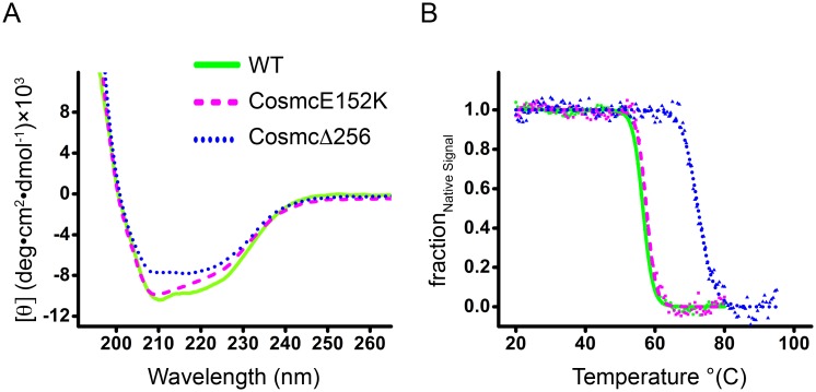
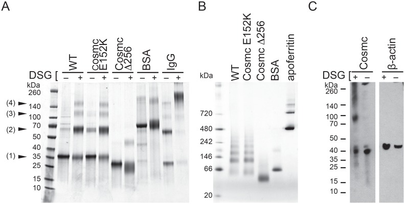
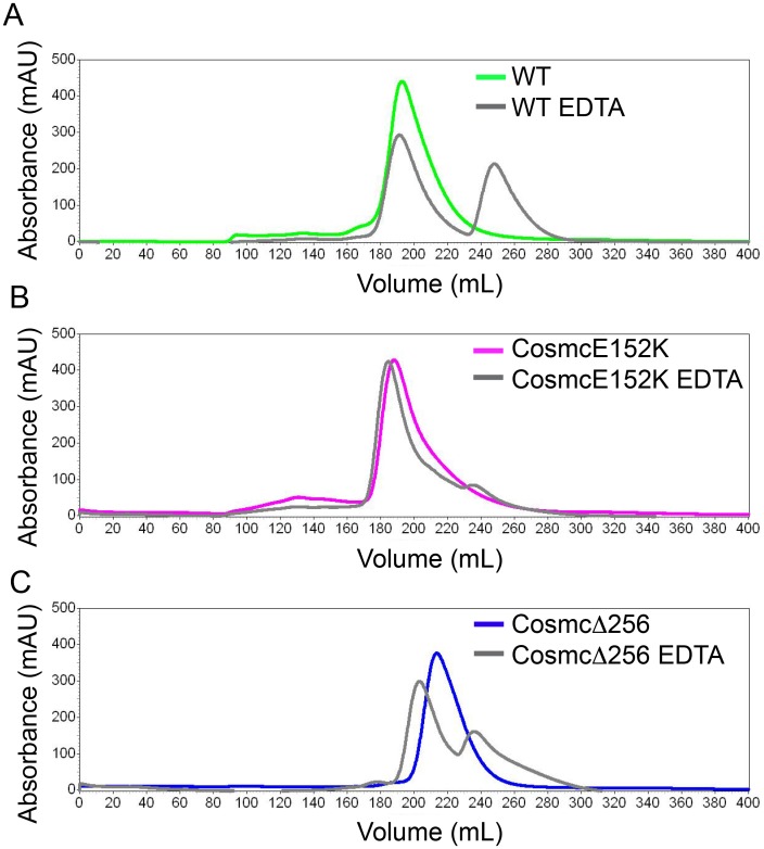
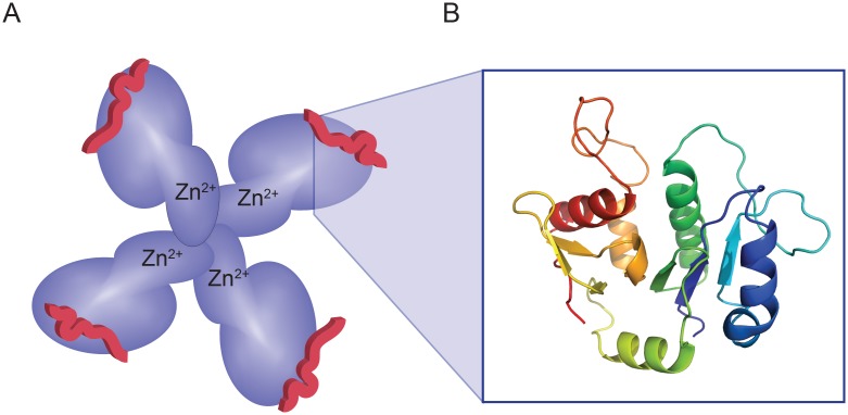
Cosmc is an endoplasmic reticulum chaperone necessary for normal protein O-GalNAc glycosylation through regulation of T-synthase, its single client. Loss-of-function of Cosmc results in expression of the Tn antigen, which is associated with multiple human diseases including cancer. Despite intense interest in dysregulated expression of the Tn antigen, little is known about the structure and function of Cosmc, including domain organization, secondary structure, oligomerization, and co-factors. Limited proteolysis experiments show that Cosmc contains a structured N-terminal domain (CosmcΔ256), and biochemical characterization of CosmcΔ256 reveals wild type chaperone activity. Interestingly, CosmcE152K, which shows loss of function in vivo, exhibits wild type-like activity in vitro. Cosmc and CosmcE152K heterogeneously oligomerize and form monomeric, dimeric, trimeric, and tetrameric species, while CosmcΔ256 is predominantly monomeric as characterized by chemical crosslinking and blue native page electrophoresis. Additionally, Cosmc selectively binds divalent cations in thermal shift assays and metal binding is abrogated by the CosmcΔ256 truncation, and perturbed by the E152K mutation. Therefore, the N-terminal domain of Cosmc mediates T-synthase binding and chaperone function, whereas the C-terminal domain is necessary for oligomerization and metal binding. Our results provide new structure-function insight to Cosmc, indicate that Cosmc behaves as a modular protein and suggests points of modulation or regulation of in vivo chaperone function.


Conflict of interest statement

Competing Interests: The authors have declared that no competing interests exist.

Figures

Fig 1
Fig 1. Sequence alignment between Cosmc and T-synthase.
A pairwise sequence alignment between human Cosmc and its client, human T-synthase, highlighting identical (dark pink) and conserved residues (light pink). The conservation at each residue position in Cosmc is shown in a yellow bar graph above the sequence. The putative transmembrane domain of Cosmc (black line), the Cosmc recognition region in T-synthase (black double line), and the galactosyltransferase domain of T-synthase (black dashed line) are shown. Additionally, inactivating point mutations, E152K, and S193P, and premature stop codons (red circles), along with a permissive substitution, D131E, (green circle) are indicated. The designed truncation mutation CosmcΔ256 is also annotated (light blue circle). Predicted secondary structure elements, helices (blue) and strands (green), are shown for Cosmc above the sequence [14]; conservation was calculated from a multiple sequence alignment of over 100 Cosmc orthologs using ConSurf [15].
Fig 2
Fig 2. Cosmc functional assays and limited proteolysis experiments.
(A) Active Cosmc can be recombinantly expressed and purified from bacterial, insect, and mammalian host cells. Cosmc was tested in a chaperone assay at three concentrations, 200nM, 600nM and 2uM. Data is reported as mean and standard deviation from quadruplicate measurements. (B) Chaperone activity assay comparing the WT and mutant Cosmc proteins, along with EDTA-treated and alkylated Cosmc. Data is reported as mean and standard deviation for quadruplicate measurements. (C) A cartoon representation of a two-domain protein, where unstructured regions at the domain boundaries are more susceptible to protease cleavage. (D) Limited proteolysis experiment in which Cosmc was digested with trypsin at 1:500 molar ratio and analyzed by SDS-PAGE (this experiment utilized the bacterially produced Cosmc). Cosmc samples were taken before trypsin addition (lane 2), immediately after trypsin addition (lane 3), and after 5min, 10min, 30min, 1hr, and 3hr (lanes 4–8). For the BSA control, samples were taken before protease addition (lane 9), after 5min, 10min, 30min, 1hr, and 3hr (lanes 10–14). (E) Limited proteolysis experiments with Cosmc purified from HEK293F cells using a broad range of proteases (i-vi). Samples were taken for SDS-PAGE before protease addition (lane 2), and after 20min, 1hr, and overnight, with α-chymotrypsin, trypsin, elastase, papain, subtilisin, and endoproteinase Glu-C.
Fig 3
Fig 3. CD spectra and thermal denaturation curves.
WT Cosmc (solid line, green), CosmcE152K (dashed line, magenta), and CosmcΔ256 (dotted line, blue) are plotted together for comparison. (A) The far UV CD spectra of Cosmc and mutants are indicative of well-folded proteins with significant helical content. (B) Thermal denaturation was followed by recording the CD signal at 222 nm with a slope of 1°C/min. Data are presented as fraction of the native signal (Ffold) as a function of temperature.
Fig 4
Fig 4. Oligomeric behavior of WT, CosmcE152K, and CosmcΔ256.
(A) Chemical crosslinking followed by SDS PAGE shows products corresponding to the mass of Cosmc monomer (1), dimer (2), trimer (3), and tetramer (4) for Cosmc and CosmcE152K, while CosmcΔ256 yields only monomer and small amounts of dimer species. Cosmc was treated with either the crosslinker DSG (+), or mock treated (-), along with negative and positive controls, BSA and IgG respectively, for protein oligomerization. (B) BN-PAGE analysis of Cosmc along with monomeric and oligomeric control proteins, BSA and apoferritin, respectively. WT and CosmcE152K show heterogeneous oligomers, while CosmcΔ256 shows a single predominant species. (C) Chemical crosslinking with DSG of full length Cosmc from crude HEK293SC lysates shows monomeric, dimeric, and trimeric species. Cosmc from cell lysates was detected by western blotting.
Fig 5
Fig 5. SEC of Cosmc, CosmcE152K, and CosmcΔ256, and EDTA treated proteins.
Cosmc was injected at 1.5 mg/ml onto a Sephacryl S300 size exclusion column (SEC) with phosphate buffered saline as the running buffer. SEC profiles are shown for untreated and EDTA treated (A) Cosmc WT; (B) CosmcE152K; (C) CosmcΔ256.
Fig 6
Fig 6. Cartoon model of Cosmc structure.
(A) A representation of Cosmc with an N-terminal domain (dark blue) that is responsible for interacting with the client (red peptide) and a smaller, C-terminal domain (light blue) that mediates oligomerization and metal binding (Zn2+). A putative tetramer is shown for illustration purposes only, however our data is consistent with heterogeneous oligomeric states. (B) An atomic model of Cosmc’s glycosyltransferase homology region based on Mfng (PDB ID 2J0B) using SWISS-MODEL. The glycosyltransferase homology region within Cosmc is restricted to the N-terminal domain.

References

    1. Ju T, Cummings RD. A unique molecular chaperone Cosmc required for activity of the mammalian core 1 beta 3-galactosyltransferase. Proc Natl Acad Sci USA. 2002;99(26):16613–8. doi: 10.1073/pnas.262438199 - DOI - PMC - PubMed
    1. Ju T, Cummings RD, Canfield WM. Purification, characterization, and subunit structure of rat core 1 Beta1,3-galactosyltransferase. J Biol Chem. 2002;277(1):169–77. doi: 10.1074/jbc.M109056200 - DOI - PubMed
    1. Bennett EP, Mandel U, Clausen H, Gerken TA, Fritz TA, Tabak LA. Control of mucin-type O-glycosylation: A classification of the polypeptide GalNAc-transferase gene family. Glycobiology. 2012;22(6):736–56. doi: 10.1093/glycob/cwr182 - DOI - PMC - PubMed
    1. Steentoft C, Vakhrushev SY, Joshi HJ, Kong Y, Vester-Christensen MB, Schjoldager KTBG, et al. Precision mapping of the human O-GalNAc glycoproteome through SimpleCell technology. EMBO J. 2013;32(10):1478–88. doi: 10.1038/emboj.2013.79 - DOI - PMC - PubMed
    1. Ju T, Brewer K, D'Souza A, Cummings RD, Canfield WM. Cloning and expression of human core 1 beta1,3-galactosyltransferase. J Biol Chem. 2002;277(1):178–86. doi: 10.1074/jbc.M109060200 - DOI - PubMed

LinkOut - more resources