

Mammographic density and ageing: A collaborative pooled analysis of cross-sectional data from 22 countries worldwide
- PMID: 28666001
- PMCID: PMC5493289
- DOI: 10.1371/journal.pmed.1002335
Mammographic density and ageing: A collaborative pooled analysis of cross-sectional data from 22 countries worldwide
Abstract
Background: Mammographic density (MD) is one of the strongest breast cancer risk factors. Its age-related characteristics have been studied in women in western countries, but whether these associations apply to women worldwide is not known.
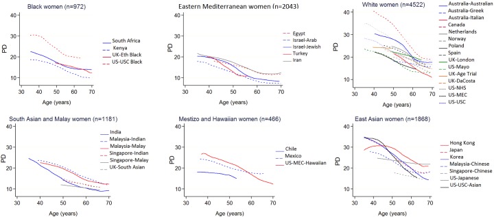
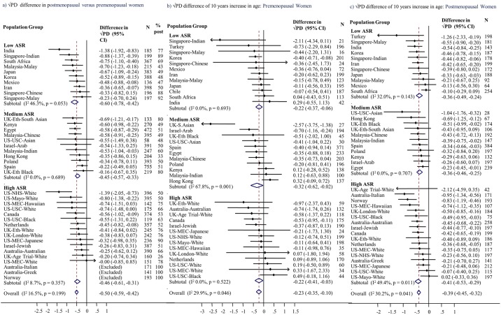
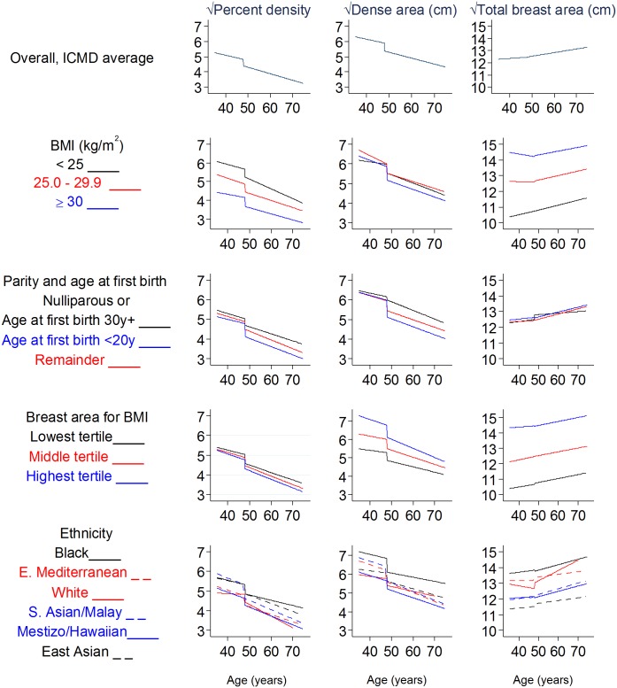
Methods and findings: We examined cross-sectional differences in MD by age and menopausal status in over 11,000 breast-cancer-free women aged 35-85 years, from 40 ethnicity- and location-specific population groups across 22 countries in the International Consortium on Mammographic Density (ICMD). MD was read centrally using a quantitative method (Cumulus) and its square-root metrics were analysed using meta-analysis of group-level estimates and linear regression models of pooled data, adjusted for body mass index, reproductive factors, mammogram view, image type, and reader. In all, 4,534 women were premenopausal, and 6,481 postmenopausal, at the time of mammography. A large age-adjusted difference in percent MD (PD) between post- and premenopausal women was apparent (-0.46 cm [95% CI: -0.53, -0.39]) and appeared greater in women with lower breast cancer risk profiles; variation across population groups due to heterogeneity (I2) was 16.5%. Among premenopausal women, the √PD difference per 10-year increase in age was -0.24 cm (95% CI: -0.34, -0.14; I2 = 30%), reflecting a compositional change (lower dense area and higher non-dense area, with no difference in breast area). In postmenopausal women, the corresponding difference in √PD (-0.38 cm [95% CI: -0.44, -0.33]; I2 = 30%) was additionally driven by increasing breast area. The study is limited by different mammography systems and its cross-sectional rather than longitudinal nature.
Conclusions: Declines in MD with increasing age are present premenopausally, continue postmenopausally, and are most pronounced over the menopausal transition. These effects were highly consistent across diverse groups of women worldwide, suggesting that they result from an intrinsic biological, likely hormonal, mechanism common to women. If cumulative breast density is a key determinant of breast cancer risk, younger ages may be the more critical periods for lifestyle modifications aimed at breast density and breast cancer risk reduction.
Conflict of interest statement
I have read the journal's policy and the authors of this manuscript have identified the following competing interests: ML: Non-restricted investigator-initiated grant from AstraZeneca and minor support from Swiss Re.
Figures
References
-
- Boyd NF, Rommens JM, Vogt K, Lee V, Hopper JL, Yaffe MJ, et al. Mammographic breast density as an intermediate phenotype for breast cancer. Lancet Oncol. 2005;6(10):798–808. doi: 10.1016/S1470-2045(05)70390-9 - DOI - PubMed
-
- Boyd N, Martin L, Stone J, Little L, Minkin S, Yaffe M. A longitudinal study of the effects of menopause on mammographic features. Cancer Epidemiol Biomarkers Prev. 2002;11(10 Pt 1):1048–53. - PubMed
-
- McCormack VA, dos Santos Silva I. Breast density and parenchymal patterns as markers of breast cancer risk: a meta-analysis. Cancer Epidemiol Biomarkers Prev. 2006;15(6):1159–69. doi: 10.1158/1055-9965.EPI-06-0034 - DOI - PubMed
-
- Pike MC, Krailo MD, Henderson BE, Casagrande JT, Hoel DG. ‘Hormonal’ risk factors, ‘breast tissue age’ and the age-incidence of breast cancer. Nature. 1983;303(5920):767–70. - PubMed
-
- Anderson WF, Matsuno R. Breast cancer heterogeneity: a mixture of at least two main types? J Natl Cancer Inst. 2006;98(14):948–51. doi: 10.1093/jnci/djj295 - DOI - PubMed
Publication types
MeSH terms
Grants and funding
- R03 CA167771/CA/NCI NIH HHS/United States
- R01 CA177150/CA/NCI NIH HHS/United States
- P50 CA116201/CA/NCI NIH HHS/United States
- UM1 CA186107/CA/NCI NIH HHS/United States
- R01 CA085265/CA/NCI NIH HHS/United States
- R01 CA131332/CA/NCI NIH HHS/United States
- N01 CA015083/CA/NCI NIH HHS/United States
- R01 CA097396/CA/NCI NIH HHS/United States
- UM1 CA176726/CA/NCI NIH HHS/United States
- 10/57/55/DH_/Department of Health/United Kingdom
- R01 CA124865/CA/NCI NIH HHS/United States
- 14565/CRUK_/Cancer Research UK/United Kingdom
- P30 CA015083/CA/NCI NIH HHS/United States
- 001/WHO_/World Health Organization/International
- R01 CA140286/CA/NCI NIH HHS/United States
- R37 CA054281/CA/NCI NIH HHS/United States
LinkOut - more resources
Full Text Sources
Other Literature Sources
Medical
