. 2017 Jun 30;12(6):e0180455.
doi: 10.1371/journal.pone.0180455. eCollection 2017.

Contribution of reactive oxygen species to the pathogenesis of pulmonary arterial hypertension

Affiliations

Contribution of reactive oxygen species to the pathogenesis of pulmonary arterial hypertension

Nikki L Jernigan et al. PLoS One. .

Abstract

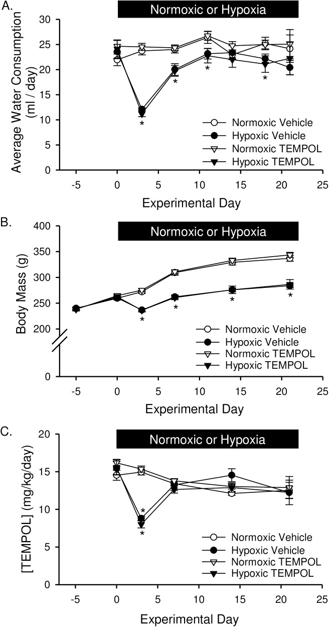
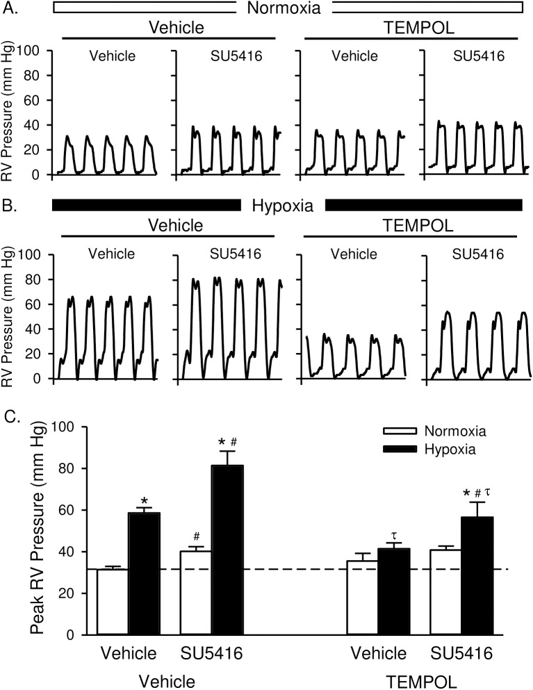
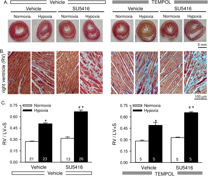
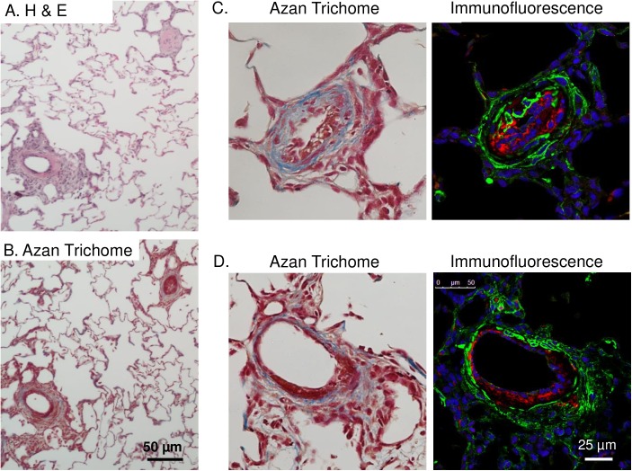
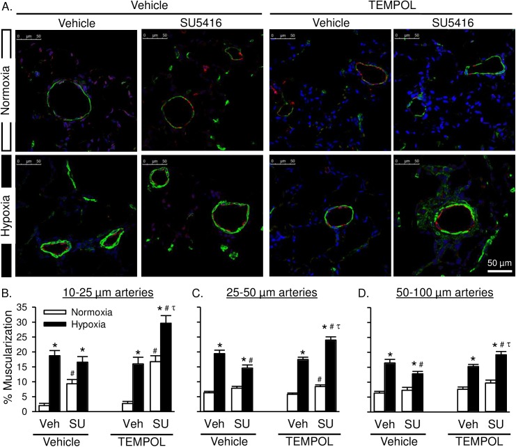
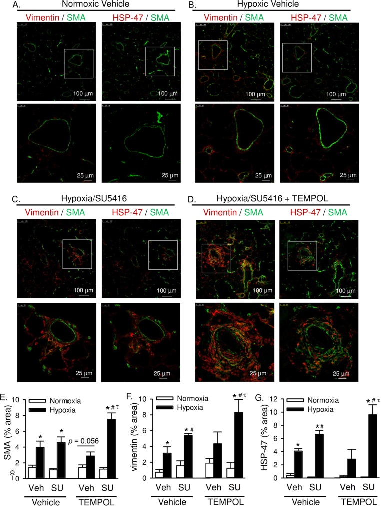
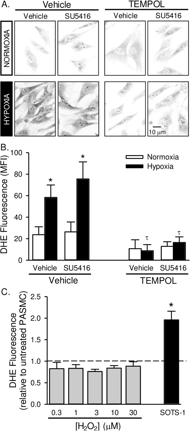
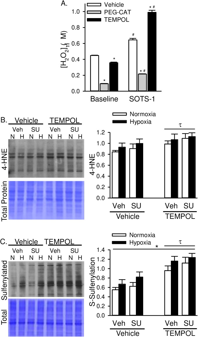
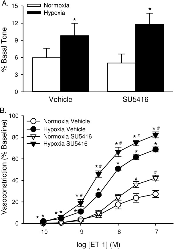
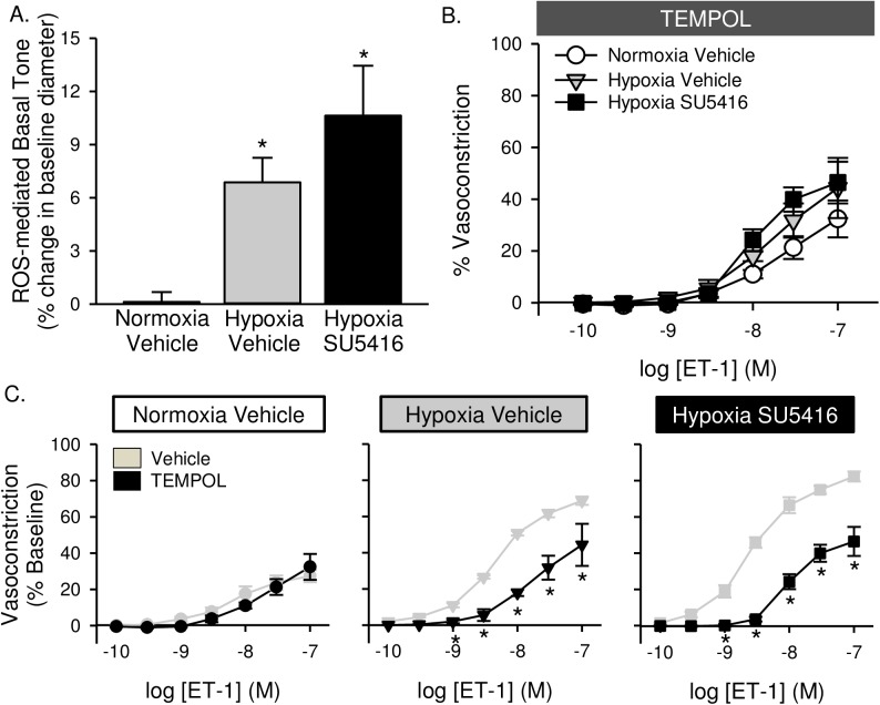
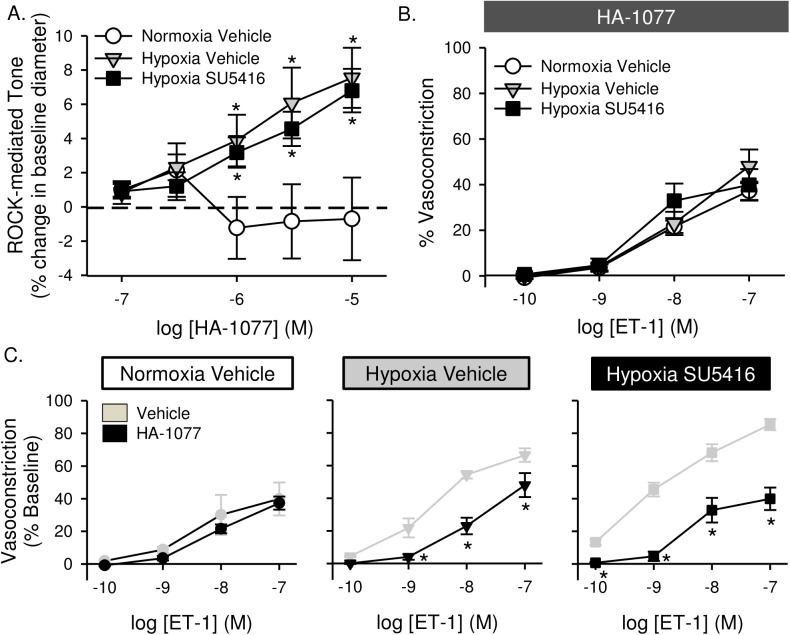
Pulmonary arterial hypertension is associated with a decreased antioxidant capacity. However, neither the contribution of reactive oxygen species to pulmonary vasoconstrictor sensitivity, nor the therapeutic efficacy of antioxidant strategies in this setting are known. We hypothesized that reactive oxygen species play a central role in mediating both vasoconstrictor and arterial remodeling components of severe pulmonary arterial hypertension. We examined the effect of the chemical antioxidant, TEMPOL, on right ventricular systolic pressure, vascular remodeling, and enhanced vasoconstrictor reactivity in both chronic hypoxia and hypoxia/SU5416 rat models of pulmonary hypertension. SU5416 is a vascular endothelial growth factor receptor antagonist and the combination of chronic hypoxia/SU5416 produces a model of severe pulmonary arterial hypertension with vascular plexiform lesions/fibrosis that is not present with chronic hypoxia alone. The major findings from this study are: 1) compared to hypoxia alone, hypoxia/SU5416 exposure caused more severe pulmonary hypertension, right ventricular hypertrophy, adventitial lesion formation, and greater vasoconstrictor sensitivity through a superoxide and Rho kinase-dependent Ca2+ sensitization mechanism. 2) Chronic hypoxia increased medial muscularization and superoxide levels, however there was no effect of SU5416 to augment these responses. 3) Treatment with TEMPOL decreased right ventricular systolic pressure in both hypoxia and hypoxia/SU5416 groups. 4) This effect of TEMPOL was associated with normalization of vasoconstrictor responses, but not arterial remodeling. Rather, medial hypertrophy and adventitial fibrotic lesion formation were more pronounced following chronic TEMPOL treatment in hypoxia/SU5416 rats. Our findings support a major role for reactive oxygen species in mediating enhanced vasoconstrictor reactivity and pulmonary hypertension in both chronic hypoxia and hypoxia/SU5416 rat models, despite a paradoxical effect of antioxidant therapy to exacerbate arterial remodeling in animals with severe pulmonary arterial hypertension in the hypoxia/SU5416 model.


Conflict of interest statement

Competing Interests: The authors have declared that no competing interests exist.

Figures

Fig 1
Fig 1. TEMPOL does not effect water consumption or body mass.
At the onset of hypoxic exposure, (A) water consumption (ml/day), (B) body mass (g), and (C) concentration of TEMPOL (mg/kg/day) dropped significantly. Water consumption and TEMPOL concentration was largely restored after 1 wk hypoxia. Body mass also increased following 1 wk chronic hypoxia in a parallel fashion to normoxic rats but remained lower due to the initial weight loss. There were no differences between SU5416- and vehicle-treated animals; therefore, data were grouped together for this analysis. Values are means ± SE; n = 10 animals/group. *P < 0.05 hypoxia vs. normoxia; there was no difference between vehicle and TEMPOL treated. Data analyzed with two-way ANOVA and individual groups compared with the Student-Newman-Keuls test.
Fig 2
Fig 2. TEMPOL attenuates the development of pulmonary hypertension.
Representative traces of right ventricular (RV) pressure measurements (mmHg) in rats treated with vehicle, SU5416, and/or TEMPOL and exposed to normoxia (A) or hypoxia (B). (C) Summary data for peak RVSP (mmHg). Dotted line equals normoxia-vehicle average. Values are means ± SE; n = 5 animals/group. *P < 0.05 vs. the normoxia group; # P < 0.05 vs. the corresponding SU5416 vehicle group; τ p < 0.05 vs. corresponding TEMPOL-vehicle group; analyzed by multiple two-way ANOVA and individual groups compared with the Student-Newman-Keuls test.
Fig 3
Fig 3. Right ventricular hypertrophy is not attenuated by TEMPOL.
Representative AZAN trichrome-stained whole heart sections (A) and higher magnification images of the right ventricle (B) from rats treated with vehicle, SU5416, and/or TEMPOL and exposed to normoxia or hypoxia. AZAN trichrome shows cell nuclei (dark red), collagen (blue) and orange-red in cytoplasm. C) Summary data showing Fulton’s index [ratio of RV to left ventricular plus septal (LV + S) heart weight] Values are means ± SE; n = animals/group (indicated in bars). *P < 0.05 vs. the normoxia group; # P < 0.05 vs. the corresponding SU5416 vehicle group; analyzed with multiple two-way ANOVA and individual groups compared with the Student-Newman-Keuls test.
Fig 4
Fig 4. Hypoxia/SU5416 treatment causes both neointimal proliferation of endothelial cells and early plexiform lesions with collagen deposition.
All images are from hypoxia/SU5416-treated rats. (A-B) Representative H&E- and Azan trichrome-stained lung sections. (C-D) Higher magnification images of AZAN-stained and fluorescently-labeled arteries showing medial fibrosis and neointimal proliferation of endothelial cells and an early plexiform lesion. AZAN trichrome shows cell nuclei (dark red), collagen (blue) and orange-red in cytoplasm. Fluorescence labeling shows smooth muscle α-actin (green), Von Willebrand factor (red), and sytox (blue).
Fig 5
Fig 5. Superoxide scavenging exacerbates arterial remodeling in rats with severe PAH.
A) Representative immunofluorescence images of lung sections from normoxic (top row) and hypoxic (bottom row) rats treated with SU5416 and/or TEMPOL. Smooth muscle α-actin (green), Von Willebrand factor (red), and sytox (blue). B) Percent muscularization calculated as percent thresholded smooth muscle α-actin area divided by total arterial wall area according to arterial diameter: 10–25 μm (left), 25–50 μm (middle), and 50–100 μm (right). Values are means ± SE; n = 4 animals/group. *P ≤ 0.05 vs. normoxic group; # P < 0.05 vs. corresponding SU5416-vehicle group; τ p < 0.05 vs. TEMPOL-vehicle group; analyzed by multiple two-way ANOVA and individual groups compared with the Student-Newman-Keuls test.
Fig 6
Fig 6. Superoxide scavenging exacerbates adventitial remodeling in rats with severe PAH.
Representative immunofluorescence images of lung sections from rats treated with normoxic vehicle (A), hypoxic vehicle (B), hypoxia/SU5416 (C), or hypoxia/SU5416/TEMPOL (D). Sections were incubated with anti-smooth muscle α-actin (SMA, green) and either vimentin (red, left) or HSP-47 (red, right). Summary data showing average percent area of positive immunofluorescence from 10 random 20x images per lung section for E) SMA, F) vimentin, and G) HSP-47. Values are means ± SE; n = 4 animals/group. *P ≤ 0.05 vs. normoxic group; # P < 0.05 vs. corresponding SU5416-vehicle group; τ p < 0.05 vs. TEMPOL-vehicle group; analyzed by multiple two-way ANOVA and individual groups compared with the Student-Newman-Keuls test.
Fig 7
Fig 7. Superoxide levels are increased in pulmonary artery smooth muscle cells from pulmonary hypertensive rats.
Representative images (A) and summary data (B) showing background-subtracted mean fluorescence intensity (MFI) of dihydroethidium (DHE) in pulmonary arterial smooth muscle cells from rats treated with SU5416, TEMPOL or vehicle and exposed to normoxia or hypoxia. Fluorescence images were digitally inverted to provide improved signal contrast. Values are means ± SE; n = 5 animals per group; *P < 0.05 vs. corresponding normoxia group; τ p < 0.05 vs. corresponding TEMPOL-vehicle group; analyzed by multiple two-way ANOVA and individual groups compared with the Student-Newman-Keuls test. C) DHE fluorescence in PASMC following pre-incubation with increasing doses of H2O2 (0.3–30 μM) or SOTS-1 (10 μM). Dotted line represents untreated cells. Values are means ± SE; n = 5 animals per group; *P < 0.05 vs. untreated cells; analyzed by one-way ANOVA.
Fig 8
Fig 8. TEMPOL increases H2O2-specific oxidative stress.
A) H2O2 levels assessed by Amplex Red Assay in control PASMC in the absence or presence of PEG-catalase (PEG-CAT; 250 U/ml) or TEMPOL (1 mM). SOTS-1 (0.01 mM) is a superoxide donor and used to stimulate increased oxidative stress. Values are means ± SE; n = 5 animals per group; *P ≤ 0.05 vs. vehicle-treated group; # P < 0.05 vs. baseline; analyzed by two-way ANOVA and individual groups compared with the Student-Newman-Keuls test. Representative western blot and summary data showing B) 4-HNE and C) S-sulfenylated proteins in whole lung homogenates from normoxic and hypoxic rats treated with SU5416 and/or TEMPOL. Values are means ± SE; n = 6 animals per group; *P ≤ 0.05 vs. normoxic group; τ p < 0.05 vs. TEMPOL-vehicle group; analyzed by multiple two-way ANOVA and individual groups compared with the Student-Newman-Keuls test.
Fig 9
Fig 9. ET-1-induced VSM Ca2+ sensitization and vasoconstriction are augmented in small pulmonary arteries from animals treated with SU5614.
(A) Basal tone (baseline diameter as a % of Ca2+-free diameter) in non-permeabilized, endothelium-disrupted, pressurized small pulmonary arteries. (B) vasoconstriction (% baseline diameter) to endothelin-1 (ET-1; 10−10 to 10−7 M) in Ca2+-permeabilized, endothelium-disrupted, pressurized small pulmonary arteries from rats treated with vehicle or SU5416 and exposed to normoxia or hypoxia. Values are means ± SE; n = 5–6 animals/group. *P ≤ 0.05 vs. normoxic group; #P <0.05 vs. corresponding vehicle treatment; analyzed by two-way ANOVA and individual groups compared with the Student-Newman-Keuls test.
Fig 10
Fig 10. Enhanced basal tone and ET-1-induced pulmonary VSM Ca2+ sensitization following hypoxia/SU5416 is mediated by reactive oxygen species.
(A) TEMPOL-mediated reversal of basal tone (% baseline diameter) in non-permeabilized, endothelium-disrupted, pressurized small pulmonary arteries. (B) Vasoconstriction (% baseline diameter) to endothelin-1 (ET-1; 10−10 to 10−7 M) in the presence of TEMPOL (1 mM) in Ca2+-permeabilized, endothelium-disrupted, pressurized small pulmonary arteries from normoxic and hypoxic rats treated with vehicle or SU5416. (C) Effect of TEMPOL on ET-1 mediated vasoconstriction in each group compared to vehicle-treated arteries (from Fig 9). Values are means ± SE; n = 4–5 animals/group; * p < 0.05 vs. vehicle-treated arteries; analyzed by two-way ANOVA and individual groups compared with the Student-Newman-Keuls test.
Fig 11
Fig 11. Enhanced basal tone and ET-1-induced pulmonary VSM Ca2+ sensitization following hypoxia/SU5416 is mediated by Rho kinase (ROK).
(A) HA-1077-mediated change in vessel diameter (% baseline diameter) in non-permeabilized, endothelium-disrupted, pressurized small pulmonary arteries. (B) Vasoconstriction (% baseline diameter) to endothelin-1 (ET-1; 10−10 to 10−7 M) in the presence of HA-1077 (10 μM) in Ca2+-permeabilized, endothelium-disrupted, pressurized small pulmonary arteries from normoxic and hypoxic rats treated with vehicle or SU5416. (C) Effect of HA-1077 on ET-1 mediated vasoconstriction in each group compared to vehicle-treated arteries (from Fig 9). Values are means ± SE; n = 4–5 animals/group; * p < 0.05 vs. vehicle-treated arteries; analyzed by two-way ANOVA and individual groups compared with the Student-Newman-Keuls test.

References

    1. Howell K, Preston RJ, McLoughlin P. Chronic hypoxia causes angiogenesis in addition to remodelling in the adult rat pulmonary circulation. The Journal of Physiology. 2003;547(1):133–45. doi: 10.1113/jphysiol.2002.030676 - DOI - PMC - PubMed
    1. Taraseviciene-Stewart L, Kasahara Y, Alger L, Hirth P, Mc Mahon G, Waltenberger J, et al. Inhibition of the VEGF receptor 2 combined with chronic hypoxia causes cell death-dependent pulmonary endothelial cell proliferation and severe pulmonary hypertension. The FASEB Journal. 2001;15(2):427–38. doi: 10.1096/fj.00-0343com - DOI - PubMed
    1. Aggarwal S, Gross CM, Sharma S, Fineman JR, Black SM. Reactive oxygen species in pulmonary vascular remodeling. Comprehensive Physiology. 2013;3(3):1011–34. doi: 10.1002/cphy.c120024 - DOI - PMC - PubMed
    1. Voelkel NF, Bogaard HJ, Husseini AA, Farkas L, Gomez-Arroyo J, Natarajan R. Antioxidants for the Treatment of Patients with Severe Angioproliferative Pulmonary Hypertension? Antioxidants & Redox Signaling. 2012;18(14):1810–7. doi: 10.1089/ars.2012.4828 - DOI - PubMed
    1. Wong C-M, Bansal G, Pavlickova L, Marcocci L, Suzuki YJ. Reactive Oxygen Species and Antioxidants in Pulmonary Hypertension. Antioxidants & Redox Signaling. 2013;18(14):1789–96. doi: 10.1089/ars.2012.4568 - DOI - PMC - PubMed

MeSH terms