Review
. 2017 Aug;58(8):1316-1329.
doi: 10.1111/epi.13829. Epub 2017 Jun 30.

High-frequency oscillations: The state of clinical research

Affiliations
Review

High-frequency oscillations: The state of clinical research

Birgit Frauscher et al. Epilepsia. 2017 Aug.

Abstract

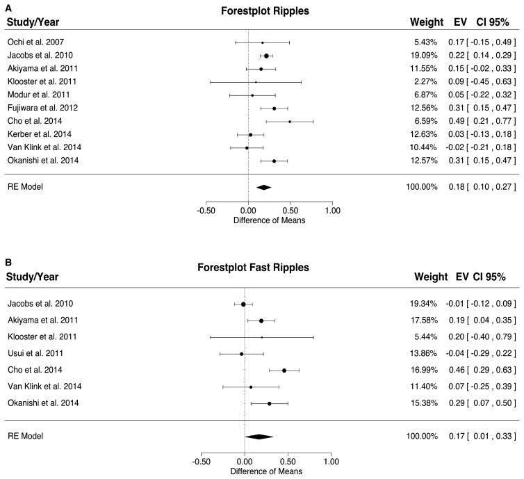
Modern electroencephalographic (EEG) technology contributed to the appreciation that the EEG signal outside the classical Berger frequency band contains important information. In epilepsy, research of the past decade focused particularly on interictal high-frequency oscillations (HFOs) > 80 Hz. The first large application of HFOs was in the context of epilepsy surgery. This is now followed by other applications such as assessment of epilepsy severity and monitoring of antiepileptic therapy. This article reviews the evidence on the clinical use of HFOs in epilepsy with an emphasis on the latest developments. It highlights the growing literature on the association between HFOs and postsurgical seizure outcome. A recent meta-analysis confirmed a higher resection ratio for HFOs in seizure-free versus non-seizure-free patients. Residual HFOs in the postoperative electrocorticogram were shown to predict epilepsy surgery outcome better than preoperative HFO rates. The review further discusses the different attempts to separate physiological from epileptic HFOs, as this might increase the specificity of HFOs. As an example, analysis of sleep microstructure demonstrated a different coupling between HFOs inside and outside the epileptogenic zone. Moreover, there is increasing evidence that HFOs are useful to measure disease activity and assess treatment response using noninvasive EEG and magnetoencephalography. This approach is particularly promising in children, because they show high scalp HFO rates. HFO rates in West syndrome decrease after adrenocorticotropic hormone treatment. Presence of HFOs at the time of rolandic spikes correlates with seizure frequency. The time-consuming visual assessment of HFOs, which prevented their clinical application in the past, is now overcome by validated computer-assisted algorithms. HFO research has considerably advanced over the past decade, and use of noninvasive methods will make HFOs accessible to large numbers of patients. Prospective multicenter trials are awaited to gather information over long recording periods in large patient samples.

Keywords: Biomarker; Scalp EEG; Seizure; Sleep; Surgical outcome.


Figures

Figure 1
Figure 1
Patient with a left central lesion, with fast ripples (FRs) in post-electrocorticography (ECoG) and recurrent seizures (auras) after surgery. (A) Spike and high-frequency oscillation events in a selection of bipolar channels (indicated as 1–5 in C). (B) Preresection photograph. (C) Post-ECoG. The resected area is delineated by the dotted white line. The area near the resection (resection margin = 1 cm) is marked with transparent white. Two ECoG recordings were performed postresection. We represent the location of the bipolar channels analyzed. Note that FRs (rate = 25/min/electrode; yellow) were present in the margin of the resected lesion (dysembryoplastic neuroepithelial tumor), in a larger region with spikes (range rate = 4–41/min/electrode; blue). Almost all electrodes showed ripples and are therefore not depicted. Based on the FRs present in the resection margin, a different surgical decision could have been made if FRs were not as close to eloquent central cortex. EEG, electroencephalogram. Source: van ‘t Klooster et al., with permission from Wolters Kluwer.
Figure 2
Figure 2
Meta-analysis results for ripples (A) and fast ripples (B). The resection ratio for both ripples and fast ripples is higher in seizure-free patients compared to non–seizure-free patients. For each study, a graphical representation of the effect (i.e., the difference of the resection ratio between the good- and bad-outcome groups) and of the confidence interval (CI) is given along with the exact values (EVs) and the weights. RE, rodent epilepsy. Source: Höller et al., published open access using a Creative Commons Attribution CC-BY licence.
Figure 3
Figure 3
Ripple oscillations in the scalp electroencephalogram (EEG) recorded from a child with Landau–Kleffner syndrome. Representative spikes (left and middle columns, arrowheads) are associated with ripple oscillation, which was largely invariant irrespective of low-cut frequency (LCF), whether of 60 or 120 Hz (EEG traces filtered at 0.5, 60, and 120 Hz are shown in green, blue, and red, respectively). The EEG was recorded during non–rapid eye movement sleep and therefore did not include muscle activity or eye movements. Identical EEG data are presented in a referential montage (top: O1 with reference to the average EEG of bilateral earlobes, indicated as O1–Aav) and a bipolar montage (bottom: P3–O1). Note that spike-related ripples with at least four consecutive oscillations are clearly observed in both montages. Each panel of time–frequency spectra shows a corresponding discrete blob (arrows) with a frequency at around 130 Hz irrespective of referential or bipolar montage. In contrast, muscle activity (right column) contamination to scalp EEG recorded during wakefulness is dominant over the temporal region (T4, F8–T4) close to muscles and has very irregular morphology and a noisy spectral pattern with no outstanding blobs. Source: Worrell et al., with permission from Elsevier.
Figure 4
Figure 4
Representative examples for the coupling of epileptic spikes and high-frequency oscillations (HFOs) across the slow wave cycle. Examples are shown of a slow wave and an epileptic spike (left panel), a slow wave and an HFO in a channel with epileptic activity (middle panel), and an HFO in a channel with normal electroencephalographic (EEG) activity (right panel). The top row shows the slow wave in a scalp channel, the second row shows the same time period for an intracranial channel with normal EEG activity, and the third row shows an intracranial channel with epileptic EEG activity. The fourth row shows the ripple band signal with a different time and amplitude scale, corresponding to the shaded periods in the intracranial channels. All channels are in the left frontal region; each example corresponds to a different patient. The scalp slow wave in the right panel is of shorter duration than the scalp slow waves in the left and middle panels. *In this example, a normal sleep slow wave with no epileptic spike is seen in a channel designated as epileptic because it has spikes at other times. Note that the spike and the HFO in the intracranial channel with epileptic activity (middle) occurs prior to the peak of the scalp negative half-wave, whereas the HFO in the channel with normal EEG activity (right) occurs after the peak of the scalp negative half-wave. Source: Frauscher et al., published open access using a Creative Commons Attribution CC-BY licence.

References

    1. Jacobs J, Staba R, Asano E, et al. High-frequency oscillations (HFOs) in clinical epilepsy. Prog Neurobiol. 2012;98:302–315. - PMC - PubMed
    1. Bragin A, Engel J, Jr, Wilson CL, et al. High-frequency oscillations in human brain. Hippocampus. 1999;9:137–142. - PubMed
    1. Zijlmans M, Worrell GA, Dümpelmann M. How to record high-frequency oscillations in epilepsy: A practical guideline. Epilepsia. 2017:58. https://doi.org/10.1111/epi.13814. - DOI - PubMed
    1. Allen PJ, Fish DR, Smith SJ. Very high-frequency rhythmic activity during SEEG suppression in frontal lobe epilepsy. Electroencephalogr Clin Neurophysiol. 1992;82:155–159. - PubMed
    1. Fisher RS, Webber WR, Lesser RP, et al. High-frequency EEG activity at the start of seizures. J Clin Neurophysiol. 1992;9:441–448. - PubMed

Publication types