Review
. 2017 Jul 25;56(29):3710-3724.
doi: 10.1021/acs.biochem.7b00346. Epub 2017 Jul 11.

Bacterial Cell Mechanics

Affiliations
Review

Bacterial Cell Mechanics

George K Auer et al. Biochemistry. .

Abstract

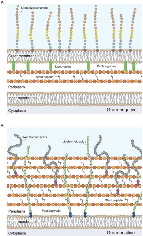
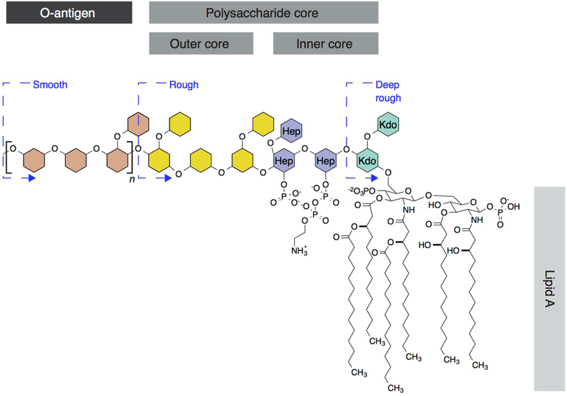
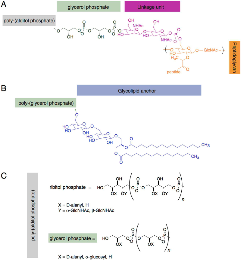
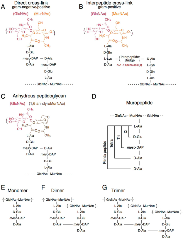
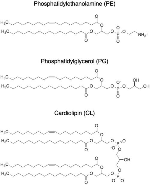
Cellular mechanical properties play an integral role in bacterial survival and adaptation. Historically, the bacterial cell wall and, in particular, the layer of polymeric material called the peptidoglycan were the elements to which cell mechanics could be primarily attributed. Disrupting the biochemical machinery that assembles the peptidoglycan (e.g., using the β-lactam family of antibiotics) alters the structure of this material, leads to mechanical defects, and results in cell lysis. Decades after the discovery of peptidoglycan-synthesizing enzymes, the mechanisms that underlie their positioning and regulation are still not entirely understood. In addition, recent evidence suggests a diverse group of other biochemical elements influence bacterial cell mechanics, may be regulated by new cellular mechanisms, and may be triggered in different environmental contexts to enable cell adaptation and survival. This review summarizes the contributions that different biomolecular components of the cell wall (e.g., lipopolysaccharides, wall and lipoteichoic acids, lipid bilayers, peptidoglycan, and proteins) make to Gram-negative and Gram-positive bacterial cell mechanics. We discuss the contribution of individual proteins and macromolecular complexes in cell mechanics and the tools that make it possible to quantitatively decipher the biochemical machinery that contributes to bacterial cell mechanics. Advances in this area may provide insight into new biology and influence the development of antibacterial chemotherapies.


Figures

Figure 1.
Figure 1.
Structure of the bacterial cell walls. (A) Cartoon depicting the structure of the Gram-negative cell wall. The peptidoglycan thickness is ~4 nm; monosaccharides in the peptidoglycan are represented as hexagons, and the colors demonstrate that this material consists of repeating disaccharide building blocks. Peptide cross-links in the peptidoglycan are depicted as gray lines. Monosaccharides in lipopolysaccharides are depicted as hexagons. Aqua and purple denote the inner polysaccharide core; yellow denotes the outer polysaccharide core, and brown denotes the O-antigen. Lipoproteins (green) connect the outer membrane to the peptidoglycan. (B) Cartoon depicting the Gram-positive bacterial cell wall. The peptidoglycan thickness is ~19–33 nm. Lipoteichoic acid is inserted into the membrane and consists of a glycolipid anchor (blue) and poly(glycerol phosphate) (green). The wall teichoic acid is directly cross-linked to the peptidoglycan through a linkage unit (red) and consists of glycerol phosphate (green) and poly(alditol phosphate).
Figure 2.
Figure 2.
Structure of LPS in Gram-negative bacteria. LPS consists of three primary regions: lipid A, the polysaccharide core (composed of an inner and outer core), and the O-antigen. The monosaccharides of LPS, polysaccharide core, and O-antigen are represented schematically as hexagons to simplify the structure of the molecule. The inner core is highly conserved among species and is composed of 3-deoxy-d-manno-octulosonic acid (Kdo) (aqua) and heptose (Hep) (purple). The outer core (yellow) and O-antigen (brown) are variable among bacteria. n represents the number of O-antigen repeats, which vary in length depending on the species and can be as large as 40 repeating units. Alterations in the length of the LPS (depicted by the blue dashed lines) result in physical alterations in colony and cell morphology that ranges from smooth to deep rough.
Figure 3.
Figure 3.
Glycopolymers of Gram-positive bacteria. (A) Wall teichoic acids (WTAs) are cross-linked to the peptidoglycan (orange) through a linkage unit (red), which is followed by two glycerol phosphate units (green) and repeating poly(alditol phosphate) units depicted in panel C. Dashed lines indicate the connection to the cross-linked peptidoglycan. (B) Lipoteichoic acids (LTAs) consist of a glycolipid anchor (blue) and repeating poly(glycerol phosphate) units. (C) The structure of poly(alditol phosphate) in WTAs and LTAs consists of glycerol phosphate or ribitol phosphate polymers that range in length from 20 to 40 repeat units (n = 20–40). X and Y indicate the location of chemical modifications to the polysaccharide chain of WTAs and LTAs.
Figure 4.
Figure 4.
Structure of the peptidoglycan. (A) Structure of cross-linked meso-DAP containing peptidoglycan found in both Gram-negative and Gram-positive bacteria. A 3–4 cross-link is depicted between meso-DAP in position 3 and d-Ala in position 4. (B) Structure of l-Lys peptidoglycan cross-linked through an interpeptide bridge ranging from one to seven amino acids that is found only in Gram-positive bacteria. A 3–4 cross-link is shown between l-Lys in position 3 and d-Ala in position 4. (C) Structure of anhydrous-terminated peptidoglycan containing 1,6-anhydroMurNAc. (D) Cartoon depicting the length of the stem peptides ranging from di (two amino acids) to tri (three amino acids) to tetra (four amino acids) to penta (five amino acids). (E) Structure of monomeric peptidoglycan containing a meso-DAP tetrapeptide. (F) Structure of dimeric peptidoglycan containing a meso-DAP tetrapeptide cross-linked at position 4–3. (G) Structure of trimeric peptidoglycan containing a meso-DAP tetrapeptide cross-linked at position 4–3.
Figure 5.
Figure 5.
Major phospholipids of Gram-negative and Gram-positive bacteria. In E. coli membranes, phosphatidylethanolamine (PE) represents 70–80% of total lipids, phosphatidlyglycerol (PG) represents 20–25% of total lipids, and cardiolipin (CL) represents 5–10% of total lipids. Phospholipids are shown with alkyl tails representing the most common degree of unsaturation found in E. coli membranes. Double bonds can be located in different positions and have different geometries (cis as shown, or trans), and alkyl tails can have multiple unsaturated bonds.
Figure 6.
Figure 6.
Techniques for measuring bacterial cell stiffness. (A) Atomic force microscopy. The top panel shows a force–distance curve of a bacterium with high bacterial stiffness (green) and low bacterial stiffness (purple). The bottom panel is an illustration of an AFM probe contacting the surface of a cell with high or low stiffness. (B) Microchannel measurements of cell bending rigidity. Bacteria are loaded and filamented in microfluidic channels, and fluid flow through the central channel applies a force on cells; cells bend when the magnitude of force is sufficient. Cartoon reproduced with permission from ref . Copyright 2014 National Academy of Sciences. (C) Transverse compression microfluidic device. The left panel is a side view of the poly(dimethylsiloxane) (PMDS) microfluidic device. The right panel is a three-dimensional view of the device. Cells are placed within a microfluidic device consisting of a glass coverslip patterned with 0.8–0.9 μm tall micropillars positioned below a polymer layer with a height that can be controlled by air pressure. Cartoon reproduced with permission from ref . Copyright 2002 American Society for Microbiology. (D) Extrusion loading microfluidic device. In the top panel, 12 parallel channels have diameters that taper between the entrance and exit; fluid flow pushes cells into channels and applies loads on them ranging from 0.0037 to 0.045 MPa. In the bottom panel, at equivalent pressures, cells that are more deformable are forced further down tapered channels than stiff cells. Cartoon reproduced with permission from ref . Copyright 1999 American Society for Microbiology. (E) Bacterial growth encapsulated in agarose. Cells are mixed with a solution of warm agarose, poured into a PDMS mold, and gelled. The cells are imaged at 1 min intervals using phase contrast microscopy to monitor cell growth. Cartoon reproduced with permission from ref . Copyright 2012 John Wiley & Sons, Inc.

References

    1. Reuter M, Hayward NJ, Black SS, Miller S, Dryden DT, and Booth IR (2014) Mechanosensitive channels and bacterial cell wall integrity: does life end with a bang or a whimper? J. R. Soc., Interface 11, 20130850. - PMC - PubMed
    1. Tuson HH, Auer GK, Renner LD, Hasebe M, Tropini C, Salick M, Crone WC, Gopinathan A, Huang KC, and Weibel DB (2012) Measuring the stiffness of bacterial cells from growth rates in hydrogels of tunable elasticity. Mol. Microbiol 84, 874–891. - PMC - PubMed
    1. Wang S, Arellano-Santoyo H, Combs PA, and Shaevitz JW (2010) Actin-like cytoskeleton filaments contribute to cell mechanics in bacteria. Proc. Natl. Acad. Sci U. S. A 107, 9182–9185. - PMC - PubMed
    1. Amir A, Babaeipour F, McIntosh DB, Nelson DR, and Jun S (2014) Bending forces plastically deform growing bacterial cell walls. Proc. Natl. Acad. Sci. U. S. A 111, 5778–5783. - PMC - PubMed
    1. Louise Meyer R, Zhou X, Tang L, Arpanaei A, Kingshott P, and Besenbacher F (2010) Immobilisation of living bacteria for AFM imaging under physiological conditions. Ultramicroscopy 110, 1349–1357. - PubMed

Publication types

MeSH terms

LinkOut - more resources