Review
. 2017 Jun 29;170(1):17-33.
doi: 10.1016/j.cell.2017.06.009.

RAS Proteins and Their Regulators in Human Disease

Affiliations
Review

RAS Proteins and Their Regulators in Human Disease

Dhirendra K Simanshu et al. Cell. .

Abstract

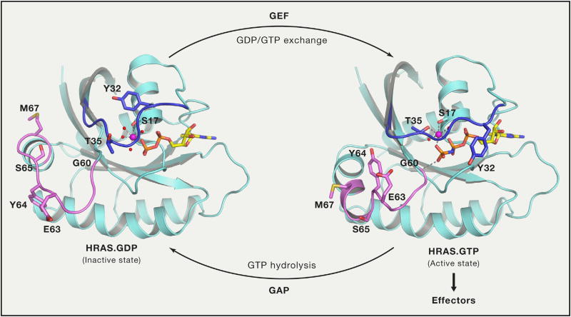
RAS proteins are binary switches, cycling between ON and OFF states during signal transduction. These switches are normally tightly controlled, but in RAS-related diseases, such as cancer, RASopathies, and many psychiatric disorders, mutations in the RAS genes or their regulators render RAS proteins persistently active. The structural basis of the switch and many of the pathways that RAS controls are well known, but the precise mechanisms by which RAS proteins function are less clear. All RAS biology occurs in membranes: a precise understanding of RAS' interaction with membranes is essential to understand RAS action and to intervene in RAS-driven diseases.

Keywords: CRAFT; KRAS; KRAS therapies; NF1; RAF1; RAS effectors; RAS in the membrane; RAS proteins; RAS-driven cancer; RASopathies.


Figures

Figure 1
Figure 1. Conformational Changes in Switch Regions when RAS Transitions from Inactive GDP-Bound State to Active GTP-Bound State
Switch I and switch II regions are colored blue and violet, respectively, and side chain atoms of residues that undergo large conformation changes during transition are shown in stick representation. Interactions formed by γ-phosphate and magnesium ions are shown using dashed lines. HRAS.GDP and HRAS.GppNHp are from PDB: 4Q21 and 5P21, respectively. RAS is activated by GDP/GTP exchange stimulated by GEFs and inactivated by GTP hydrolysis stimulated by GAPs.
Figure 2
Figure 2. The RAS Pathway
Genes highlighted in pink are frequently deleted in human cancers and RASopathies, as described in the text. Genes in green are frequently activated by mutation. This pathway is a simplified version of the pathways illustrated on http://www.cancer.gov compiled by the RAS Initiative and the RAS research community.
Figure 3
Figure 3. Schematic of the Four Human RAS Proteins
In humans, three RAS genes encode four distinct isoforms: HRAS, NRAS, and the two splice variants of KRAS gene, KRAS4a and KRAS4b, containing exons 4a and 4b, respectively. All four RAS isoforms contain identical residues in the first half of the GTPase domain (G-domain) and they share 82% sequence identity in the second half of the G-domain. The last 19–20 amino acids located at the C-terminal end exhibit significant sequence diversity among RAS isoforms and constitute the “hypervariable region (HVR)”. Unlike the other RAS isoforms, KRAS4b is not palmitoylated and contains a polybasic region (shown in blue color). In the figure, G-domains are shown in ribbon representation and membrane anchored farnesyl and palmitoyl lipid chains are shown in orange and green color.
Figure 4
Figure 4. Structure of Full-Length Farnesylated and Methylated KRAS4b in Complex with PDEδ
This structure (PDB: 5TAR) provided atomic details of the hypervariable region for the first time. In this structure, HVR residues 166–180 are extend of helix 5. In the figure, KRAS4b and PDEδ are shown in ribbon and surface representations whereas prenylated C185 and GDP are shown in sphere and stick representations, respectively.
Figure 5
Figure 5. Phylogenetic Analysis and Conservation of Switch I Region in Members of RAS Subfamily
(A) Phylogenetic analysis of members of RAS subfamily proteins from humans. (B) Multiple sequence alignment showing conservation of residues in the switch I region in different members of RAS subfamily in humans.
Figure 6
Figure 6. MRAS Recruits the Phosphatase PP1c to the Plasma Membrane and Dephosphorylates S259 on CRAF Bound to Active RAS
Amongst all RAS subfamily members, only MRAS (shown in blue color) bind SHOC2.PP1c, and it does so in a GTPdependent manner. Recruitment of this complex to the plasma membrane de-phosphorylates the negative regulatory site on CRAF, S259, and equivalent sites on ARAF and BRAF. This step is essential for efficient signal transduction from RAS to the MAPK cascade.
Figure 7
Figure 7. KRAS Residues with Higher Mutational Frequency in Cancer
(A) KRAS residues with more than 10 mutations in cBioPortal database for cancer genomics are shown along with number of mutations (in a total of 1845 mutation) in parenthesis. All these residues are in the switch regions or in the G1-G5 sequence motifs that play a critical role in recognition and binding of guanine nucleotide. (B) Position of KRAS residues (shown in green color) with more than 10 mutations in cBioPortal database are mapped on the tertiary structure of KRAS.GDP (PDB: 4EPV).

Similar articles

Cited by

References

    1. Abankwa D, Gorfe AA, Inder K, Hancock JF. Ras membrane orientation and nanodomain localization generate isoform diversity. Proc. Natl. Acad. Sci. USA. 2010;107:1130–1135. - PMC - PubMed
    1. Ahmadian MR, Stege P, Scheffzek K, Wittinghofer A. Confirmation of the arginine-finger hypothesis for the GAP-stimulated GTP-hydrolysis reaction of Ras. Nat. Struct. Biol. 1997;4:686–689. - PubMed
    1. Alvarez-Moya B, López-Alcalá C, Drosten M, Bachs O, Agell N. K-Ras4B phosphorylation at Ser181 is inhibited by calmodulin and modulates K-Ras activity and function. Oncogene. 2010;29:5911–5922. - PubMed
    1. Aoki Y, Niihori T, Inoue S, Matsubara Y. Recent advances in RASopathies. J. Hum. Genet. 2016;61:33–39. - PubMed
    1. Arafeh R, Qutob N, Emmanuel R, Keren-Paz A, Madore J, Elkahloun A, Wilmott JS, Gartner JJ, Di Pizio A, Winograd-Katz S, et al. Recurrent inactivating RASA2 mutations in melanoma. Nat. Genet. 2015;47:1408–1410. - PMC - PubMed