Review
. 2017 Oct 15;430(2):275-287.
doi: 10.1016/j.ydbio.2017.06.029. Epub 2017 Jun 28.

Coordination of cellular differentiation, polarity, mitosis and meiosis - New findings from early vertebrate oogenesis

Affiliations
Review

Coordination of cellular differentiation, polarity, mitosis and meiosis - New findings from early vertebrate oogenesis

Yaniv M Elkouby et al. Dev Biol. .

Abstract

A mechanistic dissection of early oocyte differentiation in vertebrates is key to advancing our knowledge of germline development, reproductive biology, the regulation of meiosis, and all of their associated disorders. Recent advances in the field include breakthroughs in the identification of germline stem cells in Medaka, in the cellular architecture of the germline cyst in mice, in a mechanistic dissection of chromosomal pairing and bouquet formation in meiosis in mice, in tracing oocyte symmetry breaking to the chromosomal bouquet of meiosis in zebrafish, and in the biology of the Balbiani body, a universal oocyte granule. Many of the major events in early oogenesis are universally conserved, and some are co-opted for species-specific needs. The chromosomal events of meiosis are of tremendous consequence to gamete formation and have been extensively studied. New light is now being shed on other aspects of early oocyte differentiation, which were traditionally considered outside the scope of meiosis, and their coordination with meiotic events. The emerging theme is of meiosis as a common groundwork for coordinating multifaceted processes of oocyte differentiation. In an accompanying manuscript we describe methods that allowed for investigations in the zebrafish ovary to contribute to these breakthroughs. Here, we review these advances mostly from the zebrafish and mouse. We discuss oogenesis concepts across established model organisms, and construct an inclusive paradigm for early oocyte differentiation in vertebrates.

Keywords: Animal-vegetal axis; Balbiani body; Centrosome; Chromosomal bouquet; Meiosis; Oocyte polarity; Oogenesis; Symmetry-breaking; Zebrafish.


Figures

Figure 1
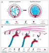
Figure 1. Early oogenesis in vertebrates
Major events in early oogenesis from germline stem cells through the formation of the primary follicle are depicted. Germline stem cells give rise to the mitotic oogonial cells. Upon the induction of meiosis, oocytes undergo the stages of prophase I: leptotene, zygotene, pachytene and diplotene. Oocytes arrest at diplotene stage (dyctate) until meiosis resumes during oocyte maturation later in oogenesis. The top row shows the cellular organization of oogonial cells, oocytes and somatic follicle cells at each stage. For the oogonia stage, a germline cyst is on the right and a presumptive cell division pattern in the cyst with cytoplasmic bridges depicted is on the left. This division pattern is based on the Drosophila model for germline cyst construction, and remains to be determined in vertebrates. On the left, five major themes in early oogenesis are depicted: mitosis, meiosis, cellular organization, apoptosis, and the Balbiani body mRNP granule. The blue bars to the right of each major process indicate their timing in the oogenesis pipeline. Within the bars, primary events of each process are depicted at each stage. Indigo bars indicate processes that occur similarly in zebrafish and mouse oocytes. Species-specific information is indicated by a dark aqua-blue bar for zebrafish, and a light aqua-blue bar for mouse. Known regulators are indicated in red (CDK2, RingoA and Tex14 were analyzed in mouse oocytes, Bucky ball in zebrafish oocytes. Retinoic acid is a known regulator in mouse and strongly implicated in zebrafish (Rodriguez-Mari et al., 2013)). Telomeres (red) indicate the bouquet configuration at zygotene, and Bb components (green) depict Bb formation in zygotene through diplotene.. The proposed scheme provides a conserved unified model for vertebrate oogenesis.
Figure 2
Figure 2. Mechanisms of telomere NE attachment and clustering during bouquet formation
(A) Telomere clustering and bouquet formation. Telomere tethering to perinuclear microtubules that emanate from the centrosome enable their rotation around the NE. Telomere movements shuffle entire chromosomes in the nucleus and allow for homology searches. Eventually, these movements lead to clustering of telomeres apposing the centrosome, and the cessation of movements then stabilizes correct homologous chromosome pairing. (B) A model for telomere attachment to the NE. (1) At the onset of meiosis, telomeres are found randomly within the nucleus. The Trf1 protein is bound as a dimer to telomeres as part of the Shelterin complex, a hexameric protein complex that binds to telomeric repeat DNA. Terb1 binds to Terb2, which can bind to Majin. Majin is anchored to the inner nuclear membrane (INM). At leptotene-zygotene stages, RingoA activates CDK2, which phosphorylates SUN1 in the nucleoplasm. This phosphorylation is required for telomere attachment to SUN1 by an unknown mechanism. It is not known if the phosphorylation is transient or persists during the following steps. (2) In response to a yet unknown signal, and perhaps stochastically, Terb1 binds one Trf1 monomer displacing the other. This recruits telomeres to the Terb1/Terb2/Majin complex. (3) Terb1 then binds telomeric DNA and a nearby SUN1 protein on the INM. SUN1 is bound to a KASH protein in the outer nuclear membrane (ONM), which in turn is associated with microtubules in the cytoplasm. This mechanism connects the intranuclear telomeres with the cytoplasmic perinuclear microtubules, allowing for telomere movements around the NE (in A).
Figure 3
Figure 3. Models for acetylated tubulin cables in the germline cyst
1. Cables extending through the CB and midbody of sister oocytes. 2. Cables curve around and remain within the cytoplasm. The acetylated cables may comprise a new type of cilium-like structure that is fully cytoplasmic or partially cytoplasmic (not shown). 3a. Cables are a primary cilium. 3b. Cables are a long subtype of primary cilia. For simplicity, pairwise CB connections between oocytes in the cyst are depicted. The number of CBs per oocyte may vary from one to four or more depending on the number of mitotic divisions and when each CB undergoes abscission.
Figure 4
Figure 4. Dynamics of oocyte polarization and early Bb formation
(A) The model depicts the size and cellular morphology of specific meiotic stages in the zebrafish and specifies events in oocyte polarization and Bb formation. Oocyte symmetry is broken during the zygotene bouquet stage, when Bb precursors first localize in the cytoplasm apposing the telomere bouquet. This is executed by the centrosome organizing center (MVC, Blue dashed circle). A nuclear cleft forms at pachytene, which is most pronounced at the onset of diplotene and then recedes until the nucleus resumes a spherical shape in ~50 m in diameter mid-diplotene oocytes. The centrosome localizes roughly to the center of the cleft and dissociates in the early ~25 m diameter diplotene oocyte. The panels below the cartoons show immunostained oocytes representative of each stage. Key steps in Bb formation are shown in green boxes. During cleft stages the aggregates are still amorphous and gradually consolidate into a single spherical granule of the mature Bb, in parallel to cleft closure. Scale bars are 5 m for oogonia-zygotene, and 10 m for pachytene and diplotene. Figure panels modified from (Elkouby et al., 2016). (B) Conservation of early Bb formation at the onset of meiosis. Left, a mouse oocyte at E14.5 shows early aggregation of Bb Golgi (white arrow, GM130, red) around the centrosome (Pericentrin, green), similar to the zebrafish Bb. Scale bar is 5 m. Image from (Lei & Spradling, 2016). Right, histological section of a Thermobia ovary showing a zygotene oocyte (dashed yellow ellipse), stained with Methylene Blue. A Bb mitochondrial aggregate (black arrow) is detected apposing the presumptive telomere cluster of the zygotene bouquet (white arrowheads, where chromosomes “touch” the NE), as also depicted in the zebrafish zygotene oocyte in (A). Scale bar is 5 m. Image modified from (Tworzydlo et al., 2016a).

References

    1. Ablain J, Durand EM, Yang S, Zhou Y, Zon LI. A CRISPR/Cas9 vector system for tissue-specific gene disruption in zebrafish. Dev Cell. 2015;32(6):756–764. doi: 10.1016/j.devcel.2015.01.032. - DOI - PMC - PubMed
    1. Akhmanova A, Hoogenraad CC. Microtubule minus-end-targeting proteins. Curr Biol. 2015;25(4):R162–171. doi: 10.1016/j.cub.2014.12.027. - DOI - PubMed
    1. Albamonte MI, Albamonte MS, Stella I, Zuccardi L, Vitullo AD. The infant and pubertal human ovary: Balbiani’s body-associated VASA expression, immunohistochemical detection of apoptosis-related BCL2 and BAX proteins, and DNA fragmentation. Hum Reprod. 2013;28(3):698–706. doi: 10.1093/humrep/des453. - DOI - PubMed
    1. Barton BR, Hertig AT. Ultrastructure of annulate lamellae in primary oocytes of chimpanzees (Pan troglodytes) Biol Reprod. 1972;6(1):98–108. - PubMed
    1. Beer RL, Draper BW. nanos3 maintains germline stem cells and expression of the conserved germline stem cell gene nanos2 in the zebrafish ovary. Dev Biol. 2013;374(2):308–318. doi: 10.1016/j.ydbio.2012.12.003. - DOI - PubMed

Publication types

MeSH terms

LinkOut - more resources