

The traumatic bone: trauma-induced heterotopic ossification
- PMID: 28668522
- PMCID: PMC6715128
- DOI: 10.1016/j.trsl.2017.06.004
The traumatic bone: trauma-induced heterotopic ossification
Abstract
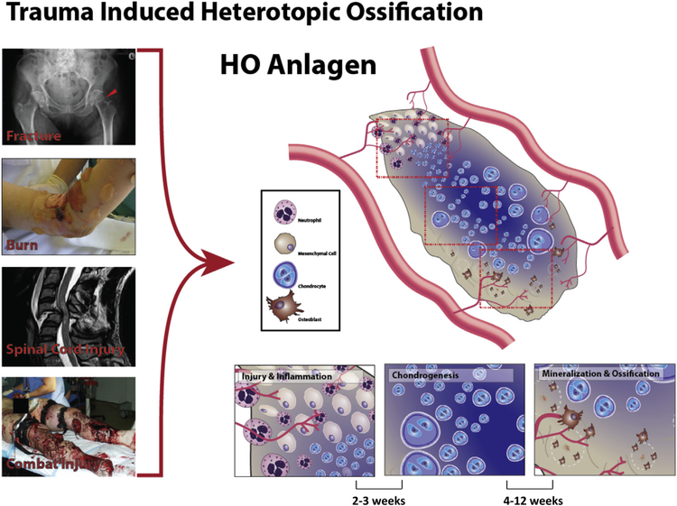
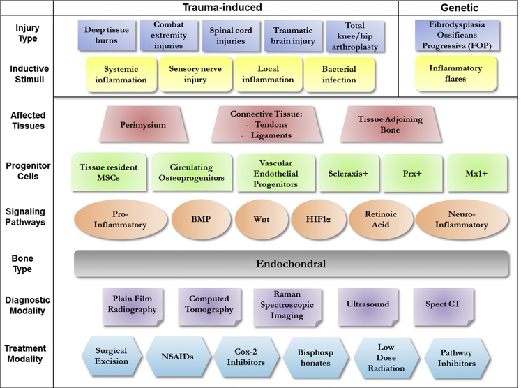
Heterotopic ossification (HO) is a common occurrence after multiple forms of extensive trauma. These include arthroplasties, traumatic brain and spinal cord injuries, extensive burns in the civilian setting, and combat-related extremity injuries in the battlefield. Irrespective of the form of trauma, heterotopic bone is typically endochondral in structure and is laid down via a cartilaginous matrix. Once formed, the heterotopic bone typically needs to be excised surgically, which may result in wound healing complications, in addition to a risk of recurrence. Refinements of existing diagnostic modalities, like micro- and nano-CT are being adapted toward early intervention. Trauma-induced HO is a consequence of aberrant wound healing, systemic and local immune system activation, infections, extensive vascularization, and innervation. This intricate molecular crosstalk culminates in activation of stem cells that initiate heterotopic endochondral ossification. Development of animal models recapitulating the unique traumatic injuries has greatly facilitated the mechanistic understanding of trauma-induced HO. These same models also serve as powerful tools to test the efficacy of small molecules which specifically target the molecular pathways underlying ectopic ossification. This review summarizes the recent advances in the molecular understanding, diagnostic and treatment modalities in the field of trauma-induced HO.
Published by Elsevier Inc.
Figures
References
-
- Ritter MA, Vaughan RB. Ectopic ossification after total hip arthroplasty. Predisposing factors, frequency, and effect on results. J Bone Joint Surg Am 1977;59:345–51. - PubMed
-
- Gear AJ, Buckley C, Kaplan F, Vanbeek A. Multifactorial refractory heterotopic ossification. Ann Plast Surg 2004;52:319–24. - PubMed
-
- Potter BK, Forsberg JA, Davis TA, et al. Heterotopic ossification following combat-related trauma. J Bone Joint Surg Am 2010;92 Suppl 2:74–89. - PubMed
-
- Fiori JL, Billings PC, de la Pena LS, Kaplan FS, Shore EM. Dysregulation of the BMP-p38 MAPK signaling pathway in cells from patients with fibrodysplasia ossificans progressiva (FOP). J Bone Miner Res 2006;21:902–9. - PubMed
Publication types
MeSH terms
Grants and funding
LinkOut - more resources
Full Text Sources
Other Literature Sources
Medical
