Review
. 2017 Aug;10(4):650-660.
doi: 10.1016/j.tranon.2017.04.007. Epub 2017 Jun 29.

Vascular Mimicry: A Novel Neovascularization Mechanism Driving Anti-Angiogenic Therapy (AAT) Resistance in Glioblastoma

Affiliations
Review

Vascular Mimicry: A Novel Neovascularization Mechanism Driving Anti-Angiogenic Therapy (AAT) Resistance in Glioblastoma

Kartik Angara et al. Transl Oncol. 2017 Aug.

Abstract

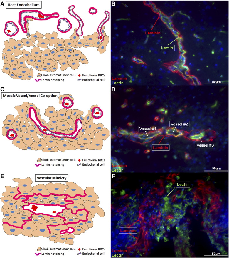
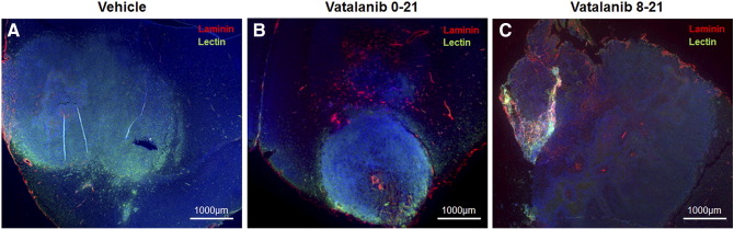
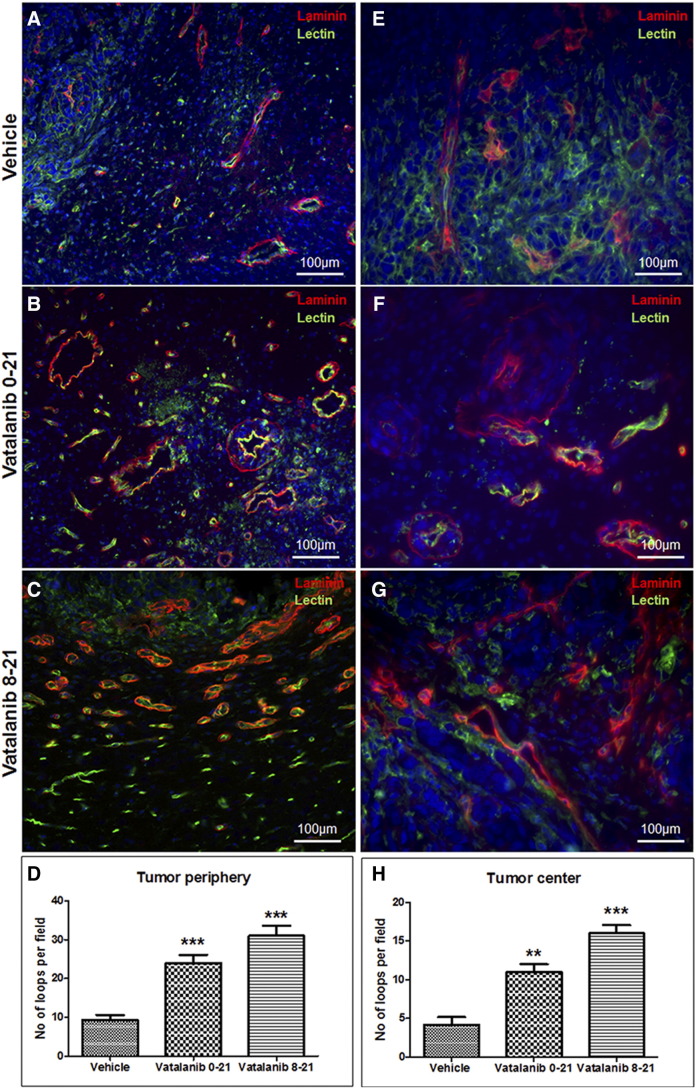
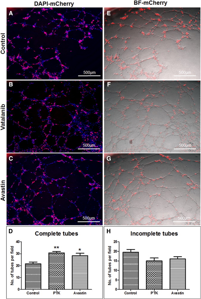
Glioblastoma (GBM) is a hypervascular neoplasia of the central nervous system with an extremely high rate of mortality. Owing to its hypervascularity, anti-angiogenic therapies (AAT) have been used as an adjuvant to the traditional surgical resection, chemotherapy, and radiation. The benefits of AAT have been transient and the tumors were shown to relapse faster and demonstrated particularly high rates of AAT therapy resistance. Alternative neovascularization mechanisms were shown to be at work in these resilient tumors to counter the AAT therapy insult. Vascular Mimicry (VM) is the uncanny ability of tumor cells to acquire endothelial-like properties and lay down vascular patterned networks reminiscent of host endothelial blood vessels. The VM channels served as an irrigation system for the tumors to meet with the increasing metabolic and nutrient demands of the tumor in the event of the ensuing hypoxia resulting from AAT. In our previous studies, we have demonstrated that AAT accelerates VM in GBM. In this review, we will focus on the origins of VM, visualizing VM in AAT-treated tumors and the development of VM as a resistance mechanism to AAT.


Figures

Figure 1
Figure 1
Schematic of vascular mimicry development driving anti-angiogenic therapy (AAT) resistance in glioblastoma. (A) Host endothelial-dependent blood vessel formation. (B) Host endothelial vessels visualized outside the tumor area. In the host tissue, the Laminin staining is present on the outside and the Lectin staining is present on the inside. (C) Formation of mosaic vessels in the tumor periphery. (D) The formation of a mosaic vessels can be evidenced here. The three host endothelial vessels are being encapsulated by the tumor cells (in red, Laminin). However, the tumors can be seen trying to form a physiological connection with the host endothelial-cell dependent vessels as observed in the vessels #1 and #2. The laminin-positive loops are seen to be attached or incorporated into host endothelial vessels and thus present as a super-imposed yellow colored structures (green from host endothelium and red from the tumor derived laminin). (E) Vascular Mimicry-Tube like vascular structures in the tumor center. Here the laminin based patterned networks are independent of the host endothelial cell contribution. (F) Vascular mimicry like patterned networks can be seen in the tumor center. In the tumor center, the Laminin staining is present independently of the Lectin staining because the tumor has begun the process of VM. The Lectin now depicts the tumor cells acquiring endothelial like properties, reminiscent of the host endothelial cells (Magnification 40×).
Figure 2
Figure 2
Accelerated VM in AAT-treated tumors in orthotopic human GBM mouse model. (B and C) Vatalanib-treated tumors show significantly higher number of laminin-positive areas that are spread over the entire tumor when compared to the vehicle-treated tumors (A). The images have been taken at a magnification of 2.5× to ease the depiction of tumor size in each treated animal of the respective group.
Figure 3
Figure 3
Mosaic vessels and Vascular Mimicry like-structures in the AAT-treated tumors. Tumor periphery showing laminin loops present in conjunction with the host-endothelial structures as visible with the green lectin staining in vehicle- (A) and vatalanib (B and C)-treated tumors. The mosaic vessels are also present at the tumor periphery, which are evident as the red-laminin and green-lectin superimposed yellow loop like structures. (D) Quantification of laminin-positive loop like-structures in the tumor periphery. In the central areas, Vatalanib-treated (F and G) tumors show significantly higher number of laminin-positive loop like structures indicative of VM, compared to the vehicle-treated tumors (E). These laminin loops are present independently of the host-endothelial structures. These laminin-positive patterned matrices are formed by the tumor cells alone, without any contribution from the host endothelial cells. (H) The quantification of the laminin-positive loops in the tumor center. Images have been taken from four different areas of the tumor. One representative image from one animal in the respective group has been shown here. The laminin-positive loops have been counted using Image J software and the statistical analyses were performed using Student's t test and P <.05 was considered significant.
Figure 4
Figure 4
Post-treated glioblastoma cells incorporate into host endothelial blood vessels. Panels A–C represent the super-imposition of red fluorescent (mCherry U251 cells) and the DAPI images. (D) Quantification of complete tubes in AAT-treated groups. Panels E–G represent the superimposition of red fluorescent (mCherry U251 cells) image over the bright field (BF) image (HUVEC cells). (H) Quantification of incomplete tubes in AAT-treated groups. mCherry U251 GBM cells and HUVEC cells were seeded in 1:2 ratio, treated with AAT post seeding and incubated for 6 h. One representative image from each treatment group has been shown here. Statistical analyses were performed using Student's t test and P < .05 was considered significant.

References

    1. Maniotis AJ, Folberg R, Hess A, Seftor EA, Gardner LM, Pe'er J, Trent JM, Meltzer PS, Hendrix MJ. Vascular channel formation by human melanoma cells in vivo and in vitro: vasculogenic mimicry. Am J Pathol. 1999;155(3):739–752. [S0002–9440(10)65173–5 [pii]] - PMC - PubMed
    1. Folberg R, Maniotis AJ. Vasculogenic mimicry. APMIS. 2004;112(7–8):508–525. [APMapm11207–0810 [pii]] - PubMed
    1. Vartanian AA. Signaling pathways in tumor vasculogenic mimicry. Biochemistry (Mosc) 2012;77(9):1044–1055. [BCM77091258 [pii]] - PubMed
    1. Clemente M, Perez-Alenza MD, Illera JC, Pena L. Histological, immunohistological, and ultrastructural description of vasculogenic mimicry in canine mammary cancer. Vet Pathol. 2010;47(2):265–274. - PubMed
    1. Chen X, Maniotis AJ, Majumdar D, Pe'er J, Folberg R. Uveal melanoma cell staining for CD34 and assessment of tumor vascularity. Invest Ophthalmol Vis Sci. 2002;43(8):2533–2539. - PubMed