Review
. 2018 May 15;12(3):227-235.
doi: 10.5009/gnl17061.

Prevalence, Pathogenesis, Diagnosis, and Management of Microscopic Colitis

Affiliations
Review

Prevalence, Pathogenesis, Diagnosis, and Management of Microscopic Colitis

Nicole Gentile et al. Gut Liver. .

Abstract

Microscopic colitis (MC), which is comprised of lymphocytic colitis and collagenous colitis, is a clinicopathological diagnosis that is commonly encountered in clinical practice during the evaluation and management of chronic diarrhea. With an incidence approaching the incidence of inflammatory bowel disease, physician awareness is necessary, as diagnostic delays result in a poor quality of life and increased health care costs. The physician faces multiple challenges in the diagnosis and management of MC, as these patients frequently relapse after successful treatment. This review article outlines the risk factors associated with MC, the clinical presentation, diagnosis and histologic findings, as well as a proposed treatment algorithm. Prospective studies are required to better understand the natural history and to develop validated histologic endpoints that may be used as end points in future clinical trials and serve to guide patient management.

Keywords: Colitis, collagenous; Colitis, lymphocytic; Colitis, microscopic.


Conflict of interest statement

CONFLICTS OF INTEREST

No potential conflict of interest relevant to this article was reported.

Figures

Fig. 1
Fig. 1
Lymphocytic colitis. Images of H&E staining showing intraepithelial lymphocytosis and a preserved colonic architecture (×20).
Fig. 2
Fig. 2
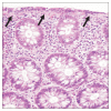
Collagenous colitis. Images of H&E staining showing a thickened subepithelial collagen band (arrows) and a preserved colonic architecture (×40).
Fig. 3
Fig. 3
Management of microscopic colitis (MC) according to symptom severity. NSAIDs, nonsteroidal anti-inflammatory drugs.

References

    1. Münch A, Langner C. Microscopic colitis: clinical and pathologic perspectives. Clin Gastroenterol Hepatol. 2015;13:228–236. doi: 10.1016/j.cgh.2013.12.026. - DOI - PubMed
    1. Fernández-Bañares F, Esteve M, Viver JM. Epidemiology of microscopic colitis. Gut. 2007;56:1033. doi: 10.1136/gut.2007.123133. - DOI - PMC - PubMed
    1. Gentile NM, Khanna S, Loftus EV, Jr, et al. The epidemiology of microscopic colitis in Olmsted County from 2002 to 2010: a population-based study. Clin Gastroenterol Hepatol. 2014;12:838–842. doi: 10.1016/j.cgh.2013.09.066. - DOI - PMC - PubMed
    1. Pardi DS, Loftus EV, Jr, Smyrk TC, et al. The epidemiology of microscopic colitis: a population based study in Olmsted County, Minnesota. Gut. 2007;56:504–508. doi: 10.1136/gut.2006.105890. - DOI - PMC - PubMed
    1. Bonderup OK, Wigh T, Nielsen GL, Pedersen L, Fenger-Grøn M. The epidemiology of microscopic colitis: a 10-year pathology-based nationwide Danish cohort study. Scand J Gastroenterol. 2015;50:393–398. doi: 10.3109/00365521.2014.940378. - DOI - PubMed

LinkOut - more resources