Review
. 2017 Oct:163:2-16.
doi: 10.1016/j.exer.2017.06.020. Epub 2017 Jun 29.

Meibomian glands, meibum, and meibogenesis

Affiliations
Review

Meibomian glands, meibum, and meibogenesis

Igor A Butovich. Exp Eye Res. 2017 Oct.

Abstract

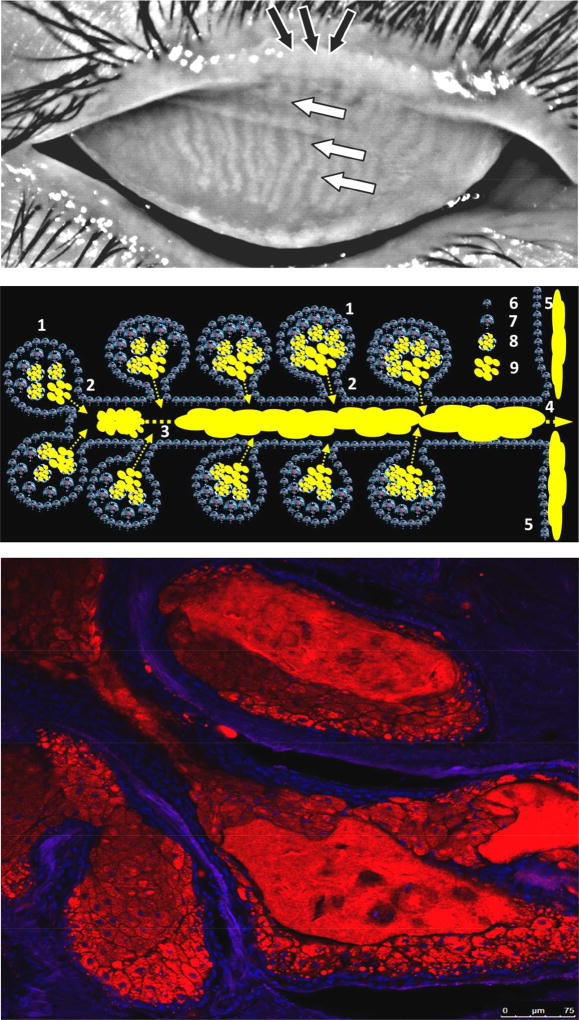
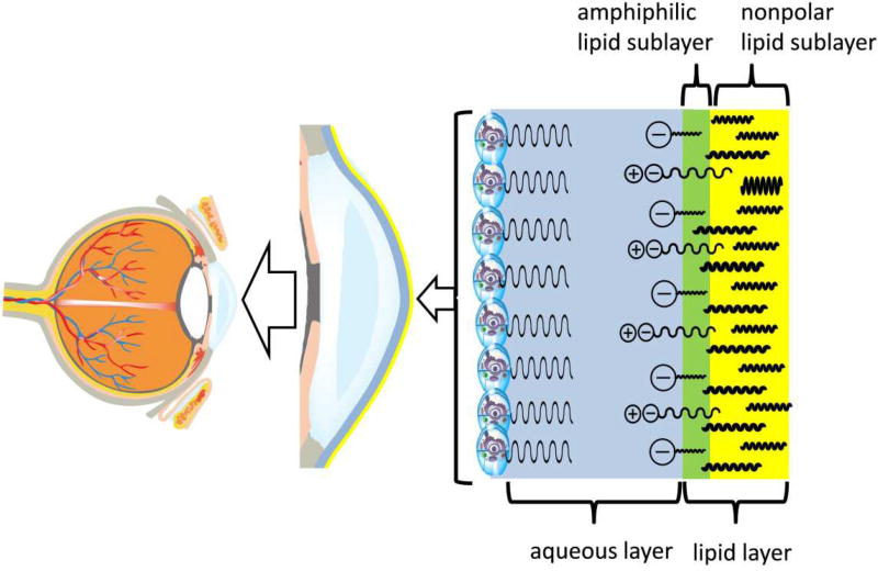
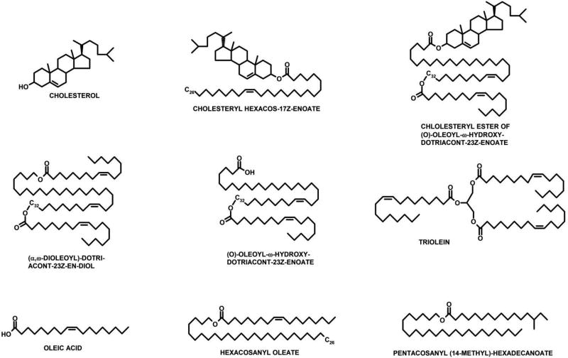
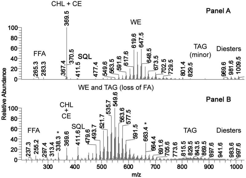
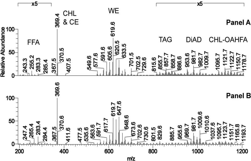
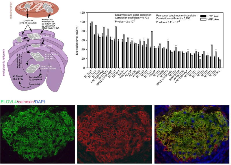
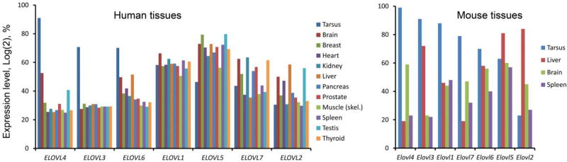
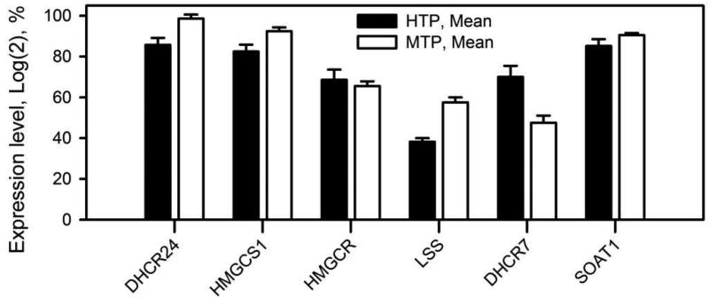
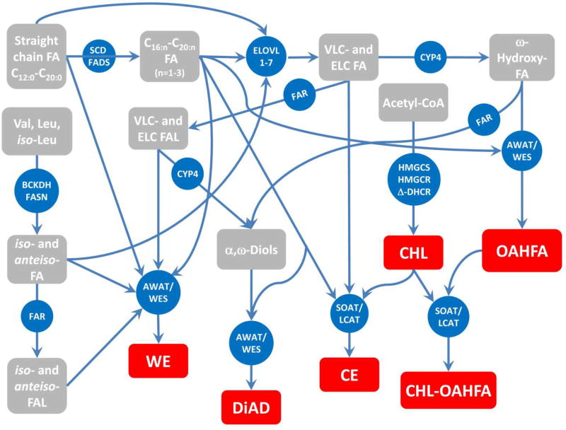
Meibum is a lipid-rich secretion that is produced by fully differentiated meibocytes in the holocrine Meibomian glands (MG) of humans and most mammals. The secretion is a part of a defense mechanism that protects the ocular surface from hazardous environmental factors, and from desiccation. Meibomian lipids that have been identified in meibum are very diverse and unique in nature. The lipid composition of meibum is different from virtually any other lipid pool found in the human body. In fact, meibum is quite different from sebum, which is the closest secretion that is produced by anatomically, physiologically, and biochemically related sebaceous glands. However, meibum of mice have been shown to closely resemble that of humans, implying similar biosynthetic mechanisms in MG of both species. By analyzing available genomic, immunohistochemical, and lipidomic data, we have envisioned a unifying network of enzymatic reactions that are responsible for biosynthesis of meibum, which we call meibogenesis. Our current theory is based on an assumption that most of the biosynthetic reactions of meibogenesis are catalyzed by known enzymes. However, the main features that make meibum unique - the ratio of identified classes of lipids, the extreme length of its components, extensive ω-hydroxylation of fatty acids and alcohols, iso- and anteiso-branching of meibomian lipids (e.g. waxes), and the presence of rather unique complex lipids with several ester bonds - make it possible that either the activity of known enzymes is altered in MG, or some unknown enzymes contribute to the processes of meibogenesis, or both. Studies are in progress to elucidate meibogenesis on molecular level.


Figures

Figure 1
Figure 1
Human eyelids, meibomian glands, and ascini. Upper panel. A high-contrast photography of a human upper eyelid with normal Meibomian glands that are visible as elongated whitish structures marked with white arrows (Wojtowicz and Butovich, unpublished). Middle panel. A schematic representation of the Meibomian gland: 1 – ascini; 2 – ductules; 3 – central duct which is being filled with meibum; 4 – orifice; 5 – secreted meibum flowing onto the lid margin and then onto ocular surface; 6 – undifferentiated Meibomian gland epithelial cells; 7 – partially differentiated meibocytes; 8 – completely differentiated, mature meibocytes with lipid droplets (yellow); 9 – lipid content released by ruptured meibocytes at the last stage of their life cycle. Note that different types of progenitor cells may exist in the glands. Lower panel. Lipid material, stored in lipid droplets, increases in amount within meibocytes as they move towards the center of the ascinus (McMahon, Wojtowicz, and Butovich, unpublished). Following cellular disintegration, the lipid (shown in red) is observed as a bulk mass. Lipids were stained with Nile Red and imaged to detect the signals from neutral lipids (excitation 488nm, emission 500–550nm). The nuclei were counterstained with DAPI (shown in blue). Note the virtual absence of neutral lipid staining in the basal layer of cells (only nuclei are visible) and a decreasing number of nuclei in the central parts of the ascinus (only lipid material is visible).
Figure 2
Figure 2
Schematic representation of the human tear film and its layers and sublayers. The inner aqueous layer is shown in blue. The layer is formed of a complex mixture of proteins, salts, and other low-molecular weight compounds. The aqueous layer covers the epithelial cells of cornea and conjunctiva, many of which express soluble (secreted, gel-forming) and bound (surface) mucins (Argueso, 2013; Hodges and Dartt, 2013; Mantelli and Argueso, 2008). The opposite side of the aqueous layer is covered with the tear film lipid layer (shown in green and yellow). The inner part of the lipid layer is enriched with amphiphilic lipids (anionic OAHFA and free fatty acids, and, possibly, zwitter-ionic species such as SM, PC, and lyso-PC). This layer is called amphiphilic (or "polar") lipid sublayer and is shown in green. The outer part of the lipid layer is formed mostly of nonpolar lipids and is called nonpolar lipid sublayer (shown in yellow). The actual tear film and tear film lipid layer are dynamic structures whose thickness and geometrical features change with time, blinking, aqueous tears in- and outflow, secretion of meibum, and tear evaporation.
Figure 3
Figure 3
Typical lipids of human meibum. The most common representatives of the corresponding classes of lipids are shown.
Figure 4
Figure 4
Mass-spectrometric comparison of human meibum and sebum. Most of ions depicted in Panels A and B are (M+H)+ adducts of the corresponding lipid species. Panel A. A representative APCI mass spectrum of a human meibum sample taken in the positive ion mode. Panel B. A representative APCI mass spectrum of a human sebum sample taken in the positive ion mode Adopted from (Butovich et al., 2016).
Figure 5
Figure 5
Observation MS profiles of mouse and human meibum. Panel A. Mass spectrum of mouse meibum in the m/z range of 200 to 1200. Normal phase HPLC-atmospheric pressure chemical ionization MS, positive ion mode. Averaged spectrum of meibomian lipids is shown. Panel B. Mass spectrum of human meibum. Same conditions as in Panel A. (from (Butovich et al., 2016)). Most of ions depicted in Panels A and B are (M+H)+ adducts of the corresponding lipid species.
Figure 6
Figure 6
Elongation of fatty acids in Meibomian glands of humans and mice. Upper left panel. Proposed sequence of FA elongation steps occurring in mitochondria and endoplasmic reticulum of meibocytes. Upper right panel. Expression patterns of major fatty acid elongation genes in tarsal plates of humans (4 donors, one sample of tarsal plate from each) and mice (12 mice; two pooled samples, 6 mice × 4 tarsal plates each) (Butovich et al., unpublished). Expression levels (Log2 values of SST-RMA-Gene-Full-Signals, varying from ~20 to ~2) were calculated using the Expression Console (built 1.4.1.46) from Affymetrix, then ranked from high to low, normalized from 100% (the highest expression level) to about 10% (the lowest expression level which was considered to be just above the noise level), and plotted as shown in the Panel (Butovich et al., 2016). Bottom panels. Immunohistochemical anti-ELOVL4 (green)/anti-calnexin (red)/DAPI (blue) co-staining of meibocytes in the human tarsal plates acini [from (Butovich et al., 2016)].
Figure 7
Figure 7
Differential expression profiles of the genes of the ELOVL family in human and mouse tissues. Left panel. Human tissues. Data for human tarsal plates is from (Butovich et al., 2016). Data for other tissues is recalculated from Affymetrix (file Human_Exon_tissues_AGCC_ARR_CHP, obtained using Affymetrix GeneChip® Human Exon 1.9 ST Array). Right panel. Mouse tissues. Data for tarsal plates of mice is from (Butovich et al., 2016).
Figure 8
Figure 8
Expression profiles of key genes of the biosynthesis of cholesterol and cholesteryl esters in Meibomian glands of humans and mice. HTP – human tarsal plates; MTP – mouse tarsal plates (recalculated from (Butovich et al., 2016)).
Figure 9
Figure 9
Meibogenesis – a network of biosynthetic reactions in meibomian glands of humans and mice that lead to biosynthesis of meibum [modified from (Butovich et al., 2016)]. Major confirmed final lipid products of meibogenesis are shown in red. Intermediate products are shown in grey. Key enzymes are shown in blue. The presence and nature of meibomian lipids have been established in GC-MS and HPLC-MS experiments. The nature of enzymes has been predicted from their respective gene expression patterns, and confirmed (for some of them) using immunohistochemical and immunocytochemical approaches [(Butovich et al., 2016) and references cited therein].

References

    1. FAR1 Gene (Protein Coding) Fatty Acyl-CoA Reductase 1. 2017a GeneCards.org.
    1. FAR2 Gene (Protein Coding) Fatty Acyl-CoA Reductase 2. 2017b GeneCards.org.
    1. Steroid Biosynthesis - Reference Pathway. Kyoto Encyclopedia of Genes and Genomes 2017c
    1. Agbaga MP, Mandal MN, Anderson RE. Retinal very long-chain PUFAs: new insights from studies on ELOVL4 protein. Journal of lipid research. 2010;51:1624–1642. - PMC - PubMed
    1. Aluru SV, Shweta A, Bhaskar S, Geetha K, Sivakumar RM, Utpal T, Padmanabhan P, Angayarkanni N. Tear Fluid Protein Changes in Dry Eye Syndrome Associated with Rheumatoid Arthritis: A Proteomic Approach. The ocular surface. 2017;15:112–129. - PubMed