Review
. 2017 Jul;10(7):265-289.
doi: 10.1177/1756285617709723. Epub 2017 May 24.

Differential diagnosis of neuromyelitis optica spectrum disorders

Affiliations
Review

Differential diagnosis of neuromyelitis optica spectrum disorders

Sung-Min Kim et al. Ther Adv Neurol Disord. 2017 Jul.

Abstract

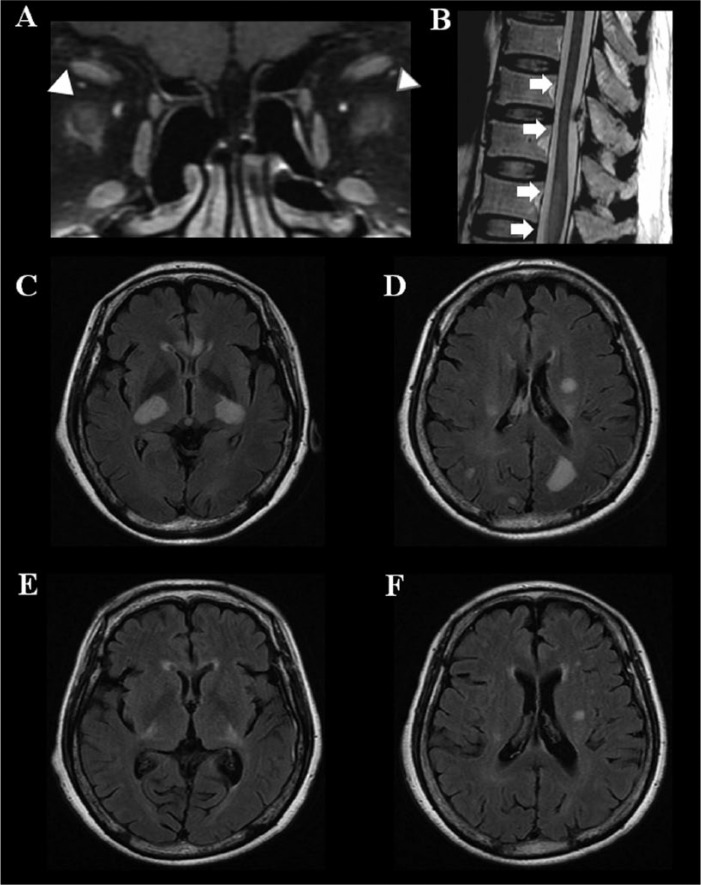
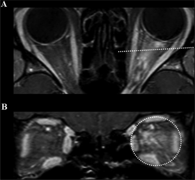
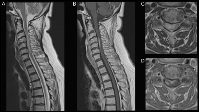
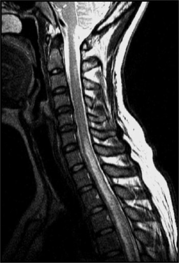
Neuromyelitis optica spectrum disorder (NMOSD) is an inflammatory disorder of the central nervous system (CNS) mostly manifesting as optic neuritis and/or myelitis, which are frequently recurrent/bilateral or longitudinally extensive, respectively. As the autoantibody to aquaporin-4 (AQP4-Ab) can mediate the pathogenesis of NMOSD, testing for the AQP4-Ab in serum of patients can play a crucial role in diagnosing NMOSD. Nevertheless, the differential diagnosis of NMOSD in clinical practice is often challenging despite the phenotypical and serological characteristics of the disease because: (1) diverse diseases with autoimmune, vascular, infectious, or neoplastic etiologies can mimic these phenotypes of NMOSD; (2) patients with NMOSD may only have limited clinical manifestations, especially in their early disease stages; (3) test results for AQP4-Ab can be affected by several factors such as assay methods, serologic status, disease stages, or types of treatment; (4) some patients with NMOSD do not have AQP4-Ab; and (5) test results for the AQP4-Ab may not be readily available for the acute management of patients. Despite some similarity in their phenotypes, these NMOSD and NMOSD-mimics are distinct from each other in their pathogenesis, prognosis, and most importantly treatment. Understanding the detailed clinical, serological, radiological, and prognostic differences of these diseases will improve the proper management as well as diagnosis of patients.

Keywords: Devic’s disease; aquaporin-4 antibody; differential diagnosis; longitudinally extensive transverse myelitis; multiple sclerosis; myelin oligodendrocyte glycoprotein antibody; neuromyelitis optica spectrum disorders; optic neuritis.


Conflict of interest statement

Conflict of interest statement: Prof. Kazuo Fujihara serves on scientific advisory boards for Bayer Schering Pharma, Biogen Idec, Mitsubishi Tanabe Pharma Corporation, Novartis Pharma, Chugai Pharmaceutical, Ono Pharmaceutical, Nihon Pharmaceutical, Merck Serono, Alexion Pharmaceuticals, Medimmune and Medical Review; has received funding for travel and speaker honoraria from Bayer Schering Pharma, Biogen Idec, Eisai Inc., Mitsubishi Tanabe Pharma Corporation, Novartis Pharma, Astellas Pharma Inc., Takeda Pharmaceutical Company Limited, Asahi Kasei Medical Co., Daiichi Sankyo, and Nihon Pharmaceutical; serves as an editorial board member of Clinical and Experimental Neuroimmunology (2009 to present) and an advisory board member of the Sri Lanka Journal of Neurology; has received research support from Bayer Schering Pharma, Biogen Idec Japan, Asahi Kasei Medical, The Chemo-Sero-Therapeutic Research Institute, Teva Pharmaceutical, Mitsubishi Tanabe Pharma, Teijin Pharma, Chugai Pharmaceutical, Ono Pharmaceutical, Nihon Pharmaceutical, and Genzyme Japan.

Figures

Figure 1.
Figure 1.
Polyfocal manifestation of acute disseminated encephalomyelitis. A = fat-suppressed T1-weighted image with gadolinium enhancement; B = T2-weighted image; C, D, E, and F = fluid-attenuated inversion recovery (FLAIR). A 66-year-old woman developed acute bilateral blindness after 2 weeks of influenza vaccination. She developed successive paraplegia and altered mental status over the following week. On admission, the MRI revealed gadolinium enhancement in both optic nerves (arrowhead, A), acute transverse myelitis (arrow) involving lower thoracic cord and conus medullaris (arrow, B), and disseminated T2 high-intensity lesions involving the cerebral white matter and deep gray matter (C and D). She was treated with high-dose methylprednisolone followed by plasmapheresis. The brain lesions almost disappeared within 11 months (E and F). She experienced no relapse during 4 years of follow-up.
Figure 2.
Figure 2.
Peri-neural enhancement pattern in orbit MRI of MOG-Ab-associated optic neuritis. A 22-year-old woman presented with recurrent bilateral optic neuritis. Her orbital MRI showed extensive enhancement patterns that were not confined to the left optic nerve, but extended to the soft tissues around the optic nerve (peri-neural enhancement, A and B). She tested negative for AQP4-Ab, but positive for MOG-Ab. After treatment with intravenous methylprednisolone followed by oral corticosteroids, her visual acuity recovered. This peri-neural enhancement pattern can be frequently found in optic neuritis associated with MOG-Ab. All MRI images are T1-weighted with gadolinium enhancement; dotted lines in (A) highlight the level of the transverse images in (B). AQP4-Ab, autoantibody against aquaporin-4; MOG-Ab, myelin oligodendrocyte glycoprotein antibody.
Figure 3.
Figure 3.
Right optic neuropathy and thoracic lymphadenopathies in patients with sarcoidosis. A 55-year-old year woman presented with right optic neuropathy. Her ophthalmologic examination revealed a relative afferent pupillary defect and disc swelling in her right eye (A). Her routine chest X-ray showed hilar enlargement (arrowhead, B), and FDG-PET showed multiple lymphadenopathies in the both mediastinal, perihilar, and subclavian areas (C). Together with bronchoscopic biopsy that revealed non-caseating granuloma and the increased level of serum angiotensin converting enzyme, she was diagnosed with neurosarcoidosis. After treatment with intravenous methylprednisolone followed by high-dose oral steroid (1 g/kg), her visual acuity improved from 0.5 to 0.8 and her lymphadenopathies were also improved (D). Interestingly, her follow-up fundus exam showed disc hemorrhage, which is uncommon in optic neuritis (E).
Figure 4.
Figure 4.
Neurosarcoidosis manifesting LETM. A 66-year-old woman with a history of ocular sarcoidosis (granulomatous uveitis) developed weakness of the right upper arm and leg, numbness and pain in her bilateral upper arm, and dysuria, which progressed for a month. Physical examination revealed no abnormalities nor lymphadenopathy. Neurological examination revealed a right hemiparesis, hyperreflexia with extensor plantar reflexes, sensory disturbance in the right C5–6 dermatome. Blood tests showed normal white blood cell count, and normal CRP and serum angiotensin converting enzyme levels. Both serum AQP4-Ab and MOG-Ab were negative. Cerebrospinal fluid examination showed a mildly elevated protein level. Chest CT revealed mediastinal lymphadenopathy. Spinal MRI exhibited a longitudinally extensive intramedullary HSI lesion at C3–6 (A) with partial contrast-enhancement (B). Axial MRI showed transverse HSI (C); and ventral and right-sided circumferential enhancements (D). She was diagnosed with sarcoid myelopathy and treated with intravenous methylprednisolone (1000 mg daily, 3 days) followed by oral prednisolone (0.5 mg/kg daily). Her symptoms began to improve. A and C = T2-weighted MRI; B and D = T1-weighted MRI with gadolinium enhancement. AQP4-Ab, autoantibody against aquaporin-4; HSI, high signal intensity; MOG-Ab, myelin oligodendrocyte glycoprotein antibody.
Figure 5.
Figure 5.
Coexistence of the neuromyelitis optic spectrum disorder and SS. A 37-year-old woman with a history of bilateral optic neuritis presented with paraparesis. Her spinal MRI showed LETM and she was positive for AQP4-Ab. Meanwhile, she also had SS, according to the symptoms (dry eye and mouth), signs (positive scintigraphy and Shirmer’s test), histopathology (lymphocytic infiltration in the salivary gland biopsy), and a positive anti-Ro antibody result. She had NMOSD-AQP4 and SS. AQP4-Ab, autoantibody against aquaporin-4; LETM, longitudinally extensive transverse myelitis; NMOSD-AQP4, neuromyelitis optica spectrum disorder with AQP4-Ab; SS, Sjogren’s syndrome.
Figure 6.
Figure 6.
Primary CNS lymphoma manifesting as a longitudinally extensive myelitis. A 70-year-old female presented with subacute paraplegia. Her initial spinal MRI showed T2 HSI lesions that were longitudinally extensive (A) and involved almost the entire width of the spinal cord in the axial plane (B). The gadolinium-enhanced MRI revealed enhancement in the peripheral white matter of the spinal cord (C and D), which is not common in NMOSD. Her brain MRI revealed multiple T2 HSI lesions in the white matter (E), external capsule of the basal ganglia, and splenium of the corpus callosum (F). Repeated assay for AQP4-Ab was negative. As her initial spinal cord biopsy did not reveal any malignant cells, she received an initial treatment of corticosteroid combined with plasmapheresis. She partially improved after the treatment, but nevertheless worsened again to develop quadriparesis. Her follow-up MRI revealed more extensive T2 HSI lesion (G) and gadolinium-enhancing lesions (H). 18F-fludeoxyglucose positron emission tomography revealed a hypermetabolic lesion in the spinal cord (I). A second spinal cord biopsy diagnosed a primary CNS lymphoma. A, B, and G = T2-weighted MRI; C, D, and H = T1-weighted MRI with gadolinium enhancement; E and F = fluid-attenuated inversion recovery (FLAIR) MRI; I = 18F-fludeoxyglucose positron emission tomography (18F-FDG-PET). Red line in A and blue line in D highlight the level where the axial images in B and C are taken, respectively. AQP4-Ab, autoantibody against aquaporin 4; HSI, high signal intensity; NMOSD, neuromyelitis optica spectrum disorder.
Figure 7.
Figure 7.
NBD manifesting LETM and progressive brain atrophy. A 25-year-old man who had been experiencing progressive emotional lability for 2 years presented with acute paraparesis. His spinal cord MRI revealed LETM involving the entire cervical and thoracic spine (A) and brain MR showed T2 HSI lesions in the cerebral white matter (B) and mild brainstem atrophy (C). He had recurrent oral ulcers, perianal ulcers, and acineform eruptions, and thereby was diagnosed with NBD. Despite combined treatment of corticosteroid and cyclophosphamide, his neurologic status worsened. In 6 months after the onset of myelitis, he became bed-ridden without any spontaneous speech. After treatment with infliximab he improved to be able to walk without assistance. His follow-up brain MRI in 4 years showed severe atrophy of the brainstem (D) and the cerebrum with moderate T2 HSI changes in the white matter (E). A = T2-weighted MRI; B–E = fluid-attenuated inversion recovery (FLAIR) MRI. HSI, high signal intensity; LETM, longitudinally extensive transverse myelitis; NBD, neuro-Behçet’s disease.
Figure 8.
Figure 8.
Two cases of SDAVF. Patient A (A and B), a 59-year-old male, presented with subacute quadriparesis that progressed to bed-ridden state over 4 months. On admission, his cervical spine MRI showed a typical signal void (arrowhead, A) and enhancing vascular structures (arrow, B) over the ventral surface of the cervical spine. He was diagnosed with SDAVF and was treated with embolization. Meanwhile, patient B (C – H), a 78-year-old male, presented with subacute progressive paraparesis over 18 months. His spinal MRI showed diffuse longitudinally extensive T2 HSI lesions in the thoracic spine without definite signal void (C and D), gadolinium enhancement of the spinal cord (E), and prominently enhanced vascular structures over the dorsal surface of the spinal cord (arrow, F). The spinal angiography of patient B, performed of the thoracic T8 spinal dorsal artery (G), showed a SDAVF and engorged/tortuous medullary veins (H). Note that the findings of conventional spinal MRI in patients with SDAVF can vary widely, therefore clinical suspicions are most important. A, C, and D = T2-weighted MRI; B, E, and F = T1-weighted MRI with gadolinium enhancement; G and H = spinal angiography. HSI, high signal intensity; SDAVF, spinal dural arteriovenous fistula; T8, thoracic vertebrae 8.
Figure 9.
Figure 9.
Bilateral optic neuropathy in neurosyphilis. A 34-year-old man experienced subacute, progressive visual loss. In 3 months he became blind in his right eye and his left vision became blurred, combined with a visual field defect. The orbit MRI revealed a diffuse T2 HSI in the right optic nerve (arrow) and also moderate T2 HSI in the left optic nerve (arrow head) (A and B). The cerebrospinal fluid revealed pleocytosis, increased level of protein, positive venereal disease research laboratory (VDRL), and fluorescent treponemal antibody absorption (FTA-ABS) test results. After treatment with intravenous penicillin, his constricted visual field in the left eye, which represented a pattern of perineuritis (C) improved over one month (D). A and B = T2-weighted image, C and D = Humphrey perimetry. HSI, high signal intensity.
Figure 10.
Figure 10.
Progressive visual field defect in a patient with LHON. A 46-year-old man had painless subacute visual loss to hand perception only in his right eye over 6 months. Eight months from symptom onset, his left eye developed a central scotoma (A) which gradually enlarged over 8 months (B–F). After two years from symptom onset, his visual acuity in the right and left eyes were hand perception and light perception, respectively. His genetic testing for LHON revealed the pathologic mitochondrial DNA 11778 (GA) point mutation. A–F = Goldmann perimetry. LHON, Leber hereditary optic neuropathy.

References

    1. Wingerchuk DM, Banwell B, Bennett JL, et al. International consensus diagnostic criteria for neuromyelitis optica spectrum disorders. Neurology 2015; 85: 177–189. - PMC - PubMed
    1. Flanagan E, Cabre P, Weinshenker B, et al. Epidemiology of aquaporin-4 autoimmunity and neuromyelitis optica spectrum. Ann Neurol. Epub ahead of print 17 February 2016. DOI: 10.1002/ana.24617. - DOI - PMC - PubMed
    1. Wingerchuk D. Neuromyelitis optica: effect of gender. J Neurol Sci 2009; 286: 18–23. - PubMed
    1. Kitley J, Leite M, Nakashima I, et al. Prognostic factors and disease course in aquaporin-4 antibody-positive patients with neuromyelitis optica spectrum disorder from the United Kingdom and Japan. Brain 2012; 135: 1834–1849. - PubMed
    1. Kim SM, Waters P, Woodhall M, et al. Gender effect on neuromyelitis optica spectrum disorder with aquaporin4-immunoglobulin G. Mult Scler. Epub ahead of print 19 October 2016. DOI: 10.1177/1352458516674366. - DOI - PubMed

LinkOut - more resources