

The crosstalk between β-catenin signaling and type I, type II and type III interferons in lung cancer cells
- PMID: 28670369
- PMCID: PMC5489881
The crosstalk between β-catenin signaling and type I, type II and type III interferons in lung cancer cells
Abstract
Background/objective: IFNs induce potent antiviral and antitumor activities. β-catenin pathway is a surviving pathway adapted by carcinogenetic mechanisms of various cancers. Crosstalk between these pathways has not been well described in lung cancer cells.
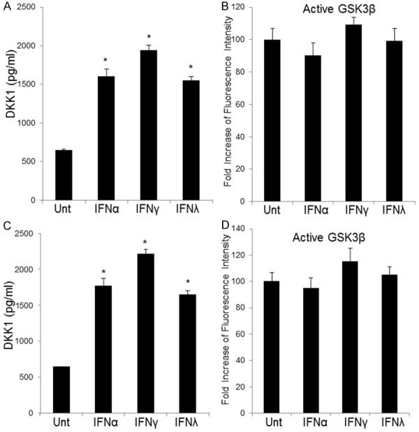
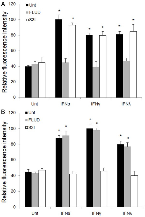
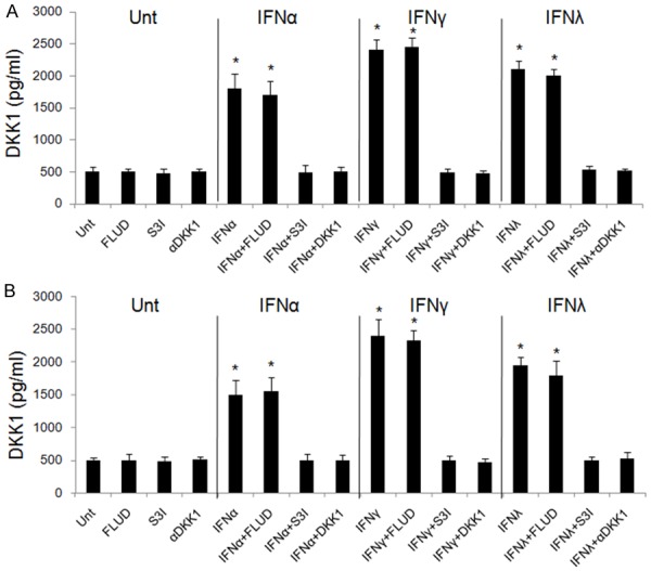
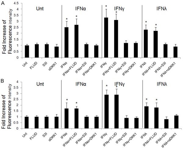
Methods: Lung cancer cell lines, A549 and Calu-3, were used in this study. β-catenin protein levels and signaling activities were tested by flow cytometry and luciferase assay. Cell proliferation was measured by counting viable cells under microscope, and apoptosis by TUNEL assay and caspase 3 activation. DKK1 and GSK3β levels were tested by flow cytometry. Secreted DKK1 was measured by ELISA. αDKK1 , FLUD and S3I were to inhibit DKK1, STAT1 and STAT3 activities, respectively.
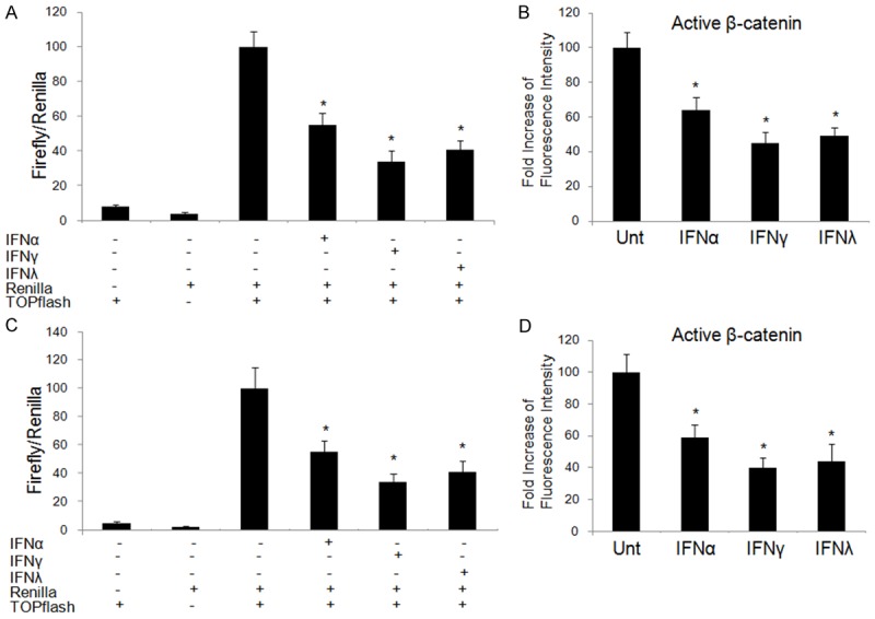
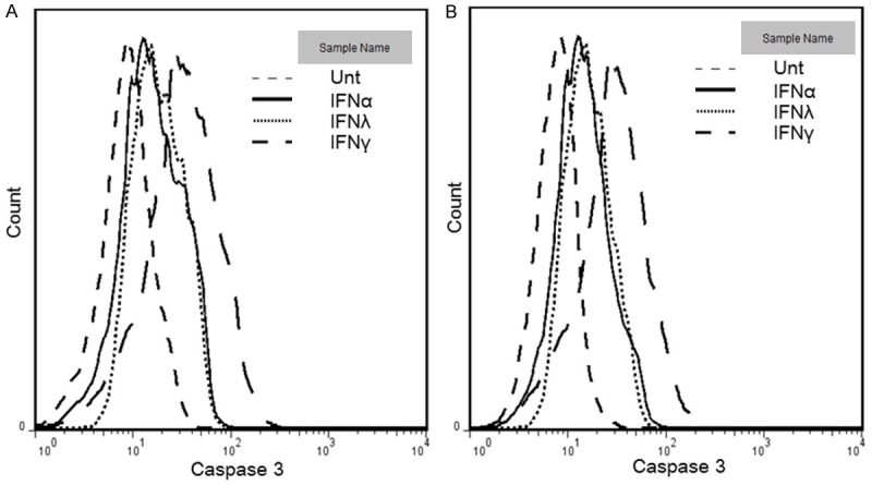
Results: All of IFNα, IFNγ and IFNλ1 suppressed β-catenin signaling in A549 and Calu-3 cells, where IFNγ was the strongest (P<0.05). They inhibited cellular proliferation and promoted apoptosis. IFNγ gave greater induction ability compared to IFNα and IFNλ1 (P<0.05). All tested IFNs promoted DKK1 activation but not GSK3β in A549 and Calu-3 cells. IFNs activated STAT1 and STAT3. But only STAT3 was vital for IFN-mediated DKK1 activation and apoptosis. Plus, DKK1 antagonist abrogated IFN-mediated apoptosis. The degree of STAT3 activation was corresponding to the level of apoptosis induced by different IFNs (P<0.05).
Conclusions: In lung cancer cells, all three types of IFNs can induce apoptosis via suppressing β-catenin signaling by a STAT3- and DKK1-dependent manner. This findings demonstrate a link between IFNs and β-catenin signaling, which may possess potentials on the development of novel therapeutic measures against lung cancer.
Keywords: crosstalk; interferon; lung cancer; β-catenin.
Conflict of interest statement
None.
Figures

Similar articles
-
Comparison of the regulation of β-catenin signaling by type I, type II and type III interferons in hepatocellular carcinoma cells.PLoS One. 2012;7(10):e47040. doi: 10.1371/journal.pone.0047040. Epub 2012 Oct 4. PLoS One. 2012. PMID: 23056571 Free PMC article.
-
STAT1 modification improves therapeutic effects of interferons on lung cancer cells.J Transl Med. 2015 Sep 8;13:293. doi: 10.1186/s12967-015-0656-0. J Transl Med. 2015. PMID: 26351076 Free PMC article.
-
STAT1-induced upregulation of LINC00467 promotes the proliferation migration of lung adenocarcinoma cells by epigenetically silencing DKK1 to activate Wnt/β-catenin signaling pathway.Biochem Biophys Res Commun. 2019 Jun 18;514(1):118-126. doi: 10.1016/j.bbrc.2019.04.107. Epub 2019 Apr 23. Biochem Biophys Res Commun. 2019. PMID: 31027730
-
Safety, Tolerability, and Immunogenicity of Interferons.Pharmaceuticals (Basel). 2010 Apr 20;3(4):1162-1186. doi: 10.3390/ph3041162. Pharmaceuticals (Basel). 2010. PMID: 27713294 Free PMC article. Review.
-
IFNs-signaling effects on lung cancer: an up-to-date pathways-specific review.Clin Exp Med. 2017 Aug;17(3):281-289. doi: 10.1007/s10238-016-0432-3. Epub 2016 Jul 14. Clin Exp Med. 2017. PMID: 27416926 Review.
Cited by
-
B7-H3 in Pediatric Tumors: Far beyond Neuroblastoma.Cancers (Basel). 2023 Jun 21;15(13):3279. doi: 10.3390/cancers15133279. Cancers (Basel). 2023. PMID: 37444389 Free PMC article. Review.
-
Age Matters: Key Contributors to Interferon Toxicity in Infants During Influenza Virus Infection.Viruses. 2025 Jul 17;17(7):1002. doi: 10.3390/v17071002. Viruses. 2025. PMID: 40733618 Free PMC article. Review.
-
Multiplexed protein profiling reveals spatial subcellular signaling networks.iScience. 2022 Aug 18;25(9):104980. doi: 10.1016/j.isci.2022.104980. eCollection 2022 Sep 16. iScience. 2022. PMID: 36093051 Free PMC article.
-
A genetic screen in macrophages identifies new regulators of IFNγ-inducible MHCII that contribute to T cell activation.Elife. 2021 Nov 8;10:e65110. doi: 10.7554/eLife.65110. Elife. 2021. PMID: 34747695 Free PMC article.
-
N-cadherin mechanosensing in ovarian follicles controls oocyte maturation and ovulation.Elife. 2025 Jul 29;13:RP92068. doi: 10.7554/eLife.92068. Elife. 2025. PMID: 40728874 Free PMC article.
References
-
- Torre LA, Bray F, Siegel RL, Ferlay J, Lortet-Tieulent J, Jemal A. Global cancer statistics, 2012. CA Cancer J Clin. 2015;65:87–108. - PubMed
-
- Hao M, Wang X, Du J, Liu L, Jiao Y, Wu H, Zheng J, Li W. Cytokine levels are associated with the severity of varicella infections. J Infect Dev Ctries. 2015;9:190–196. - PubMed
-
- Wang W, Zhu Y, Wu H, Jiao Y, Van Halm-Lutterodt N, Li W. IL-6 and IFNgamma are elevated in severe mumps cases: a study of 960 mumps patients in China. J Infect Dev Ctries. 2014;8:208–214. - PubMed
LinkOut - more resources
Full Text Sources
Research Materials
Miscellaneous