. 2017 Jun 15;9(6):2901-2910.
eCollection 2017.

Overexpression of CD47 predicts poor prognosis and promotes cancer cell invasion in high-grade serous ovarian carcinoma

Affiliations

Overexpression of CD47 predicts poor prognosis and promotes cancer cell invasion in high-grade serous ovarian carcinoma

Yinuo Li et al. Am J Transl Res. .

Abstract

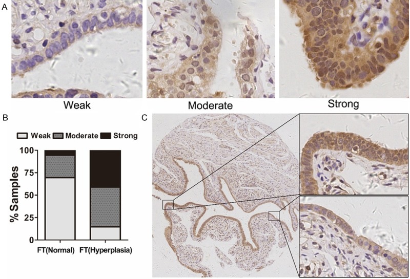
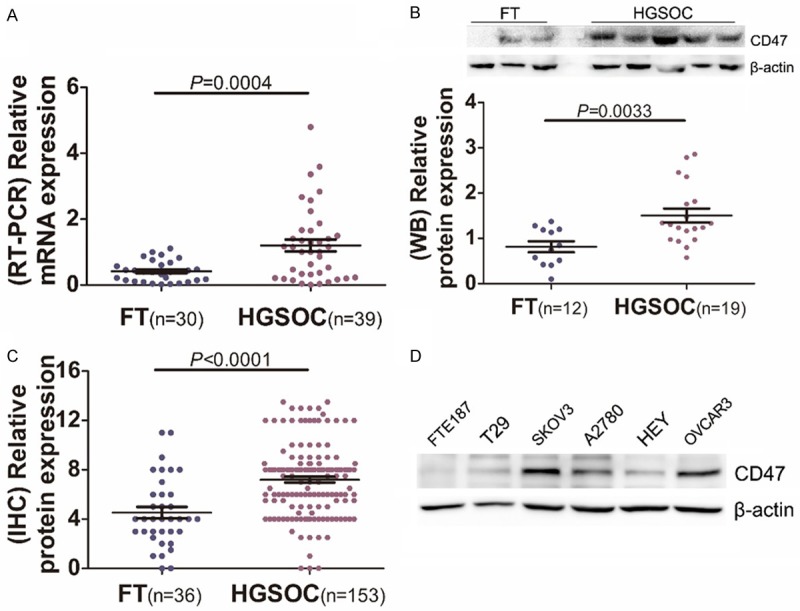
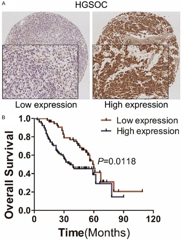
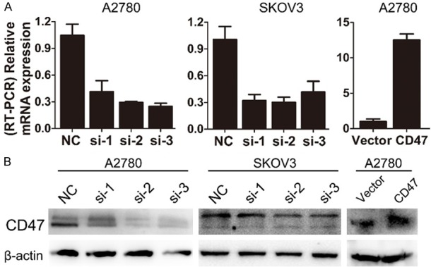
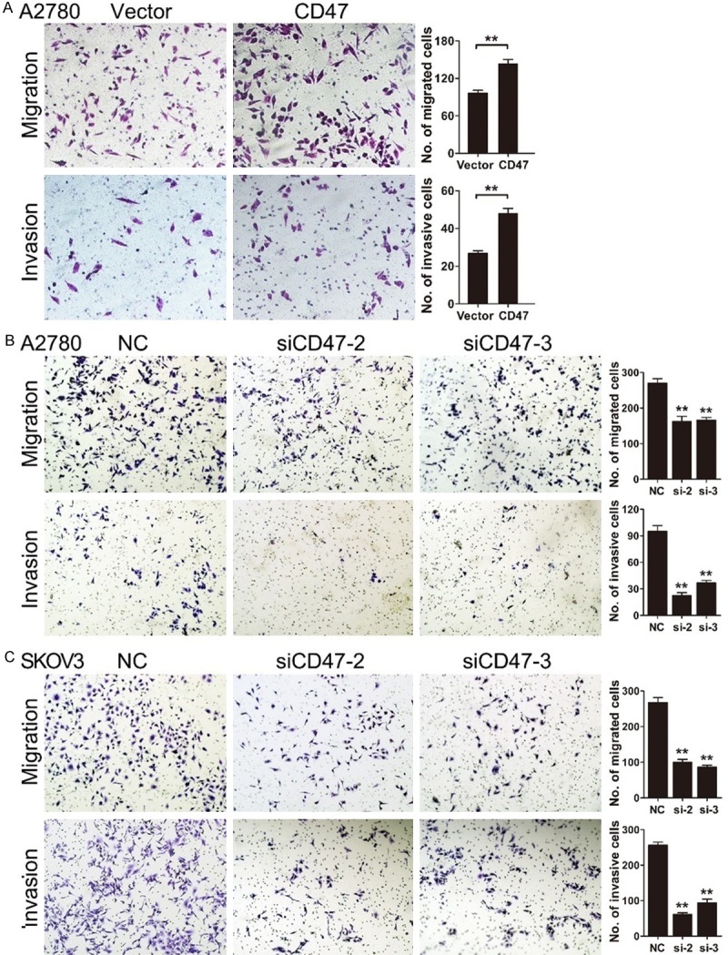
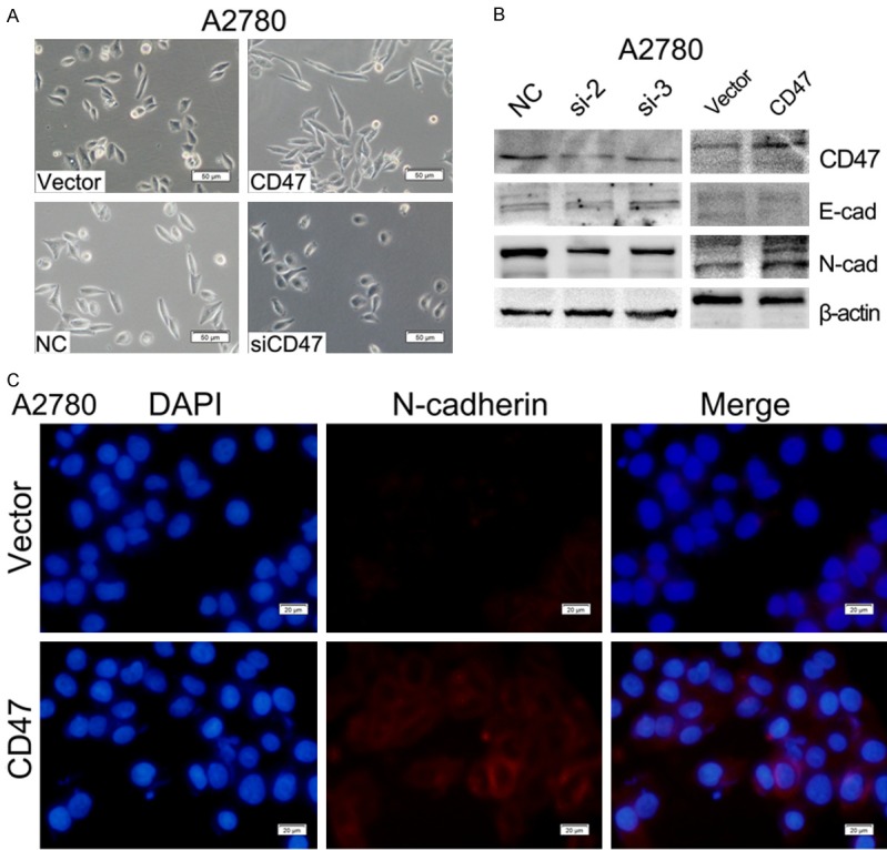
CD47 is an antiphagocytic signal that cancer cells employ to inhibit macrophage-mediated destruction. CD47 is overexpressed in various human malignancies. However, the expression and functional significance of CD47 in high-grade serous ovarian carcinoma (HGSOC) has not been completely understood. In this study, we reported that CD47 was commonly overexpressed in HGSOC. Higher CD47 expression was significantly correlated with poor prognosis of HGSOC patients. Functional investigations revealed that CD47 overexpression in ovarian cancer cells significantly promoted migration and invasion. Moreover, CD47 induced epithelial-mesenchymal transition (EMT) through modulating E-cadherin and N-cadherin. Our findings suggest that up-regulation of CD47 is correlated with ovarian cancer progression and it might be a potential biomarker for predicting clinical outcomes.

Keywords: CD47; EMT; high-grade serous ovarian carcinoma; invasion; prognosis.


Conflict of interest statement

None.

Figures

Figure 1
Figure 1
CD47 expression in epithelial hyperplasia and normal fallopian tube. A: Immunohistochemical staining of CD47 expression in normal (n=36) and hyperplasia (n=34) FTs. B: The histogram indicates the CD47 expression level distribution in normal and hyperplasia FTs. C: CD47 expression in the same FT sample including multiple layer area and single layer area.
Figure 2
Figure 2
Expression analysis of CD47 in high-grade serous ovarian carcinoma. A: Relative expression of CD47 in HGSOCs (n=39) and FTs (n=30) measured by Real-time PCR. B: Western blot analysis of CD47 in HGSOCs (n=19) compared to FTs (n=12). C: Quantitative immunohistochemical analysis of CD47 expression in HGSOCs (n=153) and FTs (n=36). D: CD47 expression in ovarian cancer cell lines and two normal cell lines measured by western blot.
Figure 3
Figure 3
Immunohistochemical analysis of CD47 expression and its correlation with prognosis of HGSOC patients. A: Representative images of immunohistochemical analysis of CD47 expression in HGSOCs. B: Kaplan-Meier survival analysis of CD47 high-expression group versus low-expression group.
Figure 4
Figure 4
CD47 expression in ovarian cancer cells with CD47 overexpression and knockdown. A: Relative CD47 mRNA level in CD47 overexpression and knockdown cell lines. B: Western blot analysis of CD47 in CD47 overexpression and knockdown cell lines.
Figure 5
Figure 5
CD47 promotes migration and invasion of ovarian cancer cells. A: Ectopic expression of CD47 enhanced migration and invasion ability in A2780 cells. B and C: A2780 and SKOV3 cell lines with CD47 knockdown showed reduced migration and invasion ability. The data shown here are from a representative experiment repeated three times with similar results. Data are presented as means ± S.D. *P<0.05, **P<0.01.
Figure 6
Figure 6
CD47 induces epithelial-mesenchymal transition (EMT) of ovarian cancer cells. A: Morphological changes in A2780 cells with CD47 overexpression or knockdown. B: E-cadherin and N-cadherin expression measured by WB in ovarian cancer cells with CD47 overexpression or knockdown. C: Immunofluorescence staining of N-cadherin in CD47-OVE-A2780 cells compared to the control.

References

    1. Chen W, Zheng R, Baade PD, Zhang S, Zeng H, Bray F, Jemal A, Yu XQ, He J. Cancer statistics in China, 2015. CA Cancer J Clin. 2016;66:115–132. - PubMed
    1. Liu Z, Gersbach E, Zhang X, Xu X, Dong R, Lee P, Liu J, Kong B, Shao C, Wei JJ. miR-106a represses the Rb tumor suppressor p130 to regulate cellular proliferation and differentiation in high-grade serous ovarian carcinoma. Mol Cancer Res. 2013;11:1314–1325. - PMC - PubMed
    1. Kurman RJ, Shih Ie M. The origin and pathogenesis of epithelial ovarian cancer: a proposed unifying theory. Am J Surg Pathol. 2010;34:433–443. - PMC - PubMed
    1. Kurman RJ, Shih Ie M. The dualistic model of ovarian carcinogenesis: revisited, revised, and expanded. Am J Pathol. 2016;186:733–747. - PMC - PubMed
    1. Brown EJ, Frazier WA. Integrin-associated protein (CD47) and its ligands. Trends Cell Biol. 2001;11:130–135. - PubMed