

Role of Anatomical Landmarks in Identifying Normal and Transitional Vertebra in Lumbar Spine Magnetic Resonance Imaging
- PMID: 28670404
- PMCID: PMC5481591
- DOI: 10.4184/asj.2017.11.3.365
Role of Anatomical Landmarks in Identifying Normal and Transitional Vertebra in Lumbar Spine Magnetic Resonance Imaging
Abstract
Study design: Retrospective study.
Purpose: Identification of transitional vertebra is important in spine imaging, especially in presurgical planning. Pasted images of the whole spine obtained using high-field magnetic resonance imaging (MRI) are helpful in counting vertebrae and identifying transitional vertebrae. Counting vertebrae and identifying transitional vertebrae is challenging in isolated studies of lumbar spine and in studies conducted in low-field MRI. An incorrect evaluation may lead to wrong-level treatment. Here, we identify the location of different anatomical structures that can help in counting and identifying vertebrae.
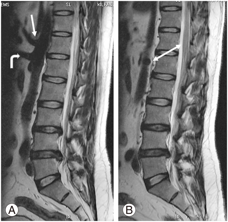
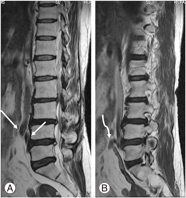
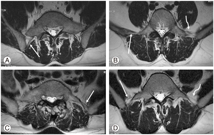
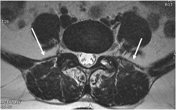
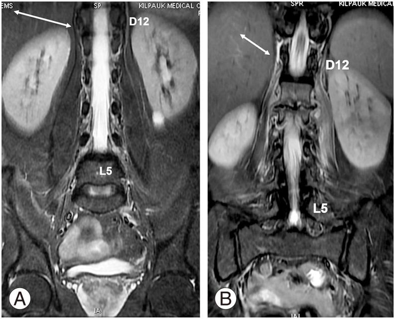
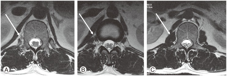
Overview of literature: Many studies have assessed the vertebral segments using various anatomical structures such as costal facets (CF), aortic bifurcation (AB), inferior vena cava confluence (IC), right renal artery (RRA), celiac trunk (CT), superior mesenteric artery root (SR), iliolumbar ligament (ILL) psoas muscle (PM) origin, and conus medullaris. However, none have yielded any consistent results.
Methods: We studied the locations of the anatomical structures CF, AB, IC, RRA, CT, SR, ILL, and PM in patients who underwent whole spine MRI at our department.
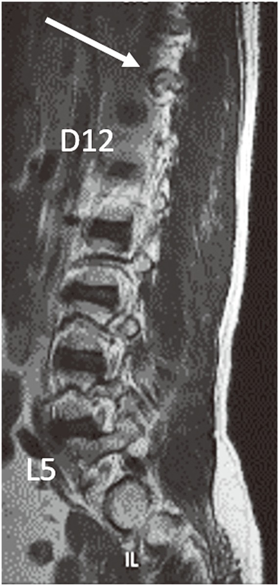
Results: In our study, 81.4% patients had normal spinal segmentation, 14.7% had sacralization, and 3.8% had lumbarization. Vascular landmarks had variable origin. There were caudal and cranial shifts with respect to lumbarization and sacralization. In 93.8% of cases in the normal group, ILL emerged from either L5 alone or the adjacent disc. In the sacralization group, ILL was commonly seen in L5. In the lumbarization group, ILL emerged from L5 and the adjacent disc (66.6%). CFs were identified at D12 in 96.9% and 91.7% of patients in the normal and lumbarization groups, respectively. The PM origin was observed from D12 or D12-L1 in most patients in the normal and sacralization groups.
Conclusions: CF, PM, and ILL were good identification markers for D12 and L5, but none were 100% accurate.
Keywords: Costal; Ligaments; Lumbar vertebrae; Magnetic resonance imaging.
Conflict of interest statement
Conflict of Interest: No potential conflict of interest relevant to this article was reported.
Figures

Similar articles
-
Identification and prediction of transitional vertebrae on imaging studies: anatomical significance of paraspinal structures.Clin Anat. 2007 Nov;20(8):905-14. doi: 10.1002/ca.20540. Clin Anat. 2007. PMID: 17879307
-
Are spinal or paraspinal anatomic markers helpful for vertebral numbering and diagnosing lumbosacral transitional vertebrae?Korean J Radiol. 2014 Mar-Apr;15(2):258-66. doi: 10.3348/kjr.2014.15.2.258. Epub 2014 Mar 7. Korean J Radiol. 2014. PMID: 24644411 Free PMC article.
-
Quantitative measurements at the lumbosacral junction are more reliable parameters for identifying and numbering lumbosacral transitional vertebrae.Eur Radiol. 2022 Aug;32(8):5650-5658. doi: 10.1007/s00330-022-08613-w. Epub 2022 Mar 8. Eur Radiol. 2022. PMID: 35258678
-
A review of lumbosacral transitional vertebrae and associated vertebral numeration.Eur Spine J. 2018 May;27(5):995-1004. doi: 10.1007/s00586-018-5554-8. Epub 2018 Mar 21. Eur Spine J. 2018. PMID: 29564611 Review.
-
Anatomical Variations That Can Lead to Spine Surgery at the Wrong Level: Part III Lumbosacral Spine.Cureus. 2020 Jul 28;12(7):e9433. doi: 10.7759/cureus.9433. Cureus. 2020. PMID: 32864257 Free PMC article. Review.
Cited by
-
Role of iliac crest tangent in correct numbering of lumbosacral transitional vertebrae.Turk J Med Sci. 2019 Feb 11;49(1):184-189. doi: 10.3906/sag-1807-258. Turk J Med Sci. 2019. PMID: 30764596 Free PMC article.
-
A Whole Spine MRI Based Study of the Prevalence, Associated Disc Degeneration and Anatomical Correlations of Lumbosacral Transitional Vertebra.Global Spine J. 2024 Sep;14(7):1952-1958. doi: 10.1177/21925682231161559. Epub 2023 Mar 3. Global Spine J. 2024. PMID: 36867110 Free PMC article.
-
Axial Spondyloarthritis: Mimics and Pitfalls of Imaging Assessment.Front Med (Lausanne). 2021 Apr 22;8:658538. doi: 10.3389/fmed.2021.658538. eCollection 2021. Front Med (Lausanne). 2021. PMID: 33968964 Free PMC article. Review.
-
Lumbosacral Transitional Vertebrae amongst the Individuals Undergoing Magnetic Resonance Imaging of the Whole Spine in a Tertiary Care Hospital: A Descriptive Cross-sectional Study.JNMA J Nepal Med Assoc. 2021 Oct 15;59(242):975-978. doi: 10.31729/jnma.6336. JNMA J Nepal Med Assoc. 2021. PMID: 35199704 Free PMC article.
-
Anterior spinal fusion (ALIF/OLIF/LLIF) with lumbosacral transitional vertebra: A systematic review and proposed treatment algorithm.Brain Spine. 2023 Jan 18;3:101713. doi: 10.1016/j.bas.2023.101713. eCollection 2023. Brain Spine. 2023. PMID: 38021000 Free PMC article. Review.
References
-
- Lee CH, Park CM, Kim KA, et al. Identification and prediction of transitional vertebrae on imaging studies: anatomical significance of paraspinal structures. Clin Anat. 2007;20:905–914. - PubMed
-
- Apazidis A, Ricart PA, Diefenbach CM, Spivak JM. The prevalence of transitional vertebrae in the lumbar spine. Spine J. 2011;11:858–862. - PubMed
LinkOut - more resources
Full Text Sources
Other Literature Sources
Research Materials
