. 2017 Jun 5;7(47):29312-29320.
doi: 10.1039/c7ra04372d.

Short-term hypoxic preconditioning promotes prevascularization in 3D bioprinted bone constructs with stromal vascular fraction derived cells

Affiliations

Short-term hypoxic preconditioning promotes prevascularization in 3D bioprinted bone constructs with stromal vascular fraction derived cells

Mitchell A Kuss et al. RSC Adv. .

Abstract

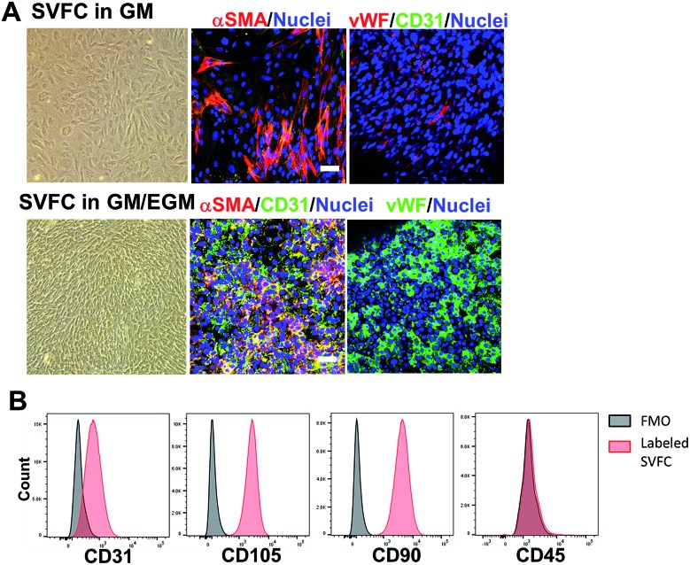
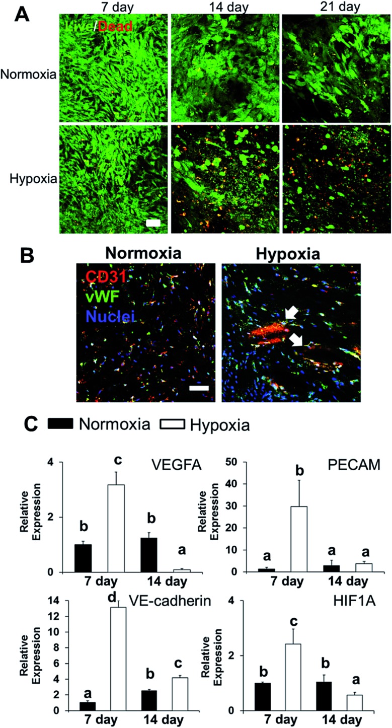
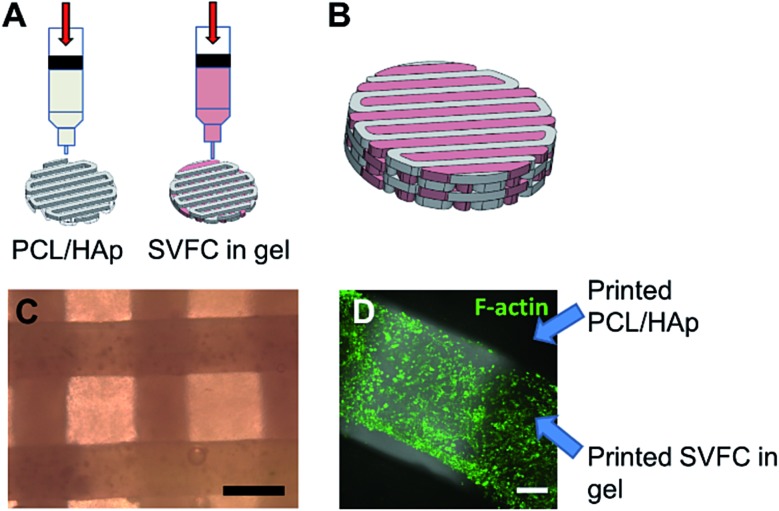
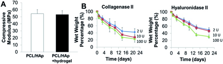
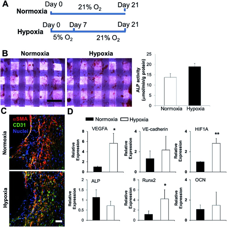
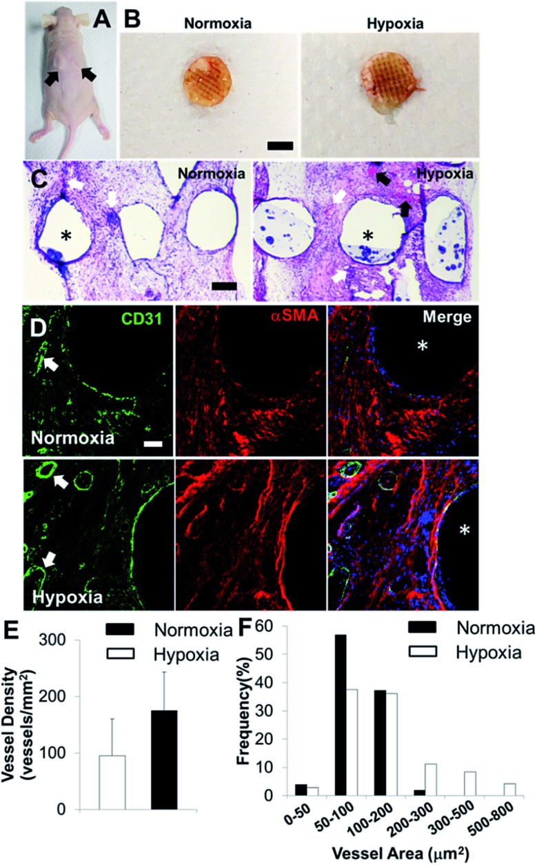
Reconstruction of complex, craniofacial bone defects often requires autogenous vascularized bone grafts, and still remains a challenge today. In order to address this issue, we isolated the stromal vascular fraction (SVF) from adipose tissues and maintained the phenotypes and the growth of endothelial lineage cells within SVF derived cells (SVFC) by incorporating an endothelial cell medium. We 3D bioprinted SVFC within our hydrogel bioinks and conditioned the constructs in either normoxia or hypoxia. We found that short-term hypoxic conditioning promoted vascularization-related gene expression, whereas long-term hypoxia impaired cell viability and vascularization. 3D bioprinted bone constructs composed of polycaprolactone/hydroxyapatite (PCL/HAp) and SVFC-laden hydrogel bioinks were then implanted into athymic mice, after conditioning in normoxic or short-term hypoxic environments, in order to determine the in vitro and in vivo vascularization and osteogenic differentiation of the constructs. Short-term hypoxic conditioning promoted microvessel formation in vitro and in vivo and promoted integration with existing host vasculature, but did not affect osteogenic differentiation of SVFC. These findings demonstrate the benefit of short-term hypoxia and the potential for utilization of SVFC and 3D bioprinting for generating prevascularized 3D bioprinted bone constructs. Furthermore, the ability to custom design complex anatomical shapes has promising applications for the regeneration of both large and small craniofacial bone defects.


Figures

Fig. 1
Fig. 1. The incorporation of EGM helped the maintenance of endothelial lineage cells and their phenotypes within SVFC. (A) 2D culture of SVFC in GM or GM/EGM. In GM, SVFC showed a spindle-like morphology, expressed αSMA and were negative for CD31 and vWF. SVFC expressed αSMA, CD31 and vWF in the culture with addition of EGM-2 medium (scale bar = 50 μm); (B) the expression of CD31, CD105, CD90 and CD45 by SVFC in GM/EGM was examined by flow cytometry. Grey histograms represent fluorescence minus one (FMO) controls, while red histograms are from SVFC that have been labelled as indicated. Graphs for CD105, CD90, and CD31 are derived from living, singlet, CD45-events. The graph for CD45 is from living, singlet events. SVFC were positive to CD31, CD105, CD90, but negative to CD45.
Fig. 2
Fig. 2. Effects of hypoxia on SVFC viability and vascularization. (A) Long-term hypoxic conditioning significantly affects SVFC viability. Cell viability for the 3D bioprinted hydrogel constructs with SVFC conditioned in GM/EGM in either hypoxia or normoxia for up to 21 days (scale bar = 50 μm); (B) IHC staining of SVFC laden constructs showed that SVFC were positive for both CD31 and vWF in both normoxia and hypoxia after 7 day culture. In hypoxia, microvessel-like structures were also observed, indicated by white arrows (scale bar = 50 μm); (C) qPCR analysis of VEGFA, PECAM1, VE-cadherin and HIF1A in SVFC laden hydrogels after 7 and 14 day culture in GM/EGM in normoxia and hypoxia. Relative gene expression is presented as normalized to 18S and expressed relative to cells in normoxia for 7 days (n = 3; bars that do not share letters are significantly different from each other; p < 0.05).
Fig. 3
Fig. 3. 3D hybrid bioprinting of SVFC laden bone constructs. (A) Schematic of 3D hybrid bioprinting using multiple cartridges. Multiple PCL/HAp frames were first printed using high temperature printing cartridge in each layer throughout the construct, then SVFC and hydrogels were deposited in between the frames by using a low temperature cartridge; (B) model of bioprinted bone constructs; (C) bright field image showed the part of bioprinted PCL/HAp and hydrogel construct (scale bar = 500 μm); (D) IHC staining of F-actin showed that the SVFC laden hydrogel was sandwiched between the two frames of PCL/HAp (scale bar = 100 μm).
Fig. 4
Fig. 4. (A) Compressive modulus of 3D printed PCL/HAp and hybrid printed PCL/HAp and hydrogel scaffolds; (B) biodegradation of bioprinted hydrogels in collagenase II or hyaluronidase II solution.
Fig. 5
Fig. 5. Effects of short-term hypoxic treatment on SVFC osteogenic differentiation and vascularization within 3D bioprinted constructs in OGM/EGM. (A) Timeline and treatment for 3D bioprinted SVFC laden constructs. Hypoxia group represented the 7 day hypoxic conditioning and 14 day normoxic conditioning, while the normoxia group represented 21 day culture in normal oxygen tension; (B) short-term hypoxia did not significantly change ALP expression and activity (scale bar = 1 mm); (C) IHC staining of αSMA and CD31 after in total 21 day culture in both groups (scale bar = 50 μm); (D) qPCR analysis of vascularization markers (i.e. VEGFA, VE-cadherin and HIF1A) and osteogenic differentiation markers (i.e. ALP, Runx2 and OCN) in 3D bioprinted constructs after 21 day culture in normoxia and hypoxia groups. Relative gene expression is presented as normalized to 18S and expressed relative to cells in normoxia for 21 days (n = 3; *p < 0.05, **p < 0.01).
Fig. 6
Fig. 6. In vivo implantation and vascularization within 3D bioprinted bone constructs. (A) Two bioprinted constructs after 21 day in vitro culture were subcutaneously implanted in the athymic mouse (as indicated by black arrows); (B) overall view of explanted constructs after 4 week subcutaneous implantation (scale bar: 5 mm); (C) representative images of H&E staining (scale bar: 200 μm); (D) IHC staining for CD31 (green), αSMA (red), and nuclei (blue) within 3D bioprinted constructs (scale bar: 50 μm). The white arrows indicate the formed microvessels with lumen structures, and the black arrows indicate the murine erythrocytes within blood vessels. The asterisks indicate the PCL/HAp scaffold strand areas; quantification of CD31 positive vascular-network within 3D bioprinted constructs in hypoxia and normoxia groups by measuring the microvessel density (E) and microvessel area distribution (F) (two images per sample from the images of five scaffold samples, 10 images in total).

References

    1. Allareddy V., Allareddy V., Nalliah R. P. J. Oral Maxillofac. Surg. 2011;69:2613–2618. - PubMed
    1. Oppenheimer A. J., Mesa J., Buchman S. R. J. Craniofac. Surg. 2012;23:30–36. - PubMed
    1. Pagni G., Kaigler D., Rasperini G., Avila-Ortiz G., Bartel R., Giannobile W. V. Adv. Drug Delivery Rev. 2012;64:1310–1319. - PMC - PubMed
    1. Hung B. P., Naved B. A., Nyberg E. L., Dias M., Holmes C. A., Elisseeff J. H., Dorafshar A. H., Grayson W. L. ACS Biomater. Sci. Eng. 2016;2:1806–1816. - PMC - PubMed
    1. Shakoori P., Zhang Q., Le A. D. Oral Maxillofac. Surg. Clin. 2017;29:19–25. - PubMed

LinkOut - more resources