. 2017 Sep;6(17):10.1002/adhm.201700500.
doi: 10.1002/adhm.201700500. Epub 2017 Jul 3.

Superoxide Dismutase-Loaded Porous Polymersomes as Highly Efficient Antioxidants for Treating Neuropathic Pain

Affiliations

Superoxide Dismutase-Loaded Porous Polymersomes as Highly Efficient Antioxidants for Treating Neuropathic Pain

Sonia Kartha et al. Adv Healthc Mater. 2017 Sep.

Abstract

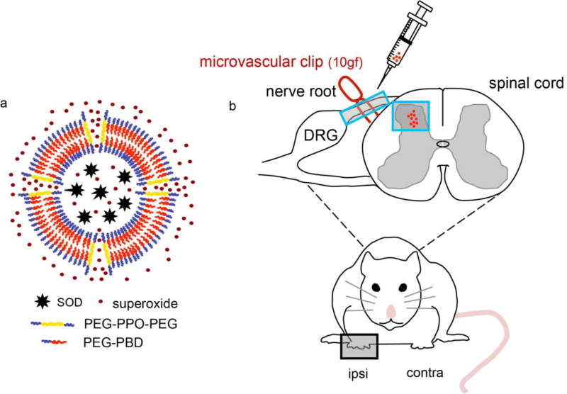
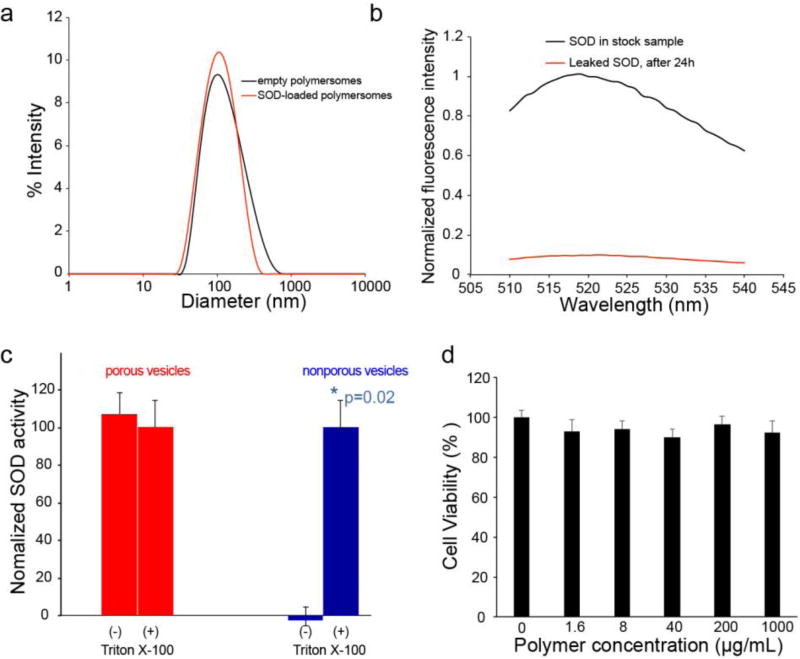
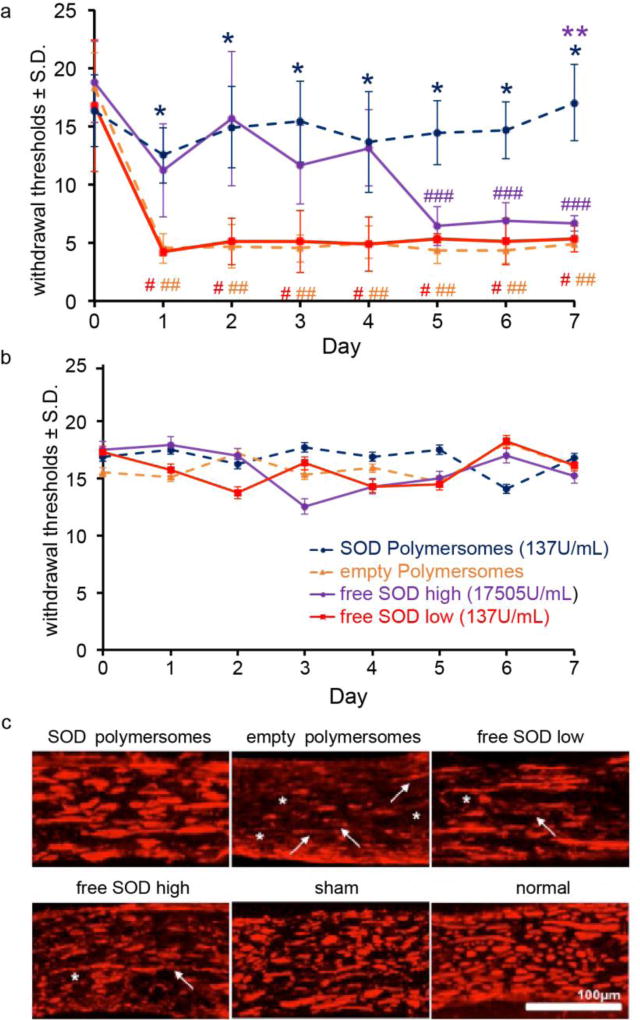
A highly efficient antioxidant is developed by encapsulating superoxide dismutase (SOD) within the aqueous interior of porous polymersomes. The porous polymersomes provide a permeable membrane that allows free superoxide radicals to pass into the aqueous interior and interact with the encapsulated antioxidant enzyme SOD. In vivo studies in the rat demonstrate that administration of SOD-encapsulated porous polymersomes can prevent neuropathic pain after nerve root compression more effectively than treatment with free antioxidant enzyme alone.

Keywords: antioxidants; neuropathic pain; porous polymersomes; superoxide dismutase.


Figures

Figure 1
Figure 1
a) Schematic diagram of SOD-loaded porous polymersomes. Porous polymersomes were formed through the co-assembly of the diblock copolymer PEG-PBD and the triblock copolymer PEG-PPO-PEG. Antioxidant enzyme SOD was loaded into the aqueous lumen of the porous polymersomes. The SOD-loaded porous polymersomes have a high membrane permeability to the small superoxide radical, while retaining SOD within their aqueous interiors. b) Nerve root compression injury was performed using a 10gf microvascular clip applied to the C7 dorsal nerve root. SOD-loaded porous polymersomes were injected directly onto the nerve root ipsilateral to injury, immediately after compression was removed.
Figure 2
Figure 2
a) Intensity-weighted size distribution of SOD-loaded porous polymersomes and empty polymersomes as measured by dynamic light scattering (DLS). b) Evaluation of SOD retention within porous polymersomes. Following 24 hours of incubation in PBS buffer (0.1 M, pH 7.4), the polymersomes were centrifuged on a Microcon filtering device with a 100 KDa MWCO membrane. The liquid that flowed through the filter was measured for fluorescence (red line). The fluorescence of unfiltered sample in the presence of Triton X-100 was also recorded (black line). The fluorescence intensity is normalized relative to the intensity of unfiltered sample at 518 nm. c) SOD activity within porous and nonporous polymersomes. SOD activity was tested before (−) and after (+) the addition of Triton-X-100. d) Cell viability in neuronal cultures was unchanged from control (0 µg/mL) following incubation with any polymersome concentration.
Figure 3
Figure 3
a) SOD porous polymersomes prevent pain developmentwith significantly higher withdrawal thresholds in the ipsilateral forepaw compared to both the empty polymersomes or free SOD low (*p<0.0001) treatments. Empty polymersome and free SOD treatment do not prevent pain with thresholds significantly lower than baseline on all days (#,## p<0.002). Pain develops after free SOD treatment after day 5 from baseline (### p<0.012) with thresholds significantly lower from SOD porous polymersomes on day 7 (**p<0.01). b) Withdrawal thresholds in the contralateral forepaw were not significantly different between groups on any day tested. c) NF200 labeling in the ipsilateral nerve roots after SOD porous polymersome treatment was similar to both sham and normal roots, yet treatment with empty polymersomes displayed the greatest evidence of axonal thinning (asterisk) and discontinuous labeling (arrows), with lesser axonal damage observed in both free SOD groups.

References

    1. Hogg-Johnson S, van der Velde G, Carroll LJ, Holm LW, Cassidy JD, Guzman J, Cote P, Haldeman S, Ammendolia C, Carragee E, Hurwitz E, Nordin M, Peloso P. Bone, P. Joint Decade - Task Force on Neck, D. Its Associated, Spine (Phila Pa 1976) 2008;33:S39. - PubMed
    2. Waters TR. J Electromyogr Kinesiol. 2004;14:7. - PubMed
    3. Cote P, Cassidy JD, Carroll LJ, Kristman V. Pain. 2004;112:267. - PubMed
    1. Yawn BP, Wollan PC, Weingarten TN, Watson JC, Hooten WM, Melton LJ., 3rd Pain Med. 2009;10:586. - PMC - PubMed
    2. Rutkowski MD, Winkelstein BA, Hickey WF, Pahl JL, DeLeo JA. Spine (Phila Pa 1976) 2002;27:1604. - PubMed
    1. Moalem G, Tracey DJ. Brain Res Rev. 2006;51:240. - PubMed
    2. Harden N, Cohen M. J Pain Symptom Manage. 2003;25:S12. - PubMed
    1. Scholz J, Woolf CJ. Nat Neurosci. 2007;10:1361. - PubMed
    1. Radhakrishnan K, Litchy WJ, O'Fallon WM, Kurland LT. Brain. 1994;117(Pt 2):325. - PubMed