. 2017 Jul 3;6(7):e353.
doi: 10.1038/oncsis.2017.57.

JWA regulates TRAIL-induced apoptosis via MARCH8-mediated DR4 ubiquitination in cisplatin-resistant gastric cancer cells

Affiliations

JWA regulates TRAIL-induced apoptosis via MARCH8-mediated DR4 ubiquitination in cisplatin-resistant gastric cancer cells

Q Wang et al. Oncogenesis. .

Abstract

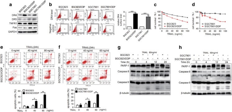
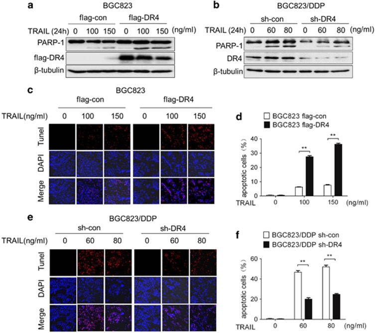
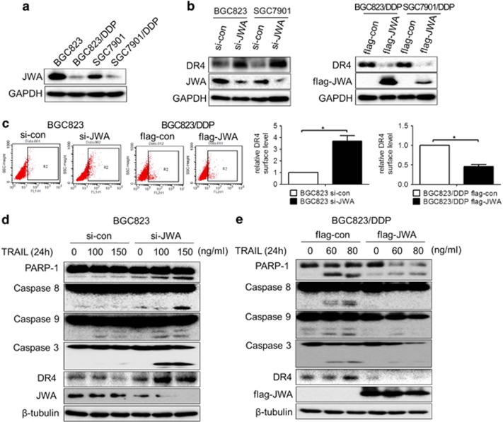
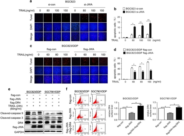
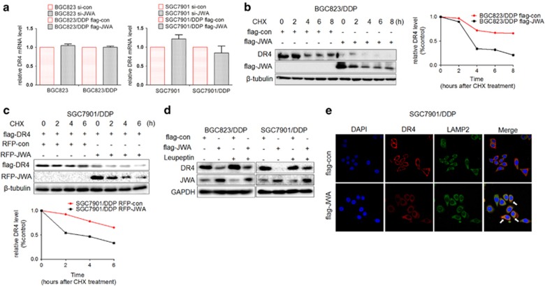
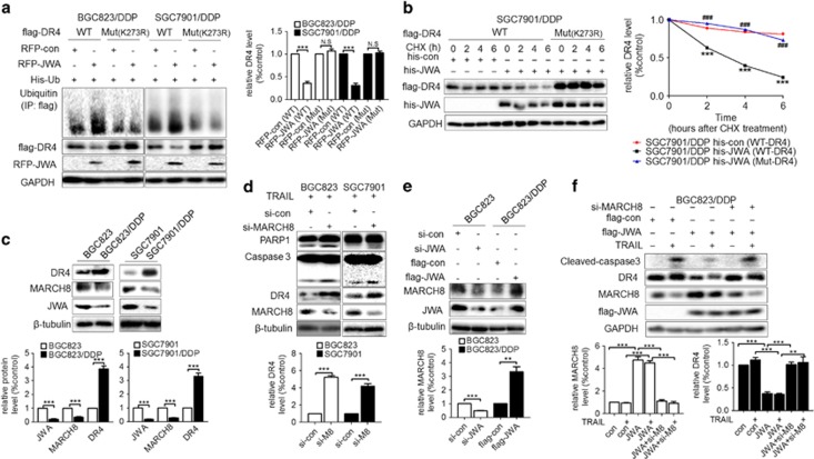
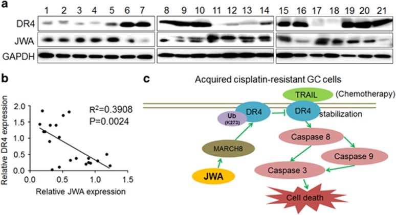
Platinum chemotherapeutics are widely used to treat solid malignant tumors, including gastric cancer (GC). Drug resistance to platinum compounds may result in cancer relapse and decreased survival. The identification and development of novel agents to reactivate apoptosis pathways in platinum-resistant cancer cells is therefore necessary. Here we report that cisplatin-resistant human GC cells (BGC823/DDP and SGC7901/DDP) but not their parental cells (BGC823 and SGC7901) exhibit high sensitivity to tumor necrosis factor-related apoptosis-inducing ligand (TRAIL) as a result of overexpression of death receptor 4 (DR4). Furthermore, we found that JWA, a molecule that promotes cisplatin-induced apoptosis in GC cells, suppressed TRAIL-induced apoptosis via negative regulation of DR4. Mechanistically, JWA promoted the ubiquitination of DR4 at K273 via upregulation of the ubiquitin ligase membrane-associated RING-CH-8 (MARCH8). In human GC tissues, JWA and DR4 protein levels were negatively correlated. Thus TRAIL may serve as an auxiliary treatment for cisplatin-resistant GC, and JWA may be a potential predictive marker of TRAIL sensitivity and may improve personalized therapeutics for treating human GC.


Conflict of interest statement

The authors declare no conflict of interest.

Figures

Figure 1
Figure 1
Acquired cisplatin-resistant human GC cells are sensitive to TRAIL. (a) The expression of DR4, DR5, TNFR1 and Fas in human GC cells BGC823 and SGC7901 and their cisplatin-resistant variants was determined by western blotting. (b) Left: Cell surface DR4 expression in BGC823 and SGC7901 cells and their variants was analyzed by flow cytometry. Right: Comparison of relative DR4 surface expression between different groups. Respective DR4 surface expression was estimated by examining the difference between the values (percentage of positive cells) of PE-DR4 and its corresponding IgG-PE control from at least three experiments in the same group. After 24 h of TRAIL treatments at the indicated concentrations in (c) BGC823 and its variant and (d) SGC7901 and its variant, the cell viability was determined by Cell Counting Kit-8 (CCK8) assays. After 24 h of TRAIL treatments at the indicated concentrations in (e) BGC823 and its variant and in (f) SGC7901 and its variant, cell apoptosis was determined by flow cytometry, and the quantification of apoptotic cells is indicated below. After exposure to TRAIL at the indicated concentrations and time points, the cleaved-PARP-1 and cleaved-caspase-8 and -9 levels were determined by western blotting analyses in BGC823 and its variant (g) or SGC7901 and its variant (h). The experiments were independently performed at least three times, and representative data are shown. For flow cytometry and CCK8 assays, the results are shown as the means±s.e.m., *P<0.05, **P<0.01, ***P<0.001.
Figure 2
Figure 2
GC cells are sensitized to TRAIL by DR4. Flag-DR4-transfected BGC823 cells (a) and sh-DR4-transfected BGC823/DDP cells (b) were exposed for 24 h to the indicated TRAIL concentrations and cleaved-PARP-1, and DR4 was then determined by western blotting analyses. Apoptosis was determined by TUNEL assay after exposure to the indicated TRAIL concentrations for 24 h in Flag-DR4-transfected BGC823 cells (c) and sh-DR4-transfected BGC823/DDP cells (e); the quantitative apoptosis ratios of (c, e) are shown in (d, f), respectively. The experiments were independently performed at least three times, and representative data are shown. For the TUNEL assay, the data are expressed as the means±s.e.m., **P<0.01.
Figure 3
Figure 3
JWA negatively regulates DR4 expression and sensitivity of GC cells to TRAIL. (a) JWA expression in GC cells BGC823 and SGC7901 and their variants was determined by western blotting analyses. (b) GC cells BGC823 and SGC7901 and their variants were transfected with si-JWA or Flag-JWA or a corresponding control for 48 h; DR4 and JWA expression in these cells was determined by western blotting analyses. (c) Left: Si-JWA or Flag-JWA GC cells or a corresponding control were transfected into BGC823 or its variant for 48 h; the DR4 surface expression was then analyzed by flow cytometry. Right: Comparison of relative DR4 surface expression between different groups. Respective DR4 surface expression was estimated as depicted in Figure 1b. Si-JWA transfected-BGC823 (d) and Flag-JWA transfected-BGC823/DDP cells (e) were exposed to the indicated TRAIL concentrations for another 24 h; JWA and DR4 expression and the apoptotic biomarkers cleaved-PARP-1 and cleaved-caspase-8, -9 and -3 were determined by western blotting analyses. The experiments were independently performed at least three times, and representative data are shown. For the flow cytometric assay, the data are shown as the means±s.e.m., *P<0.05.
Figure 4
Figure 4
JWA via DR4 inhibits TRAIL-induced apoptosis in GC cells. The apoptosis of si-JWA-transfected BGC823 cells (a) and Flag-JWA-transfected BGC823/DDP cells (c) was determined by TUNEL assays after exposure to the indicated TRAIL concentrations for 24 h; quantitative apoptosis ratios of (a, c) are shown in (b, d), respectively. Flag-con or Flag-JWA, or Flag-JWA plus Flag-DR4 plasmids were transfected into GC cells BGC823/DDP and SGC7901/DDP for 48 h and then exposed to 60 ng/ml TRAIL for an additional 24 h. The levels of Flag-JWA, DR4 and cleaved-caspase-9 and -3 were determined by western blotting analyses (e), and DR4 surface expression was analyzed by flow cytometry and by the quantification and comparison of relative DR4 surface expression among different groups, as depicted in Figure 1b (f). The experiments were independently performed at least three times, and representative data are shown. For the TUNEL and flow cytometric assays, the data are expressed as the means±s.e.m., *P<0.05, **P<0.01.
Figure 5
Figure 5
JWA inhibits DR4 expression by promoting its lysosomal degradation. (a) GC cells BGC823 and SGC7901 or their variants were transfected with si-JWA or Flag-JWA or a corresponding control for 48 h, and the DR4 mRNA in these cells was analyzed by reverse transcriptase–PCR (RT–PCR). (b) Left: Flag-JWA or Flag-con was transfected into GC cells BGC823/DDP for 48 h, and cells were then exposed to cycloheximide (CHX) at 50 μg/ml for the indicated times. DR4 protein stability was determined by western blotting. Right: Quantification curve of the DR4 protein level in BGC823/DDP cells. (c) Above: RFP-JWA and Flag-DR4 were cotransfected into SGC7901/DDP GC cells for 48 h, and cells were then exposed to 50 μg/ml CHX for the indicated times. Flag-DR4 protein stability was determined by western blotting. Below: Quantification curve of the Flag-DR4 protein level in SGC7901/DDP cells. (d) Flag-JWA or a corresponding control was transfected into cisplatin-resistant GC cells (SGC7901/DDP, BGC823/DDP) for 36 h; the cells were then exposed to another 20 h of leupeptin (5 μM), and the DR4 levels were determined by western blotting. (e) SGC7901/DDP GC cells were transfected with Flag-con or Flag-JWA for 48 h. A representative picture shows DR4 (red), the lysosome marker Lamp2 (green), the 4,6-diamidino-2-phenylindole-labeled nucleus (blue) and the colocalization of the three signals (merge), as determined by immunofluorescence assays. The experiments were independently performed at least three times, and representative data are shown. For the quantitative RT–PCR assay, the data are presented as the means±s.e.m.; NS, no significance.
Figure 6
Figure 6
JWA promotes ubiquitination of DR4 at K273 via upregulation of MARCH8 in GC cells. (a) Left: GC cells (BGC823/DDP, SGC7901/DDP) were cotransfected RFP-JWA and His-Ub with Flag-DR4 (wild type (WT)) or Flag-DR4 (mutant K273R) for 48 h. Immunoprecipitation was used to determine the ubiquitination of DR4; and the expression of ubiquitin, Flag-DR4 and RFP-JWA was determined by western blotting analyses. Right: Quantification of the Flag-DR4 protein level in the two GC cell lines. (b) Left: GC cells SGC7901/DDP were cotransfected with his-JWA and Flag-DR4 (WT) or Flag-DR4 (Mut K273R) for 48 h and then were treated with cycloheximide (CHX) at 50 μg/ml for the indicated times, and the Flag-DR4 protein stability was determined by western blotting analyses. Right: Quantification curves of the Flag-DR4 protein level in SGC7901/DDP cells. His-con (WT-DR4) vs His-JWA (WT-DR4), ***P<0.001; His-JWA (Mut-DR4) vs His-JWA (WT-DR4), ###P<0.001 (c) Above: The expression of DR4, MARCH8 and JWA in GC cells BGC823 and SGC7901 and their variants were determined by western blotting analyses. Below: Quantifications of the JWA, MARCH8 and DR4 protein level in the GC cells. (d) Above: Si-MARCH8 or its control was transfected into GC cells BGC823 and SGC7901 for 48 h and then exposed to TRAIL (BGC823: 150 ng/ml, SGC7901: 100 ng/ml) for an additional 24 h. Western blotting analyses determined the expression of MARCH8, DR4, cleaved-caspase-3 and PARP-1. Below: Quantification of DR4 protein level in the two GC cell lines. (e) Above: BGC823 cells were transfected with si-JWA or BGC823/DDP cells were transfected with Flag-JWA for 48 h. The expression of MARCH8 and JWA was then determined by western blotting analyses. Below: Quantifications of MARCH8 protein level in the two GC cell lines. (f) Above: Flag-con, Flag-JWA or Flag-JWA with MARCH8 siRNA were transfected into BGC823/DDP cells for 48 h, and the cells were then exposed to TRAIL at 80 ng/ml for another 24 h. Western blotting analyses determined the expression of Flag-JWA, MARCH8, DR4 and cleaved-caspase-3. Below: Quantification of MARCH8 and DR4 protein level in BGC823/DDP cells. The experiments were independently performed at least three times, and representative data are shown. **P<0.01, ***P<0.001; NS, no significance.
Figure 7
Figure 7
Protein levels of JWA and DR4 in human GC tissues. (a) Fresh human GC tissues from 21 patients were analyzed for JWA and DR4 protein expression by western blotting analyses. The loading control was GAPDH. (b) Correlation analysis between JWA and DR4 protein expression in the 21 GC tissues from (a). (c) Diagram illustrating the proposed molecular mechanism of how JWA is involved in DR4 degradation and decreases TRAIL-induced apoptosis in cisplatin-resistant GC cells. (1) In acquired cisplatin-resistant GC cells, substantially inhibited JWA expression results in the suppression of MARCH8. (2) Accordingly, suppressed MARCH8 further decreases ubiquitination of DR4 at K273. (3) The decreased DR4 ubiquitination confers greater stability in GC cells. (4) As a result, the DR4-based proapoptotic effect of TRAIL is dramatically enhanced, owing to the lower JWA expression in cisplatin-resistant GC cells.

References

    1. Zheng R, Zeng H, Zhang S, Chen T, Chen W. National estimates of cancer prevalence in China, 2011. Cancer Lett 2016; 370: 33–38. - PubMed
    1. Tian T, Zhang Y, Wang S, Zhou J, Xu S. Sox2 enhances the tumorigenicity and chemoresistance of cancer stem-like cells derived from gastric cancer. J Biomed Res 2012; 26: 336–345. - PMC - PubMed
    1. Torre LA, Bray F, Siegel RL, Ferlay J, Lortet-Tieulent J, Jemal A. Global cancer statistics, 2012. CA Cancer J Clin 2015; 65: 87–108. - PubMed
    1. Ferlay J, Steliarova-Foucher E, Lortet-Tieulent J, Rosso S, Coebergh JW, Comber H et al. Cancer incidence and mortality patterns in Europe: estimates for 40 countries in 2012. Eur J Cancer 2013; 49: 1374–1403. - PubMed
    1. Seah S, Low IC, Hirpara JL, Sachaphibulkij K, Kroemer G, Brenner C et al. Activation of surrogate death receptor signaling triggers peroxynitrite-dependent execution of cisplatin-resistant cancer cells. Cell Death Dis 2015; 6: e1926. - PMC - PubMed