. 2017 Aug;20(8):1172-1179.
doi: 10.1038/nn.4593. Epub 2017 Jun 26.

Engineered AAVs for efficient noninvasive gene delivery to the central and peripheral nervous systems

Affiliations

Engineered AAVs for efficient noninvasive gene delivery to the central and peripheral nervous systems

Ken Y Chan et al. Nat Neurosci. 2017 Aug.

Abstract

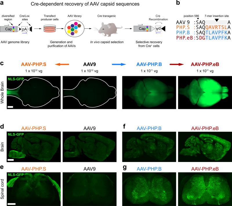
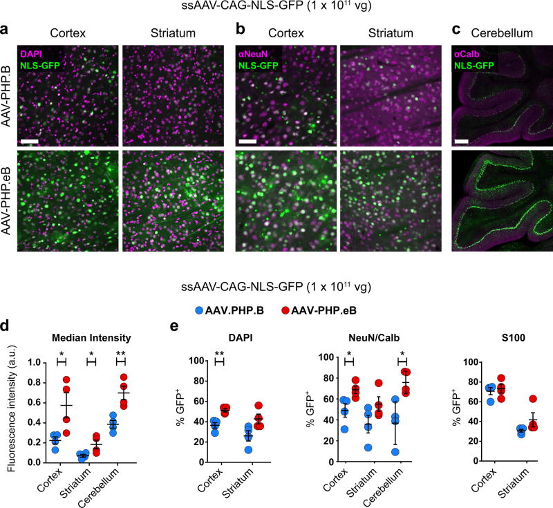
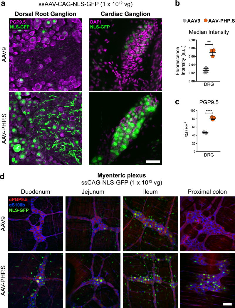
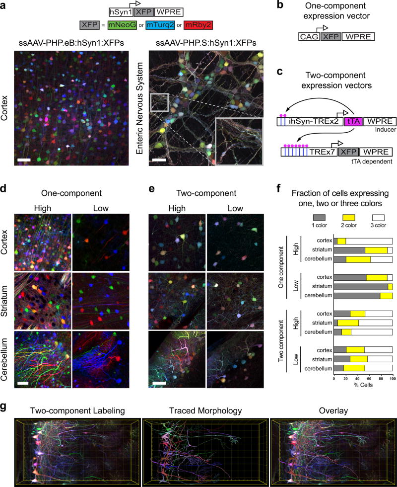
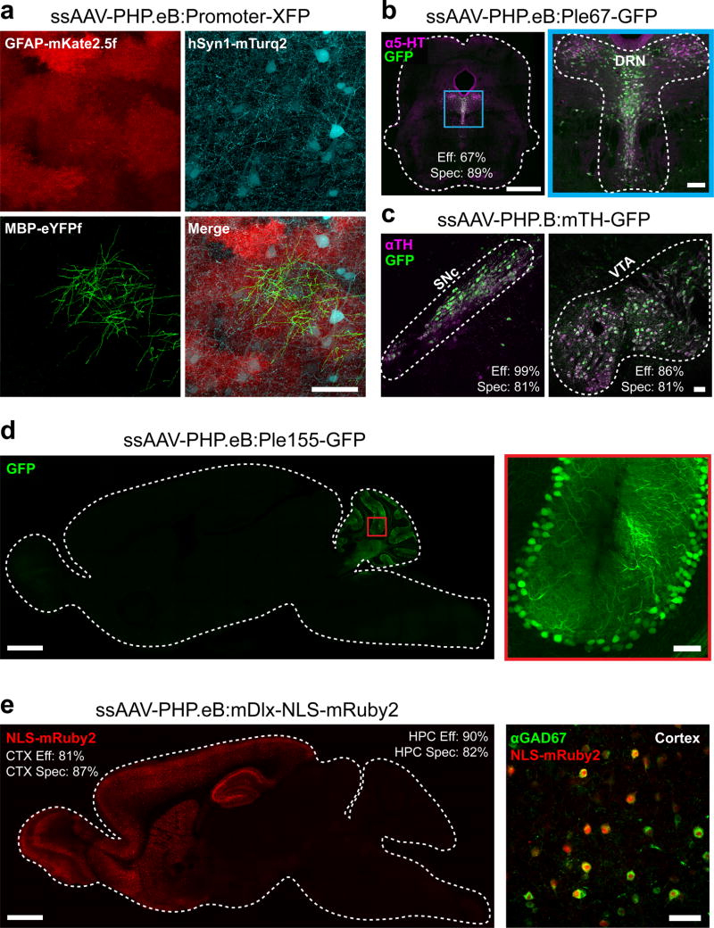
Adeno-associated viruses (AAVs) are commonly used for in vivo gene transfer. Nevertheless, AAVs that provide efficient transduction across specific organs or cell populations are needed. Here, we describe AAV-PHP.eB and AAV-PHP.S, capsids that efficiently transduce the central and peripheral nervous systems, respectively. In the adult mouse, intravenous administration of 1 × 10¹¹ vector genomes (vg) of AAV-PHP.eB transduced 69% of cortical and 55% of striatal neurons, while 1 × 10¹² vg of AAV-PHP.S transduced 82% of dorsal root ganglion neurons, as well as cardiac and enteric neurons. The efficiency of these vectors facilitates robust cotransduction and stochastic, multicolor labeling for individual cell morphology studies. To support such efforts, we provide methods for labeling a tunable fraction of cells without compromising color diversity. Furthermore, when used with cell-type-specific promoters and enhancers, these AAVs enable efficient and targetable genetic modification of cells throughout the nervous system of transgenic and non-transgenic animals.


Conflict of interest statement

Competing Financial Interest Statement

The California Institute of Technology has filed patent applications related to this work with B.E.D, K.Y.C. and V.G. listed as inventors. B.E.D. and V.G. receive research support from Voyager Therapeutics, which was not used in preparation of this manuscript.

Figures

Figure 1
Figure 1. Engineered AAV capsids for efficient transduction across the peripheral and central nervous systems
(a) Schematic of the CREATE selection method. (b) The amino acid (AA) sequences for the 7-mer insertions and flanking sequences for AAV-PHP.S, .B and .eB; the 7-mer and adjacent substitutions are highlighted in colored text. (c–g) ssAAV-CAG-NLS-GFP was packaged into the indicated capsid and intravenously injected into adult mice at 1 × 10¹² vg/mouse (AAV9 and AAV-PHP.S; c–e) or 1 × 10¹¹ vg/mouse (AAV-PHP.B and AAV-PHP.eB; c, f, and g). (c) Representative whole-brain fluorescence images after three weeks of expression. (d–g) Representative confocal images of native GFP fluorescence from sagittal brain sections (d and f) and transverse spinal cord sections (e and g) are shown for the indicated capsids. For (d–g), all imaging and display conditions are matched across panel pairs to allow for comparisons. Panels (d) and (f) are 40 µm maximum intensity projections (MIPs) and panels (e) and (g) are 300 µm MIPs. Scale bars for (c–e) are 1 mm.
Figure 2
Figure 2. AAV-PHP.eB transduces several CNS regions more efficiently than AAV-PHP.B
ssAAV-PHP.B:CAG-NLS-GFP or ssAAV-PHP.eB:CAG-NLS-GFP was intravenously injected into adult mice at 1 × 10¹¹ vg/mouse. Native GFP fluorescence was assessed after three weeks of expression. (a and b) Representative images of native GFP fluorescence (green) and DAPI staining (a, magenta) or NeuN staining (b, magenta) in the cortex and striatum. (c) Representative images of Calbindin immunohistochemistry (IHC) (magenta) in the cerebellum. (d) Mean GFP intensity in all DAPI+ nuclei in the cortex (t6 = 2.688; P = 0.0361) and striatum (t6 = 2.536; P = 0.0443) or in Calbindin+ cells in the cerebellum (t6 = 4.007; P = 0.0071). (e) Quantification of cell transduction. From left to right: The percentage of DAPI+ cells that express GFP in the cortex (t6 = 4.669; P = 0.0034) and striatum (t6 = 2.390; P = 0.0541). The percentage of NeuN+ cells that express GFP in the cortex (t6 = 2.662; P = 0.037) and in the striatum (t6 = 1.764; P = 0.128). The percentage of Calbindin+ cerebellar Purkinje cells that express GFP (t6 = 3.328; P = 0.039). The percentage of S100+ cells that express GFP in the cortex (t6 = 0.4422; P = 0.6738) and striatum (t6 = 1.512; P = 0.1814). (d and e) For quantification: n = 4 mice per group, mean ± SEM, unpaired, two-tailed t-test (***P ≤ 0.001; **P ≤ 0.01; *P ≤ 0.05; n.s., P ≥ 0.05). All images were single-plane confocal images of native GFP fluorescence. Scale bars are 50 µm (a and b) and 100 µm (c).
Figure 3
Figure 3. AAV-PHP.S efficiently transduces peripheral neurons
ssAAV-PHP.S:CAG-NLS-GFP or ssAAV9:CAG-NLS-GFP was intravenously injected into adult mice at 1 × 10¹² vg/mouse. Native GFP fluorescence was assessed after three weeks of expression. (a) Representative images of GFP expression and neuronal PGP9.5 (left, magenta) or DAPI (right, magenta) staining in a dorsal root ganglion (DRG, left) and cardiac ganglion (right). (b) Quantification of the mean GFP fluorescence intensity per cell with AAV-PHP.S or AAV9 (t4 = 7.814; P = 0.0014) (c) Quantification of the percentage of PGP9.5+ cells transduced (t4 = 18.29; P < 0.0001) (d) Representative images of GFP expression, with neuronal PGP9.5 (red) and astrocyte S100b (blue) staining in the myenteric plexus of the duodenum, jejunum, lleum and proximal colon. In (b and c) n = 3 independent animals per group, mean ± SEM, unpaired, two-tailed t-test. For representative images (a; n = 3 animals) and (d; n = 2 animals). All imaging and display conditions are matched in the GFP channel across panel pairs. Scale bars in (a and d) are 50 µm.
Figure 4
Figure 4. AAV-PHP.eB and AAV-PHP.S transfer multiple genomes per cell and enable tunable multicolor labeling
(a) Co-administration of a cocktail of three ssAAV vectors that each express one of three XFPs (top, schematic) from the hSyn1 promoter labels individual cells with a range of hues. Representative images of the cortex (left, AAV-PHP.eB; total dose: 4 × 10¹¹ vg) and the enteric nervous system (right, AAV-PHP.S; total dose: 1 × 10¹² vg). The inset highlights the dense network of neurites labeled with a range of hues. Schematics show one-component (b) and tTA-TRE-based two-component systems (c). Expression from the TRE-XFP vectors is dependent on co-transduction with a tTA (tet-off) inducer vector that contains a positive feedback loop to increase tTA expression (ssAAV-PHP.eB:ihSyn-tTA). (d) Representative images using the one-component expression system with a 3XFP cocktail expressed from the CAG promoter at an equal dose per XFP (high: total dose = 2 × 10¹¹ vg; low: total dose = 1 × 10¹⁰ vg). (e) Representative images using the two-component system: 1 × 10¹² total of the TRE-3XFP cocktail and a high (1 × 10¹¹ vg) or low (1 × 10¹⁰ vg) dose of the tTA inducer. (f) Percentages of individual cells that express one, two, or all three XFPs. (g) MIP (left) of a coronal section from the olfactory bulb of a Tbx21-Cre mouse transduced with a cocktail of ssAAV-PHP.eB:TRE-DIO-3XFP (1 × 10¹² vg total dose) and sAAV-PHP.eB:ihSyn-tTA (1 × 10¹⁰ vg). The morphologies of local neuronal dendritic arbors were traced with NeuTube and imported into Imaris v8.3 for 3D rendering (middle) and overlay (right). Scale bars (d and e) are 50 µm and are MIPs of 40 µm z-stacks. The total volume in (g) is 0.042mm3. All images were acquired on tissue extracted three weeks after intravenous AAV delivery in adult mice, except for (a) where the expression was assessed after 11 days.
Figure 5
Figure 5. AAV-PHP.eB can be used with gene regulatory elements to achieve cell type-restricted gene expression throughout the brain
(a) Images show co-transduction of the cortex 3 weeks after co-administration of 3 vectors with 3 different promoters driving distinct XFPs (intravenous injection of AAV-PHP.eB at 1 × 10¹² vg per viral vector). (b) A vector providing GFP expression driven by a FEV/serotoninergic neuron-specific promoter (ssAAV-PHP.eB:Ple67-GFP) was intravenously delivered at 1 × 10¹² vg and co-localized to serotonin (5-hydroxytryptamine, 5-HT, magenta) expressing cells in the dorsal raphe nucleus (DRN) outlined in blue and expanded for detail (right). (c) A vector providing GFP expression from a mouse tyrosine hydroxylase (TH) promoter (ssAAV-PHP.B:mTH-GFP) was intravenously injected at 1 × 10¹² vg, and imaging with IHC for TH (magenta) was performed after 2 weeks of expression. Images show the substantia nigra pars compacta (SNc, left) and ventral tegmental area (VTA, right). (d) A vector with a Purkinje cell-selective promoter (Ple155, Pcp2) driving GFP (ssAAV-PHP.eB:Ple155-GFP) was intravenously injected at 1 × 10¹² vg and expression was examined at 4 weeks. A whole sagittal section (left) shows native GFP fluorescence (green) in the cerebellum (left) in cells with the morphology of Purkinje cells (see higher resolution of the red boxed region, right). (e) A vector with a forebrain GABAergic interneuron-specific enhancer driving nuclear-localized mRuby2 (AAV-PHP.eB:mDlx-NLS-mRuby2) was intravenously injected at 3 × 10¹¹ vg and expression was examined at 8 weeks (CTX: cortex, HPC: hippocampus). Native mRuby2 expression within the forebrain (red, left). Co-localization was assessed by IHC for GABAergic cells (GAD67+, green, right). The scale bars in (a; b, right; c; d, right; e, right) are 50 µm. For (b and d, left) the scale bars are 1 mm. All panels are confocal images of native XFP fluorescence. The efficiency (Eff) and specificity (Spec) values for transduction by FEV/Ple67, mTH, mDlx vectors are giving in the DRN, VTA, SNc, cortex (CTX), and hippocampus (HPC).

Comment in

  • CREATEd viruses go global.
    Yonehara K, Roska B. Yonehara K, et al. Nat Neurosci. 2017 Jul 26;20(8):1041-1042. doi: 10.1038/nn.4600. Nat Neurosci. 2017. PMID: 28745726 No abstract available.

References

    1. Hastie E, Samulski RJ. Adeno-associated virus at 50: a golden anniversary of discovery, research, and gene therapy success--a personal perspective. Hum Gene Ther. 2015;26:257–265. - PMC - PubMed
    1. Suzuki K, et al. In vivo genome editing via CRISPR/Cas9 mediated homology-independent targeted integration. Nature. 2016;540:144–149. - PMC - PubMed
    1. Yang Y, et al. A dual AAV system enables the Cas9-mediated correction of a metabolic liver disease in newborn mice. Nat Biotechnol. 2016;34:334–338. - PMC - PubMed
    1. Rajasethupathy P, Ferenczi E, Deisseroth K. Targeting Neural Circuits. Cell. 2016;165:524–534. - PMC - PubMed
    1. Urban DJ, Roth BL. DREADDs (designer receptors exclusively activated by designer drugs): chemogenetic tools with therapeutic utility. Annu Rev Pharmacol Toxicol. 2015;55:399–417. - PubMed