. 2017 Jul 3;13(7):e1006483.
doi: 10.1371/journal.ppat.1006483. eCollection 2017 Jul.

Checkpoints of apicomplexan cell division identified in Toxoplasma gondii

Affiliations

Checkpoints of apicomplexan cell division identified in Toxoplasma gondii

Carmelo A Alvarez et al. PLoS Pathog. .

Abstract

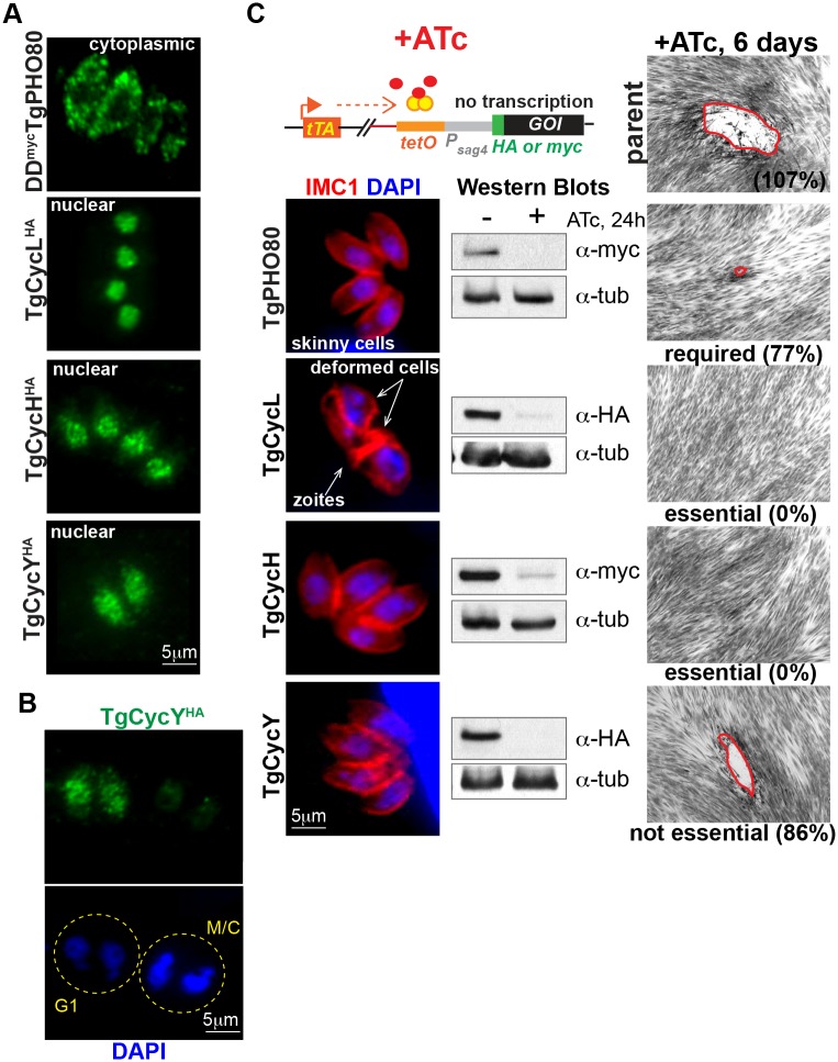
The unusual cell cycles of Apicomplexa parasites are remarkably flexible with the ability to complete cytokinesis and karyokinesis coordinately or postpone cytokinesis for several rounds of chromosome replication, and are well recognized. Despite this surprising biology, the molecular machinery required to achieve this flexibility is largely unknown. In this study, we provide comprehensive experimental evidence that apicomplexan parasites utilize multiple Cdk-related kinases (Crks) to coordinate cell division. We determined that Toxoplasma gondii encodes seven atypical P-, H-, Y- and L- type cyclins and ten Crks to regulate cellular processes. We generated and analyzed conditional tet-OFF mutants for seven TgCrks and four TgCyclins that are expressed in the tachyzoite stage. These experiments demonstrated that TgCrk1, TgCrk2, TgCrk4 and TgCrk6, were required or essential for tachyzoite growth revealing a remarkable number of Crk factors that are necessary for parasite replication. G1 phase arrest resulted from the loss of cytoplasmic TgCrk2 that interacted with a P-type cyclin demonstrating that an atypical mechanism controls half the T. gondii cell cycle. We showed that T. gondii employs at least three TgCrks to complete mitosis. Novel kinases, TgCrk6 and TgCrk4 were required for spindle function and centrosome duplication, respectively, while TgCrk1 and its partner TgCycL were essential for daughter bud assembly. Intriguingly, mitotic kinases TgCrk4 and TgCrk6 did not interact with any cyclin tested and were instead dynamically expressed during mitosis indicating they may not require a cyclin timing mechanism. Altogether, our findings demonstrate that apicomplexan parasites utilize distinctive and complex mechanisms to coordinate their novel replicative cycles.


Conflict of interest statement

The authors have declared that no competing interests exist.

Figures

Fig 1
Fig 1. Phylogenetic analysis of T. gondii Crks.
Protein sequences of Cdk-related kinases from T. gondii (Tg), P. falciparum (Pf), T. annulata (TA), C. parvum (Cdg), C. velia (Cvel), and Cdks from human cells (Hs) were analyzed in MEGA 7. Branch support was determined in 100 bootstrap replicates and the nodes supported by higher than 50% value are indicated with a blue circle. The results show that three Cdk families are shared between Alveolates and higher eukaryotes: T. gondii TgCrk2 was grouped with cell cycle Cdk5 family kinases (blue shadow), TgCrk1 was clustered with transcriptional kinases of Cdk11 family (green shadow) and TgCrk7 appeared distantly related to the CAK complex component Cdk7 (yellow shadow). Note that T. gondii does not encode canonical G1 Cdk4/6 family (teal shadow). Clusters that lack higher eukaryotic counterparts were labeled as ancestral/novel and are indicated with a pink shadow. The majority of T. gondii Crks were clustered in ancestral groups.
Fig 2
Fig 2. Conditional expression of T. gondii Crks.
The upper schematic illustrates anhydrotetracycline (ATc) mediated control of gene expression in the tet-OFF system. In the absence of ATc the tetracycline transactivator (tTA) binds to the tet-operator (tetO) maintaining the active transcription of the gene of interest (GOI). Downregulation of the GOI product is achieved by transcriptional repression with ATc that prevents tTA binding to tet-operators incorporated into the tet-OFF promoter of the GOI. IFA images on the left show predominant localization of the epitope-tagged kinases (α-HA, green) relative to nuclear staining (DAPI, blue) and IMC compartment (IMC1, red). Downregulation of TgCrk expression after 24 h treatment with 1μg/ml ATc was verified for seven kinases by IFA and Western Blot analysis (α-Tubulin A staining was used as a loading control). The essentiality of each kinase was tested by the ability of TgCrk tet-OFF mutant parasites to form plaques after 6 days with 1μg/ml ATc (representative DIC images on the right). Parent Tati-RHΔKu80 strain was included as positive control (top DIC image). Percentage number indicated below each DIC image represents the number of plaques relative to the -ATc condition for each tet-OFF mutant. All analyzed kinases were either essential or required for tachyzoite growth, with the exception of TgCrk8. Downregulation of TgCrk2, resulted in growth arrest of morphologically normal looking parasites, while knockdown of TgCrk1, TgCrk4 and TgCrk6 led to major morphological abnormalities that are indicated in the IFA images and quantified in the S2 Fig.
Fig 3
Fig 3. Conditional expression of T. gondii cyclins in the tet-OFF model.
(A) IFA analysis of TgCyclins localization. Tagged at the genomic locus TgCycHHA, TgCycLHA and TgCycYHA were visualized using α-HA antibody (green). Due to low native expression level, localization of ectopically expressed DDmycTgPHO80 is shown after a 3 h induction with 100nM Shield1. (B) TgCycYHA is dynamically expressed in G1 stage. Yellow dotted line indicates individual vacuoles. The cell cycle stage was determined based on intensity of the nuclear staining (DAPI, blue). (C) Transgenic tet-OFF clones of TgCyclins were established with alternative 3xHA or myc tags as described in Material and Methods and in the S1D Fig. IFA images show a representative vacuole after 24 h growth with 1μg/ml ATc. Parasite shape and nucleus were visualized with α-IMC1 (red) and DAPI (blue) staining, respectively. ATc-induced downregulation of the TgCyclins was confirmed by Western blot analysis using α-Tubulin A staining as a loading control. Results of the plaque assays are shown after 6 days growth with 1μg/ml ATc (DIC panel on the right). The percentage is the number of plaques formed relative to no ATc condition. The phenotype of TgPHO80 and TgCycL deficient parasites suggested the factors' involvement in the cell cycle regulation.
Fig 4
Fig 4. G1 kinase TgCrk2 predominantly interacts with cytoplasmic TgPHO80 cyclin.
(A) Downregulation of TgCrk2 led to growth arrest in G1 phase prior to centrosome duplication. The TgCrk2 tet-OFF mutant was grown for 24 h with or without 1μg/ml ATc, and co-stained with α-human Centrin1 (red, centrosome), α-IMC1 (green, parasite cytoskeleton and internal buds) and DAPI (blue, nucleus). Vacuoles with duplicated (red column) or single centrosomes (blue column) were randomly selected and counted in -ATc and +ATc populations. The average numbers and standard deviations of three independent experiments are plotted. (B) TgCrk2/cyclin complexes were immunoisolated from the soluble fraction [In] of parasites co-expressing endogenous TgCrk2HA and ectopic myc-tagged TgPHO80, TgCycL, TgCycY (upper panels), and endogenous TgCycHHA co-expressed with ectopic allele of myc-tagged TgCrk2 (lower panels). Ectopic expression was regulated by destabilization domain (DD). Beads with precipitated complexes [IP] and depleted soluble fraction [DF] were probed with α-myc and α-HA antibodies to detect cyclins and to confirm efficient pulldown of TgCrk2 (the IP panels on the top). TgCrk2 formed stable complexes with TgPHO80 cyclin, and showed weak interaction with TgCycH, while no complexes were detected with TgCycL or TgCycY, confirming specificity of interactions.
Fig 5
Fig 5. Loss of TgCrk1 that forms a stable sub-nuclear complex with TgCycL leads to abnormal assembly of the daughter cytoskeleton.
(A) Cytological defects of TgCrk1 tet-OFF mutant were analyzed and quantified (B) using antibodies against the basal complex marker MORN1 (red), cytoskeletal marker IMC1 (green) and nuclear staining DAPI (blue). The guide panel shows schematics of the parasites captured in IFA images (#) and parasite features are summarized in the description panel. During normal mitosis depicted in the –ATc images, membrane protein MORN1 localizes to the mother basal complex, intranuclear spindle compartment centrocone, and to the attached MORN rings (image a). Downregulation of TgCrk1 with 1 μg/ml ATc for 16 h resulted in incomplete assembly (image c) or fragmentation (image e) of the MORN rings and associated accumulation of IMC sacs (image f). Fully arrested vacuoles shown in the bottom panel (24 h +ATc) exhibit a catastrophic phenotype; the chaotic daughter cytoskeletal mass (image h) disconnected from parasite cytoplasm. (B) Vacuoles undergoing proper budding (normal buds) or containing parasites displaying cytological defects depicted in the images c-f (piled IMC1/MORN1), or in the images g and h (cytological defects) were quantified in the TgCrk1 tet-OFF populations grown with or without 1 μg/ml ATc for 24 h. Results are plotted on the graph. Statistically different values are indicated with asterisks (** ≤ 0.01; *** ≤ 0.001). (C) TgCrk1 tet-OFF mutant was stained with α-ISP1 antibodies to monitor changes in the apical complex. Image on the left illustrates proper expression and localization of the marker in S/M tachyzoites (-ATc). Downregulation of TgCrk1 (+ATc) caused severe morphological changes in the apical cone and reduced expression of ISP1 protein. Dotted line indicates boundary of the mis-shaped parasite. Major abnormalities are labeled and indicated by double-headed arrows. (D) TgCrk1-TgCyclin complexes were immunoisolated from parasites co-expressing TgCrk1HA and myc-tagged TgPHO80, TgCycL, TgCycY and TgCycH (used transgenic strains are listed in S1 Table). The soluble fraction before [In] and after immunoprecipitation [DF], and protein complexes on the beads [IP] were probed with α-myc antibody to detect TgCyclins and with α-HA antibody to verify TgCrk1 pulldown (top panel). The results revealed a dominant TgCrk1-TgCycL complex. (E) Dual-tagged parasites expressing TgCrk1HA and TgCycLmyc were stained with a α-HA (green) and α-myc (red) antibodies. Proteins display similar localization patterns with particular accumulation in the nuclear sub-compartment (insert, arrow).
Fig 6
Fig 6. Nuclear TgCrk6 regulates spindle biology in tachyzoites.
(A) TgCrk6 tet-OFF mutant parasites were grown in the absence (upper row) or presence of 1 μg/ml ATc for 16 h (bottom row) and analyzed by IFA using α-MORN1 (red, centrocone and basal complex) and α-IMC1 (green, parasite shape and internal buds) antibody. Chromosome dynamics was detected by DAPI staining (blue). Downregulation of TgCrk6 (+ATc) led to an inability of the centrocone compartment to duplicate during mitosis. The guide panel includes a cartoon and a description of the analyzed structures and observed deficiencies. Single (red) and duplicated (blue) centrocones (α-MORN1) were quantified in 25–50 randomly selected budding vacuoles (α-IMC1) (for raw data, see S1 Table) revealing a significantly larger number of the vacuoles with a single centrocone when TgCrk6 tet-OFF mutant was treated with ATc for 24 h (+ATc). P-value ≤ 0.0001 (***) was calculated using unpaired two-tail t-test. (B) Kinetochore dynamics were analyzed in the TgCrk6 tet-OFF mutant parasites co-expressing TgNdc80myc using α-myc antibody. To identify vacuoles in cytokinesis, microtubules of the growing internal buds were stained with antibody against acetylated Tubulin A (green). Kinetochore dynamics in TgCrk6 tet-OFF mutant after 24 h treatment with or without 1μg/ml ATc is summarized in the guide panel on the right. TgCrk6-deficient parasites (+ATc) retained a single assembled kinetochore (image g) positioned between two internal daughters (image h).
Fig 7
Fig 7. Loss of cytoplasmic TgCrk4 leads to abnormal centrosome duplication.
(A) TgCrk4 deficiency affected duplication of the structures localized in the cytoplasm. TgCrk4 tet-OFF mutant parasites were treated with 1μg/ml ATc for 24 h and stained with α-IMC1 (surface), DAPI (DNA) and the markers of the following structures: centromere (α-TgCenH3), outer core of the centrosome (α-Centrin1) and apicoplast (α-TgAtrx1). To visualize inner core of centrosome, we introduced recombinant TgCEP250myc protein on the fosmid in TgCrk4 tet-OFF mutant and stained parasites with α-myc antibody. Dynamics of organelles and structures are indicated with red arrows and summarized in the Guide panel. General deficiencies caused by the loss of TgCrk4 are indicated with white arrows. (B) Quantification of the morphologically abnormal vacuoles (blue, determined by α-IMC1 and DAPI co-staining) containing under-duplicated (green) or over-duplicated (red) centrosomes (visualized by α-Centrin1 staining) in the TgCrk4 tet-OFF mutant grown without or with ATc for 20 h is shown on the bar graph (for raw data see S1 Table). Significant difference between two indicated conditions was verified by unpaired t-test that returned the p-value 0.02 (*). Note that centrosome re-duplication is a predominant defect and together with centrosome under-duplication affected about 30% of the TgCrk4 deficient population. (C) The reduplicated centrosome of TgCrk4-deficient parasites preserved internal integrity. TgCrk4 tet-OFF mutant parasites co-expressing TgCEP250myc were stained with α-Centrin1 (outer centrosomal core) and α-myc (TgCEP250myc, inner centrosomal core) after 16 h growth with or without 1μg/ml ATc. White arrows indicate re-duplication of both centrosome cores in TgCrk4 deficient parasite. The enlarged merged image shows that only one of the centrosomes remains connected to the nucleus. (D) Reduplicated centrosomes are associated with assembled kinetochores in TgCrk4 deficient parasites. Transgenic TgCrk4 tet-OFF mutant expressing kinetochore marker TgNdc80myc was grown with or without 1μg/ml ATc for 16 h and co-stained with α-Centrin1 (centrosome), α-myc (TgNdc80myc, kinetochore), and DAPI. In mitotic cells expressing TgCrk4 (-ATc), centrosome duplication and kinetochore assembly occurs once per chromosome cycle. Kinetochores are assembled after centrosome segregation (2 centrosomes:1 kinetochore, images a, b), duplicated after the spindle break (2 centrosomes: 2 kinetochores, images c, d) and segregated/disassembled early in the budding (2 centrosomes: 0 kinetochores, images e, f). Lower panel (+ATc) shows vacuole of four TgCrk4-deficient parasites with three cells (white arrows) containing reduplicated centrosomes associated with assembled kinetochores (4 centrosomes: 2 kinetochores, images g, h). Number of structures per parasite is shown in each insert. Parasite shapes are outlined in the merge panel.
Fig 8
Fig 8. A roadmap of putative checkpoints of the tachyzoite cell cycle.
Cartoon summaries findings of the current study. The tachyzoite cell cycle consists of G1, S-phase, and mitosis that overlaps and is coordinated with cytokinesis (Black and white circle). Binary division of tachyzoites continues for 5–6 rounds within the same vacuole, and then, parasites lyse the host cell and egress. We identified several potential stopping points (checkpoints) regulated by Cdk-related kinases of T. gondii. Red hexagons indicate the timing of the synchronized growth arrest or retardation caused by dis-regulation of the major cellular pathway. It appears that T. gondii retained three conserved stop points in the cell cycle. The restriction point in G1 that is related to cell differentiation and dormancy and is regulated by TgCrk2 kinase in complex with TgPHO80 cyclin (orange arrow). Licensing of DNA replication is likely under control of atypical TgCrk5 (blue arrow) (Naumov and White, personal communication), while novel TgCrk6 (dark green arrow) might operate the spindle assembly checkpoint in metaphase. Specialized mitosis of apicomplexan parasites seems to acquire two additional points of cell cycle control. We showed that coccidian-specific kinase TgCrk4 was required to maintain proper stoichiometry of the novel bipartite centrosome (light green arrow). And distantly related to higher eukaryotic Cdk11, TgCrk1 appears to control a vital parasite-specific process of the zoite assembly (purple arrow).

References

    1. Gubbels MJ, White M, Szatanek T. The cell cycle and Toxoplasma gondii cell division: tightly knit or loosely stitched? Int J Parasitol. 2008;38(12):1343–58. doi: 10.1016/j.ijpara.2008.06.004 - DOI - PubMed
    1. Francia ME, Striepen B. Cell division in apicomplexan parasites. Nature reviews Microbiology. 2014;12(2):125–36. doi: 10.1038/nrmicro3184 - DOI - PubMed
    1. Suvorova ES, Francia M, Striepen B, White MW. A novel bipartite centrosome coordinates the apicomplexan cell cycle. PLoS Biol. 2015;13(3):e1002093 doi: 10.1371/journal.pbio.1002093 - DOI - PMC - PubMed
    1. Behnke MS, Wootton JC, Lehmann MM, Radke JB, Lucas O, Nawas J, et al. Coordinated progression through two subtranscriptomes underlies the tachyzoite cycle of Toxoplasma gondii. PLoS One. 2010;5(8):e12354 doi: 10.1371/journal.pone.0012354 - DOI - PMC - PubMed
    1. Blader IJ, Coleman BI, Chen CT, Gubbels MJ. Lytic Cycle of Toxoplasma gondii: 15 Years Later. Annu Rev Microbiol. 2015;69:463–85. doi: 10.1146/annurev-micro-091014-104100 - DOI - PMC - PubMed

LinkOut - more resources