. 2017 Oct;11(10):1380-1398.
doi: 10.1002/1878-0261.12106. Epub 2017 Aug 8.

Sphingosine-1-phosphate suppresses chondrosarcoma metastasis by upregulation of tissue inhibitor of metalloproteinase 3 through suppressing miR-101 expression

Affiliations

Sphingosine-1-phosphate suppresses chondrosarcoma metastasis by upregulation of tissue inhibitor of metalloproteinase 3 through suppressing miR-101 expression

Chun-Hao Tsai et al. Mol Oncol. 2017 Oct.

Abstract

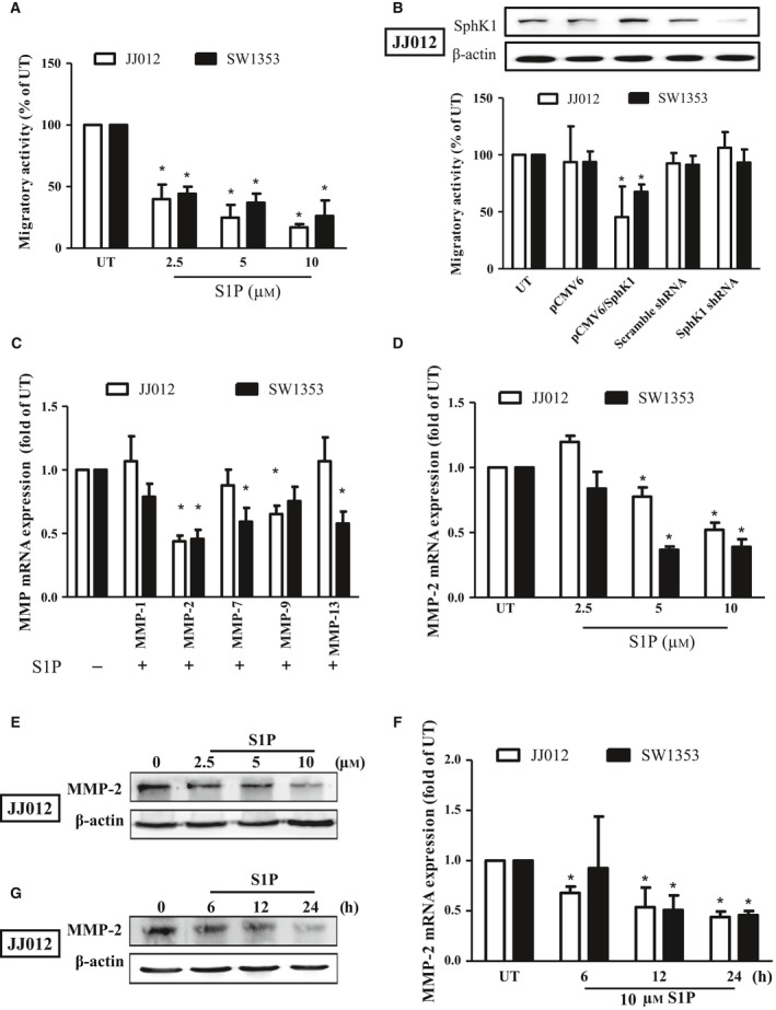
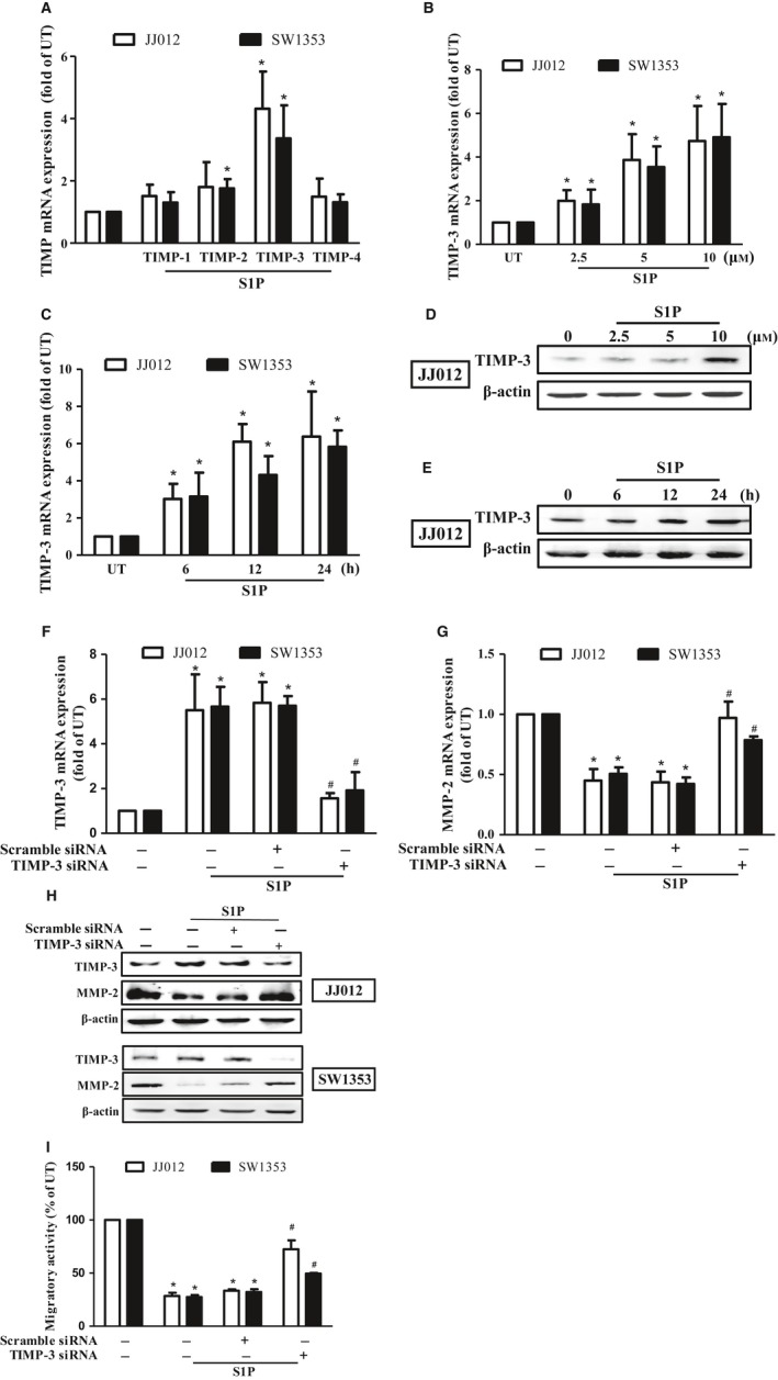
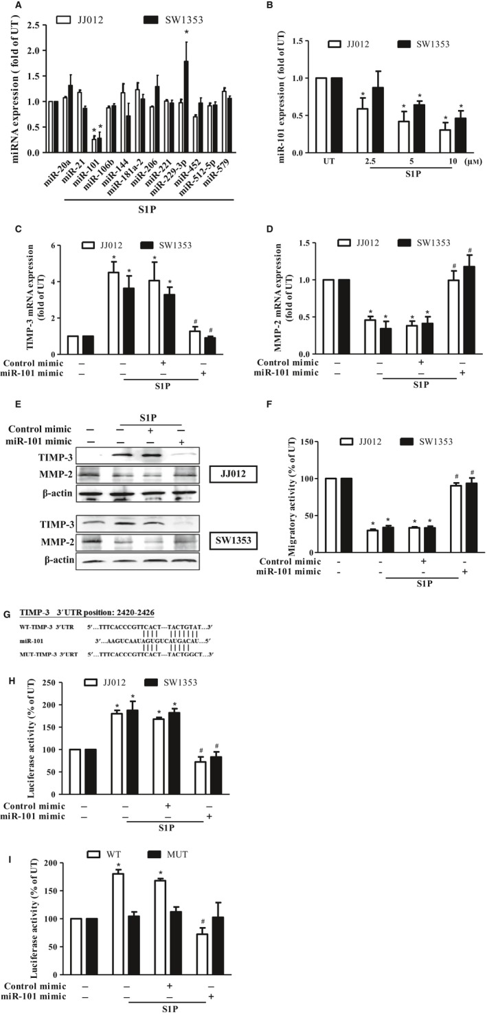
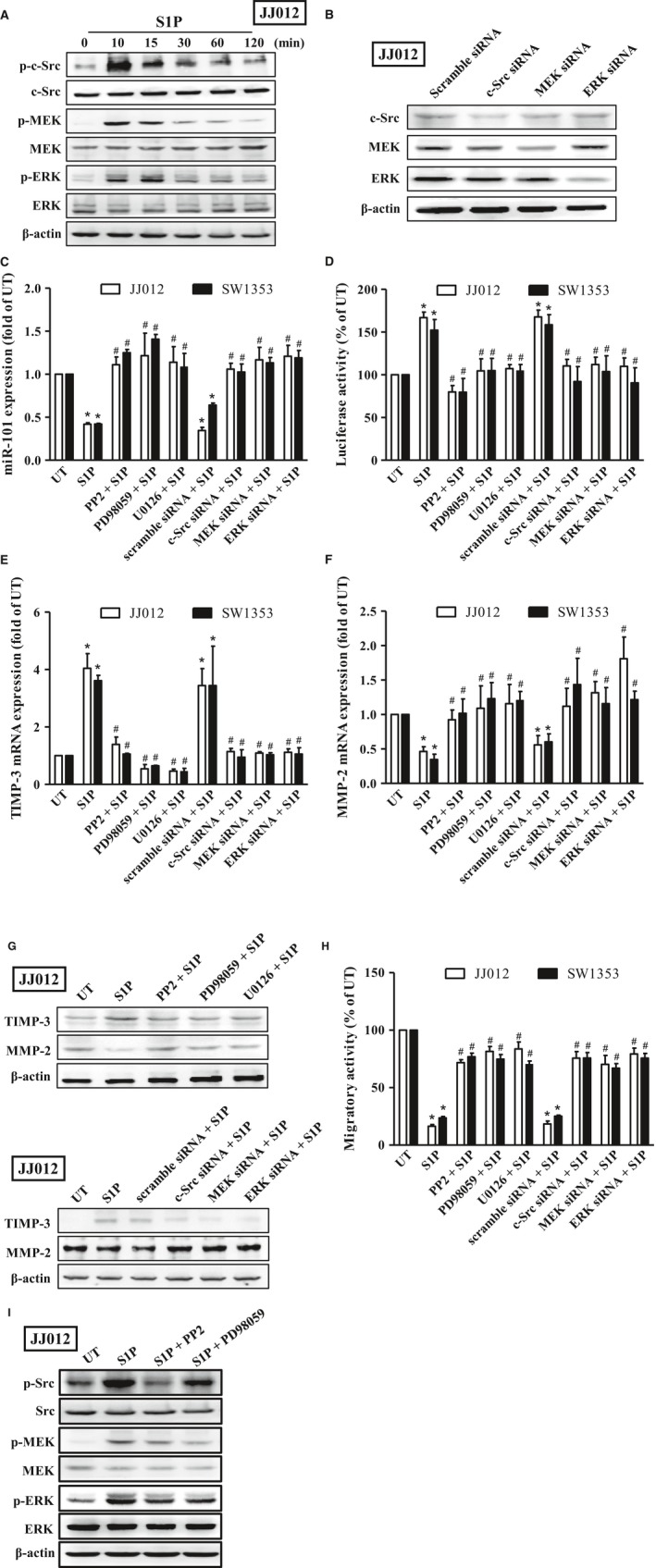
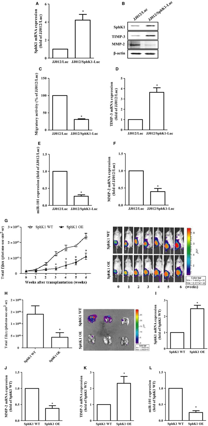
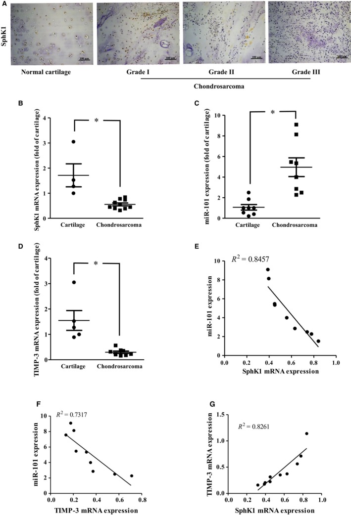
Chondrosarcoma is the second most common primary malignancy form of bone cancer, exhibiting resistance to chemotherapy and radiation therapy as well as developing high metastasis ability in late-stage tumors. Thus, understanding the metastatic processes of chondrosarcoma is considered a strategy for the treatment of this disease. Sphingosine 1-phosphate (S1P), a bioactive sphingolipid, is produced intracellularly by sphingosine kinase (SphK) and is regarded as a second signaling molecule that regulates inflammation, proliferation, angiogenesis, and metastasis. However, the effect of S1P on chondrosarcoma remains uncertain. As demonstrated by the transwell, immunoblotting, and real-time PCR analyses, we found that S1P inhibited cell migration and MMP-2 expression through the upregulation of the tissue inhibitor of metalloproteinase-3 (TIMP-3) expression in human chondrosarcoma cells. Additionally, we also showed that microRNA (miRNA)-101, which targets the 3' untranslated region (3'UTR) of TIMP-3, decreased significantly following S1P treatment. After transfection with miR-101 mimics, the S1P-regulated cell migration and TIMP-3 expression were both reversed. Furthermore, we also showed that the S1P-inhibited cell migration is mediated through the c-Src/MEK/ERK signaling axis. Meanwhile, the in vivo study indicated that overexpression of SphK1 decreases chondrosarcoma metastasis to the lungs. Our results illustrate the clinical significance between SphK1, TIMP-3, and miR-101 in human chondrosarcoma patients. Taken together, our results suggest that S1P and miR-101 may prove to be potential therapeutic targets for future chondrosarcoma treatment.

Keywords: chondrosarcoma; metastasis; microRNA; sphingosine-1-phosphate; tissue inhibitor of metalloproteinase.


Figures

Figure 1
Figure 1
S1P inhibits migration through the downregulation of MMP‐2 in human chondrosarcoma cells. Starved JJ012 (open bar) and SW1353 (closed bar) cells were incubated with S1P (2.5–10 μm) for 24 h (A), or transfected with plasmids alone or plasmids containing SphK1 coding sequence or SphK1 shRNA (B), and the migratory ability was measured by the transwell assay. The overexpression and knockdown efficiencies of SphK1 were then determined by immunoblotting in JJ012 cells (B). (C) The JJ012 and SW1353 cells were incubated with 10 μm S1P for 24 h, and the mRNA expressions of MMP‐1, MMP‐2, MMP‐7, MMP‐9, and MMP‐13 were measured by real‐time PCR. JJ012 and SW1353 cells were treated with various concentrations of S1P for 24 h (D and E) or with 10 μm of S1P for different time periods (F and G); the MMP‐2 mRNA expressions were then determined by real‐time PCR. The MMP‐2 protein expressions following S1P stimulation were determined by immunoblotting in JJ012 cells (E and G). The results are expressed as mean ± SEM. *P < 0.05 compared with untreated control (n ≧ 3). UT, untreated control.
Figure 2
Figure 2
S1P inhibits migration through upregulating TIMP‐3 expression in human chondrosarcoma cells. (A) Starved JJ012 (open bar) and SW1353 (closed bar) cells were incubated with 10 μm of S1P, and the mRNA expressions of TIMP‐1–4 were determined by real‐time PCR. JJ012 and SW1353 cells were treated with various concentrations of S1P for 24 h (B and D) or with 10 μm of S1P for different time periods (C and E); the TIMP‐3 mRNA expressions were then determined by real‐time PCR. The TIMP‐3 protein expressions following S1P stimulation were determined by immunoblotting in JJ012 cells (D and E). The JJ012 and SW1353 cells were pretransfected with scramble siRNA or TIMP‐3 siRNA, and the cell migratory activity (I), TIMP‐3 mRNA (F), MMP‐2 mRNA (G), and their protein expressions (H) were determined by transwell, real‐time PCR, and immunoblotting analyses, respectively. The results are expressed as mean ± SEM. *P < 0.05 compared with untreated control; # P < 0.05 compared with the S1P‐treated group (n ≧ 3). UT, untreated control.
Figure 3
Figure 3
S1P‐induced TIMP‐3 expression is mediated through suppressing microRNA (miR)‐101 expressions in human chondrosarcoma cells. (A) Starved JJ012 (open bar) and SW1353 (closed bar) cells were incubated with 10 μm S1P for 24 h, and the expressions of miR‐20a, miR‐21, miR‐101, miR‐106b, miR‐144, miR‐181a‐2, miR‐206, miR‐221, miR‐229‐3p, miR‐452, miR‐512‐5p, and miR‐579 were measured by real‐time PCR. (B) JJ012 and SW1353 cells were treated with various concentrations (2.5–10 μm) of S1P for 24 h, and the expressions of miR‐101 were determined by real‐time PCR. The JJ012 and SW1353 cells were pretransfected with control or miR‐101 mimics for 24 h, followed by incubating with 10 μm of S1P for another 24 h, and the mRNA expressions of TIMP‐3 (C) and MMP‐2 (D) as well as their protein expressions (E) and the migratory activity (F) were then determined by real‐time PCR, immunoblotting, and transwell analyses, respectively. (G) The luciferase reporter vectors harboring the WT 3′ UTR (WTTIMP‐3 3′ UTR) and nucleotides mutated in this region (MUTTIMP‐3 3′ UTR) of TIMP‐3 were constructed. (H) JJ012 and SW1353 cells were co‐transfected with the luciferase plasmid harboring WTTIMP‐3 3′ UTR and miR‐101 or control mimics for 24 h, followed by treatment with 10 μm of S1P for another 24 h, and the relative luciferase activity was then determined. (I) JJ012 cells were co‐transfected with the luciferase plasmids harboring either WT or MUTTIMP‐3 3′ UTR and the mimics of control or miR‐101 for 24 h, followed by treatment with 10 μm of S1P for another 24 h, and the relative luciferase activity was then determined. Cells without treatment were used as the untreated control (set to 1 or 100), and data were shown as multiples of that. The results are expressed as mean ± SEM. *< 0.05 compared with UT; # < 0.05 compared with the S1P‐treated group (n ≧ 3). UT, untreated control; 3′ UTR, 3′ untranslated region; WT, wild‐type; MUT, mutant.
Figure 4
Figure 4
S1P‐induced TIMP‐3 expression is mediated through the c‐Src/MAPK signaling axis in human chondrosarcoma cells. (A) Starved JJ012 cells were incubated with 10 μm of S1P for 10, 15, 30, 60, and 120 min, and the phosphorylation of c‐Src, MEK, and ERK was examined by immunoblotting using anti‐phospho c‐Src, MEK, or ERK antibodies, followed by reprobing against total c‐Src, MEK, ERK, and β‐actin to show the equal loading amounts. (B) The JJ012 cells were transfected with siRNA of scramble, c‐Src, MEK, and ERK for 24 h, and the protein expressions of c‐Src, MEK, and ERK were then determined to show the knockdown efficiency. Starved JJ012 (open bar) and SW1353 (closed bar) cells were pretreated with 10 μm of PP2, PD98059, and U0126 for 1 h or transfected with siRNA of scramble, c‐Src, MEK, and ERK for 24 h, followed by treatment with 10 μm of S1P for another 24 h, and expressions of miR‐101 (C), TIMP‐3 (E), and MMP‐2 (F) mRNA, as well as the protein expressions of TIMP‐3 and MMP‐2 (G), and migratory activity (H), were then determined by real‐time PCR, immunoblotting, and transwell analyses, respectively. (D) JJ012 and SW1353 cells were transfected with plasmids harboring with WTTIMP‐3 3′UTR, followed by treatment with 10 μm of PP2, PD98059, and U0126 for 1 h or transfected with siRNA of scramble, c‐Src, MEK, and ERK for 24 h prior to incubation with 10 μm of S1P for another 24 h. The promoter activity of TIMP‐3 was then determined by luciferase activity assay. (I) Starved JJ012 cells were pretreated with 10 μm of PP2 and PD98059 for 1 h, followed by treatment with 10 μm of S1P for another 10 min, and the phosphorylation of Src, MEK, and ERK was then determined by immunoblotting analysis. Cells without treatment were used as the untreated control (set to 1 or 100), and data were shown as multiples of that. The results are expressed as mean ± SEM. *< 0.05 compared with UT; # < 0.05 compared with the S1P‐treated group (n ≧ 3). UT, untreated control; 3′ UTR, 3′ untranslated region; WT, wild‐type.
Figure 5
Figure 5
Overexpression of SphK1 decreases cell migration and chondrosarcoma metastasis in vitro and in vivo. The constitutively expressed pLenti CMV V5‐Luc JJ012 cells were transfected with pCMV6 plasmid alone (JJ012/Luc) or harboring with human SphK1 ORF cDNA (JJ012/Sphk1‐Luc), followed by the determination of the protein (B) and mRNA expressions of SphkK1 (A), TIMP‐3 (D), MMP‐2 (F), as well as miR‐101 expression (E) and the migratory activity (C) by immunoblotting, real‐time PCR, and the transwell analyses, respectively. (G) JJ012/Luc or JJ012/SphK1‐Luc cells representing the wild‐type or overexpression of SphK1 were injected into the lateral tail vein of severe combined immunodeficient mice, and the development of lung metastasis was monitored by bioluminescence imaging at the time intervals. These images were then quantified (photons/s of lung region). After six weeks, these mice were humanely sacrificed, and the lung tissues were excised, photographed, and quantified (H). The mRNA expressions of SphK1 (I), MMP‐2 (J), and TIMP‐3 (K), as well as miR‐101 (L) on these tumors, were then assessed by real‐time PCR analysis. Cells without overexpression of SphK1 were used as the control (set to 1 or 100), and data were shown as multiples of that. The results are expressed as mean ± SEM. *< 0.05 compared with control (n ≧ 3). SphK1, sphingosine kinase 1; ORF, open reading frame; WT, wild‐type; OE, overexpression.
Figure 6
Figure 6
Clinical importance of SphK1, TIMP‐3, and miR‐101 in chondrosarcoma. (A) The protein expression of SphK1 was determined in the tissue array by IHC staining. The mRNA expressions of SphK1 (B) and TIMP‐3 (D), as well as miR‐101 (C) in normal cartilage and chondrosarcoma tissues, were examined by real‐time PCR. The correlation between miR‐101 and SphK1 (E), miR‐101 and TIMP‐3 (F), and TIMP‐3 and SphK1 (G) gene expressions was then determined. Quantitative results are expressed as the mean ± SEM. *< 0.05 compared with cartilage tissue samples (n ≧ 3).

Similar articles

Cited by

References

    1. Arpino V, Brock M and Gill SE (2015) The role of TIMPs in regulation of extracellular matrix proteolysis. Matrix Biol 44–46, 247–s254. - PubMed
    1. Baker AH, Edwards DR and Murphy G (2002) Metalloproteinase inhibitors: biological actions and therapeutic opportunities. J Cell Sci 115, 3719–3727. - PubMed
    1. Bartke N and Hannun YA (2009) Bioactive sphingolipids: metabolism and function. J Lipid Res 50, S91–S96. - PMC - PubMed
    1. Blaho VA and Hla T (2014) An update on the biology of sphingosine 1‐phosphate receptors. J Lipid Res 55, 1596–1608. - PMC - PubMed
    1. Chang CL, Ho MC, Lee PH, Hsu CY, Huang WP and Lee H (2009) S1P(5) is required for sphingosine 1‐phosphate‐induced autophagy in human prostate cancer PC‐3 cells. Am J Physiol Cell Physiol 297, C451–C458. - PubMed

Publication types

MeSH terms