Association Between Linear Growth and Bone Accrual in a Diverse Cohort of Children and Adolescents
- PMID: 28672287
- PMCID: PMC5632753
- DOI: 10.1001/jamapediatrics.2017.1769
Association Between Linear Growth and Bone Accrual in a Diverse Cohort of Children and Adolescents
Abstract
Importance: Prevention of osteoporosis in adulthood begins with optimizing bone health in early life. The longitudinal association between growth and bone accretion during childhood is not fully understood.
Objectives: To assess the acquisition of whole-body (WB) and skeletal site-specific bone mineral content (BMC) relative to linear growth in a healthy, diverse, longitudinal cohort of children, adolescents, and young adults and to test for differences related to sex and African American race.
Design, setting, and participants: This investigation was a mixed longitudinal study with annual assessments for up to 7 years at 5 US clinical centers. Participants were healthy children, adolescents, and young adults. The study dates were July 2002 through March 2010. The dates of the analysis were June through December 2016.
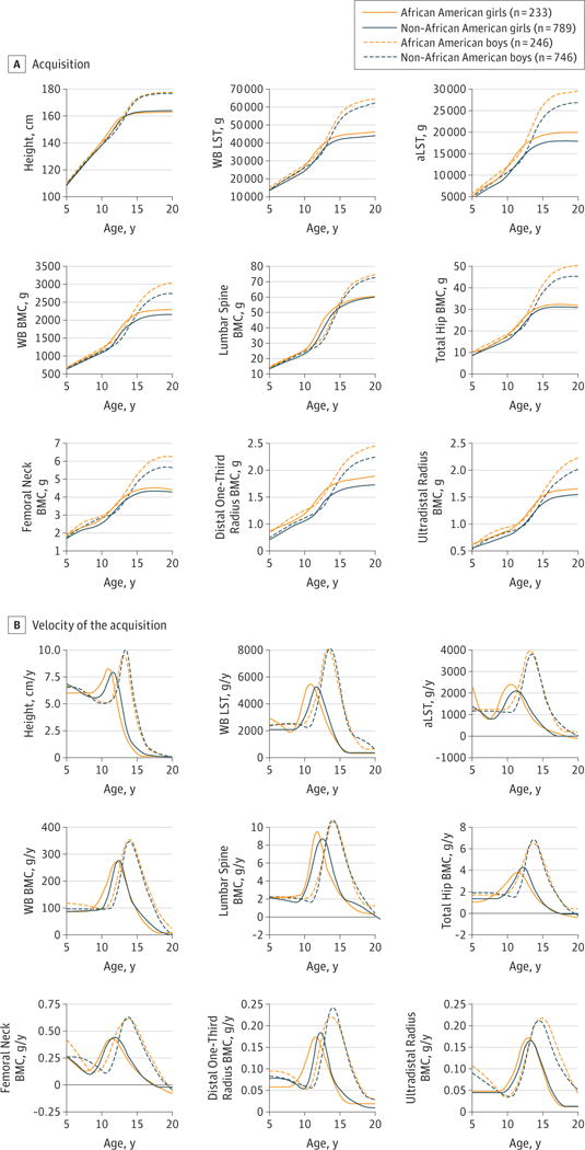
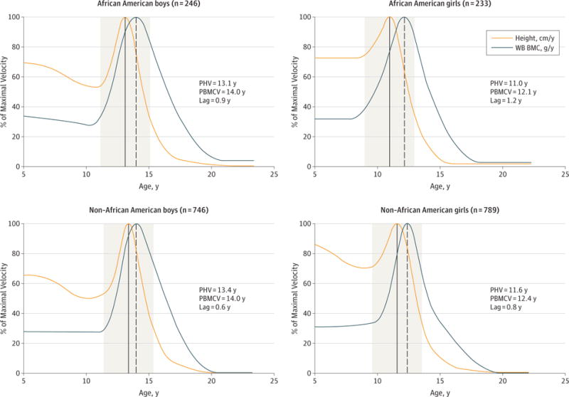
Main outcomes and measures: Anthropometrics, BMC, and body composition via dual-energy x-ray absorptiometry. The superimposition by translation and rotation (SITAR) analysis method was used to define the mean trajectories for height, WB lean soft tissue, appendicular lean soft tissue, and WB and skeletal site-specific BMC acquisition and to measure the age and magnitude of peak velocity for each parameter. The SITAR modeling was performed separately by sex and self-reported race.
Results: Among 2014 healthy children, adolescents, and young adults (1022 [50.7%] female and 479 [23.8%] African American) aged 5 to 19 years at study entry, the mean age of peak height velocity was 13.1 years (95% CI, 13.0-13.2 years) in African American boys vs 13.4 years (95% CI, 13.3-13.4 years) in non-African American boys (difference, -0.3 years; 95% CI, -0.4 to -0.1 years) and 11.0 years (95% CI, 10.8-11.1 years) in African American girls vs 11.6 years (95% CI, 11.5-11.6 years) in non-African American girls (difference, -0.6 years; 95% CI, -0.7 to -0.5 years). Age of peak acquisition of WB BMC was 14.0 years (95% CI, 13.8-14.1 years) in African American boys vs 14.0 years (95% CI, 13.9-14.1 years) in non-African American boys (difference, -0.0 years; 95% CI, -0.2 to 0.2 years) and 12.1 years (95% CI, 12.0-12.3 years) in African American girls vs 12.4 years (95% CI, 12.3-12.5 years) in non-African American girls (difference, -0.3 years; 95% CI, -0.4 to -0.1 years). At age 7 years, children had acquired 69.5% to 74.5% of maximal observed height but only 29.6% to 38.1% of maximal observed WB BMC. Adolescents gained 32.7% to 35.8% of maximal observed WB BMC during the 2 years before and 2 years after peak height velocity. Another 6.9% to 10.7% of maximal observed WB BMC occurred after linear growth had ceased. In the group at highest risk for fracture, non-African American boys, peak fracture incidence occurred approximately 1 year before peak height velocity.
Conclusions and relevance: In this longitudinal study, height gains substantially outpaced gains in BMC during childhood, which could contribute to fracture risk. A significant proportion of bone is accrued after adult height is achieved. Therefore, late adolescence represents a potentially underrecognized window of opportunity to optimize bone mass.
Conflict of interest statement
None reported.
Figures
Comment in
-
Adaptation of Bone to Mechanical Strain.JAMA Pediatr. 2018 Feb 1;172(2):196. doi: 10.1001/jamapediatrics.2017.4657. JAMA Pediatr. 2018. PMID: 29228099 No abstract available.
-
Adaptation of Bone to Mechanical Strain-Reply.JAMA Pediatr. 2018 Feb 1;172(2):196-197. doi: 10.1001/jamapediatrics.2017.4654. JAMA Pediatr. 2018. PMID: 29228153 Free PMC article. No abstract available.
References
-
- Faulkner RA, Davison KS, Bailey DA, Mirwald RL, Baxter-Jones AD. Size-corrected BMD decreases during peak linear growth: implications for fracture incidence during adolescence. J Bone Miner Res. 2006;21(12):1864–1870. - PubMed
-
- Jackowski SA, Faulkner RA, Farthing JP, Kontulainen SA, Beck TJ, Baxter-Jones AD. Peak lean tissue mass accrual precedes changes in bone strength indices at the proximal femur during the pubertal growth spurt. Bone. 2009;44(6):1186–1190. - PubMed
-
- Jackowski SA, Lanovaz JL, Van Oort C, Baxter-Jones AD. Does lean tissue mass accrual during adolescence influence bone structural strength at the proximal femur in young adulthood? Osteoporos Int. 2014;25(4):1297–1304. - PubMed
-
- Zemel BS, Kalkwarf HJ, Gilsanz V, et al. Revised reference curves for bone mineral content and areal bone mineral density according to age and sex for black and non-black children: results of the Bone Mineral Density in Childhood Study [published correction appears in J Clin Endocrinol Metab. 2013;98(1):420] J Clin Endocrinol Metab. 2011;96(10):3160–3169. - PMC - PubMed
Publication types
MeSH terms
Grants and funding
LinkOut - more resources
Full Text Sources
Other Literature Sources
Medical