Review
. 2017 Jun 24;9(7):653.
doi: 10.3390/nu9070653.

Sugars, Sweet Taste Receptors, and Brain Responses

Affiliations
Review

Sugars, Sweet Taste Receptors, and Brain Responses

Allen A Lee et al. Nutrients. .

Abstract

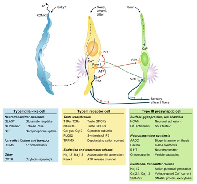
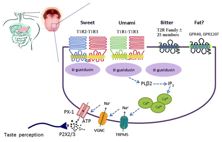
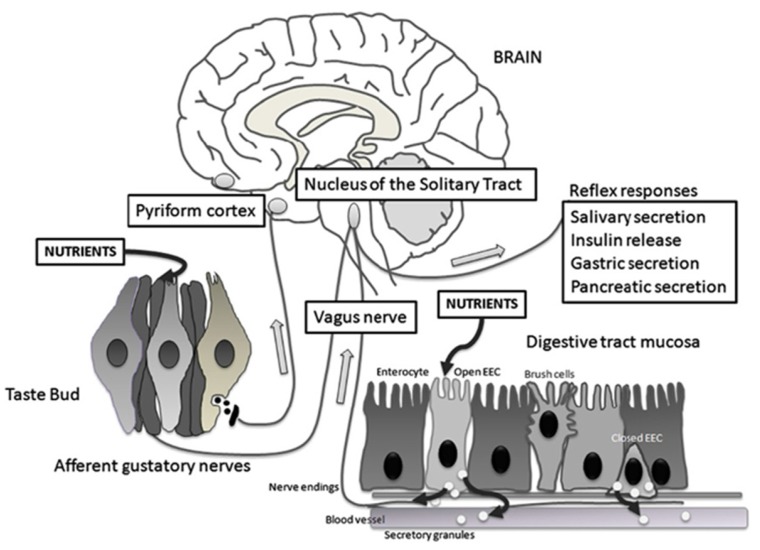
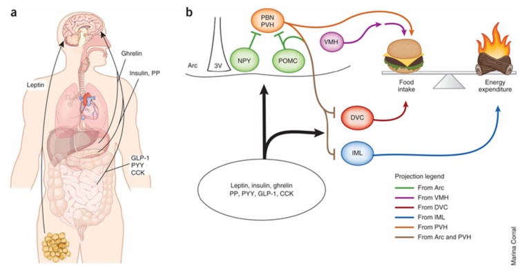
Sweet taste receptors are composed of a heterodimer of taste 1 receptor member 2 (T1R2) and taste 1 receptor member 3 (T1R3). Accumulating evidence shows that sweet taste receptors are ubiquitous throughout the body, including in the gastrointestinal tract as well as the hypothalamus. These sweet taste receptors are heavily involved in nutrient sensing, monitoring changes in energy stores, and triggering metabolic and behavioral responses to maintain energy balance. Not surprisingly, these pathways are heavily regulated by external and internal factors. Dysfunction in one or more of these pathways may be important in the pathogenesis of common diseases, such as obesity and type 2 diabetes mellitus.

Keywords: glucose sensing; hypothalamus; leptin; nutrient sensing; sweet taste receptors.


Conflict of interest statement

The authors declare no conflict of interest.

Figures

Figure 1
Figure 1
The four major classes of taste cells. This classification incorporates ultrastructural features, patterns of gene expression, and the functions of each Types I, II (receptor), III (presynaptic), and IV (progenitor, not depicted) taste cells. Type I cells (blue) degrade or absorb neurotransmitters. They also may clear extracellular K+ that accumulates after action potentials (shown as bursts) in receptor (yellow) and presynaptic (green) cells. K+ may be extruded through an apical K channel such as renal outer medullary potassium channel (ROMK). Salty taste may be transduced by some Type I cells, but this remains uncertain. Sweet, bitter, and umami taste compounds activate receptor cells, inducing them to release ATP through pannexin 1 (Panx1) hemichannels. The extracellular ATP excites ATP receptors (P2X, P2Y) on sensory nerve fibers and on taste cells. Presynaptic cells, in turn, release serotonin (5-HT), which inhibits receptor cells. Sour stimuli (and carbonation, not depicted) directly activate presynaptic cells. Only presynaptic cells form ultrastructurally identifiably synapses with nerves. Tables below the cells list some of the proteins that are expressed in a cell type-selective manner. AADC, aromatic L-amino acid decarboxylase; Ca, voltage-gated calcium channel; Gα-gus, alpha-gustducin; Gγ13, Gγ13 subunit; GAD, glutamate decarboxylase; GLAST, glutamate aspartate transporter; 5-HT, 5-hydroxytryptamine; mGluRs, metabotropic glutamate receptor; Na, voltage-gated sodium channel; NCAM, neural cell adhesion molecule; NET, norepinephrine transporter; NTPDase; nucleoside triphosphate diphosphohydrolase; OXTR, oxytoxin receptor; Panx1, pannexin 1; PKD, polycystic kidney disease-like channel; PLCβ2, phospholipase C β2; ROMK, renal outer medullary potassium channel; SNAP, synaptosomal-associated protein; T1R, taste 1 receptor family; T2R, taste 2 receptor family; TRPM5, transient receptor potential cation channel M5; Adapted with permission from [6].
Figure 2
Figure 2
Simplified model of the taste GPCR signaling pathways involved in chemosensing by taste receptors of the tongue. Subtypes of the T1R family heterodimerize to detect sweet (T1R2-T1R3) and umami (T1R1-T1R3). Bitter is detected by the T2R family. Medium-chain and long-chain fatty acids are detected by FFAR1 and GPR120. Taste receptor binding leads to activation of gustatory G-proteins, release of intracellular Ca2+, activation of TRPM5, depolarization, activation of voltage-gated Na+ channels (VGNC), and release of ATP which activates purinergic receptors on afferent fibers leading to taste perception. ATP, adenosine triphosphate; FFAR1, free fatty acid receptor 1; GPCR, G-protein coupled receptor; PX-1, pannexin 1-hemichannel; T1R, taste receptor type 1; T1R1, taste receptor type 1 member 1; T1R2, taste receptor type 1 member 2; T1R3, taste receptor type 1 member 3; T2R, taste receptor type 2; TRPM5, transient receptor potential cation channel M5; VGNC, voltage-gated Na+ channel. Reproduced with permission from [10].
Figure 3
Figure 3
Schematic representation of the taste circuitry. The gustatory system is represented by taste cells in taste buds and their gustatory nerves. Corresponding to the gastrointestinal system, there are two enteroendocrine cells (EEC), one that is open to the lumen releasing cholecystokinin (CCK) and glucagon-like peptide 1 (GLP-1) in response to luminal nutrients and one that is closed. Vagal fibers are located underneath the GI mucosa in close contact with hormone secretions. The signals from the gustatory system reach the rostral nucleus of the solitary tract whereas visceral impulses terminate at the caudal nucleus of the solitary tract. From the nucleus of the solitary tract, gustatory and visceral information projects to several brain regions including the amygdala, the hypothalamus, and the ventral posterior nucleus of the thalamus. These regions are involved with ingestive motivation, physiological reflexes, and energy homeostasis. Reproduced with permission from [50].
Figure 4
Figure 4
(a) Multiple peripheral factors have been shown to modify food intake and energy expenditure through direct effects on the central nervous system. Leptin, which likely acts on sweet taste receptor cells, is the primary regulator of energy balance and food intake in the hypothalamus. (b) Evidence suggests that melanocortin signaling regulates these physiological processes by means of distinct projection patterns originating from pro-opiomelanocortin (POMC) neurons in the arcuate nucleus (Arc). Sweet taste receptors likely act on neuropeptide Y (NPY) neurons in the hypothalamus to regulate energy homeostasis. Ultimately, MC4 receptor (MC4R)-expressing neurons downstream of POMC neurons act to suppress food intake and increase energy expenditure. Hypothalamic NPY/AgRP, paraventricular nucleus of the hypothalamus (PVH) and VMH neurons, as well as hindbrain dorsal vagal complex (DVC), parabrachial nucleus (PBN) and spinal cord intermediolateral cell column (IML) neurons, also regulate or counter-regulate these activities. PP, pancreatic polypeptide; PYY, peptide YY; 3V, third ventricle. Adapted with permission from [60].

References

    1. Laugerette F., Passilly-Degrace P., Patris B., Niot I., Febbraio M., Montmayeur J.-P., Besnard P. CD36 involvement in orosensory detection of dietary lipids, spontaneous fat preference, and digestive secretions. J. Clin. Investig. 2005;115:3177–3184. doi: 10.1172/JCI25299. - DOI - PMC - PubMed
    1. Janssen S., Depoortere I. Nutrient sensing in the gut: New roads to therapeutics? Trends Endocrinol. Metab. 2013;24:92–100. - PubMed
    1. Finger T.E., Danilova V., Barrows J., Bartel D.L., Vigers A.J., Stone L., Hellekant G., Kinnamon S.C. Neuroscience: ATP signalling is crucial for communication from taste buds to gustatory nerves. Science. 2005;310:1495–1499. doi: 10.1126/science.1118435. - DOI - PubMed
    1. Bo X., Alavi A., Xiang Z., Oglesby I., Ford A., Burnstock G. Localization of ATP-gated P2X2 and P2X3 receptor immunoreactive nerves in rat taste buds. NeuroReport. 1999;10:1107–1111. doi: 10.1097/00001756-199904060-00037. - DOI - PubMed
    1. Yang R., Montoya A., Bond A., Walton J., Kinnamon J.C. Immunocytochemical analysis of P2X2 in rat circumvallate taste buds. BMC Neurosci. 2012;13:51. doi: 10.1186/1471-2202-13-51. - DOI - PMC - PubMed

LinkOut - more resources