. 2017 Jun 26;18(7):1359.
doi: 10.3390/ijms18071359.

Immune Modulating Topical S100A8/A9 Inhibits Growth of Pseudomonas aeruginosa and Mitigates Biofilm Infection in Chronic Wounds

Affiliations

Immune Modulating Topical S100A8/A9 Inhibits Growth of Pseudomonas aeruginosa and Mitigates Biofilm Infection in Chronic Wounds

Hannah Trøstrup et al. Int J Mol Sci. .

Abstract

Pseudomonas aeruginosa biofilm maintains and perturbs local host defense, hindering timely wound healing. Previously, we showed that P. aeruginosa suppressed S100A8/A9 of the murine innate host defense. We assessed the potential antimicrobial effect of S100A8/A9 on biofilm-infected wounds in a murine model and P. aeruginosa growth in vitro. Seventy-six mice, inflicted with a full-thickness burn wound were challenged subcutaneously (s.c.) by 10⁶ colony-forming units (CFUs) of P. aeruginosa biofilm. Mice were subsequently randomized into two treatment groups, one group receiving recombinant murine S100A8/A9 and a group of vehicle controls (phosphate-buffered saline, PBS) all treated with s.c. injections daily for up to five days. Wounds were analyzed for quantitative bacteriology and contents of key inflammatory markers. Count of blood polymorphonuclear leukocytes was included. S100A8/A9-treatment ameliorated wound infection, as evaluated by quantitative bacteriology (p ≤ 0.05). In vitro, growth of P. aeruginosa was inhibited dose-dependently by S100A8/A9 in concentrations from 5 to 40 μg/mL, as determined by optical density-measurement (OD-measurement) and quantitative bacteriology. Treatment slightly augmented key inflammatory cytokine Tumor Necrosis Factor-α (TNF-α), but dampened interferon-γ (IFN-γ) levels and blood polymorphonuclear count. In conclusion, topical S100A8/A9 displays remarkable novel immune stimulatory and anti-infective properties in vivo and in vitro. Importantly, treatment by S100A8/A9 provides local infection control. Implications for a role as adjunctive treatment in healing of chronic biofilm-infected wounds are discussed.

Keywords: Pseudomonas aeruginosa; S100A8/A9; biofilm infection; chronic wounds; host defense.


Conflict of interest statement

The authors declare no conflict of interest.

Figures

Figure 1
Figure 1
Induction of local wound infection control. Log10 colony-forming units (CFUs) of P. aeruginosa/wound as expression of time in the intervention group (S100A8/A9) and control group (phosphate-buffered saline, PBS). S100A8/A9-treatment induced infection control. The number of colony-forming units (CFUs) after 5 days of treatment was significantly reduced as compared to the non-treated control group (** p < 0.05). Increase in growth over 5 days in the control group only (* p < 0.0001). Individual values indicate mean of duplicates with overall mean ± standard deviation. Black squares, PBS-treated; black circles, S100A8/A9-treated.
Figure 2
Figure 2
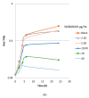
S100A8/A9 inhibits growth of Pseudomonas aeruginosa. (A) Growth inhibitory effect of S100A8/A9 on PAO1, shown by OD-curve (read at OD600 at time points indicated). (B) Growth inhibitory effect of S100A8/A9 on PAO1, shown by bacterial CFU count (1 h, 6 h, 24 h, at concentrations 40, 20, 10, 5, 2.5, or 0 μg/mL). Growth inhibitory effect of S100A8/A9 on PAO1, shown by (A) OD-curve (read at OD600 at time points indicated) and (B) bacterial count as log10 CFU (1 h, 6 h, 24 h at concentrations 40, 20, 10, 5, 2.5, or 0 μg/mL).
Figure 2
Figure 2
S100A8/A9 inhibits growth of Pseudomonas aeruginosa. (A) Growth inhibitory effect of S100A8/A9 on PAO1, shown by OD-curve (read at OD600 at time points indicated). (B) Growth inhibitory effect of S100A8/A9 on PAO1, shown by bacterial CFU count (1 h, 6 h, 24 h, at concentrations 40, 20, 10, 5, 2.5, or 0 μg/mL). Growth inhibitory effect of S100A8/A9 on PAO1, shown by (A) OD-curve (read at OD600 at time points indicated) and (B) bacterial count as log10 CFU (1 h, 6 h, 24 h at concentrations 40, 20, 10, 5, 2.5, or 0 μg/mL).
Figure 3
Figure 3
Modulation of proinflammatory cytokines. (A) Tumor Necrosis Factor-α (TNF-α) levels were increased in the S100A8/A9 group from day 1 to 5 (p < 0.0261) and day 3 to 5 (p < 0.001). No such increase was observed in the PBS group; (B) Interferon-γ (IFN-γ) levels were increased in the PBS from day 1 to 5 (p < 0.0023), 2 to 5 (p < 0.0008), and day 3 to 5 (p < 0.0054). No increase was observed in the S100A8/A9 group; (C) IL-10. No significant changes were observed in either of the two groups, but there was a tendency to an attenuated response in the S100A8/A9 group; (D) in the PBS group, Keratinocyte-derived chemokine (KC) increased from day 1 to 5 (p < 0.0156). In the S100A8/A9 group, KC increased from day 1 to 5 (p < 0.0139) and day 2 to 5 (p < 0.0017). (E) In both groups, IL-1β increased from day 1 to 5 (p < 0.0005 in the PBS group and p < 0.0482 in the S100A8/A9 group); (F) In both groups, Granulocyte-Colony Stimulating Factor (G-CSF) increased with time for all time points evaluated. (G) In both groups, Il-17 increased with time for all time points evaluated. Modulation of inflammatory response by box plots of inflammatory cytokines (AE): Levels of proteins are expressed in ρg/mL wound.
Figure 3
Figure 3
Modulation of proinflammatory cytokines. (A) Tumor Necrosis Factor-α (TNF-α) levels were increased in the S100A8/A9 group from day 1 to 5 (p < 0.0261) and day 3 to 5 (p < 0.001). No such increase was observed in the PBS group; (B) Interferon-γ (IFN-γ) levels were increased in the PBS from day 1 to 5 (p < 0.0023), 2 to 5 (p < 0.0008), and day 3 to 5 (p < 0.0054). No increase was observed in the S100A8/A9 group; (C) IL-10. No significant changes were observed in either of the two groups, but there was a tendency to an attenuated response in the S100A8/A9 group; (D) in the PBS group, Keratinocyte-derived chemokine (KC) increased from day 1 to 5 (p < 0.0156). In the S100A8/A9 group, KC increased from day 1 to 5 (p < 0.0139) and day 2 to 5 (p < 0.0017). (E) In both groups, IL-1β increased from day 1 to 5 (p < 0.0005 in the PBS group and p < 0.0482 in the S100A8/A9 group); (F) In both groups, Granulocyte-Colony Stimulating Factor (G-CSF) increased with time for all time points evaluated. (G) In both groups, Il-17 increased with time for all time points evaluated. Modulation of inflammatory response by box plots of inflammatory cytokines (AE): Levels of proteins are expressed in ρg/mL wound.
Figure 4
Figure 4
S100A8/A9 dampens systemic polymorphonuclear count. Systemic polymorphonuclear (PMN) count in S100A8/A9 (+) treated group versus PBS (−). Bars are expressed with mean + standard deviation. No significant increase was seen in the S100A8/A9 group. In contrast, in the PBS group, an increase in PMN count was seen from day 1 to 3 (p < 0.03) and day 1 to 5 (p < 0.003). Abbreviations: b: background (n = 6), b0: burned, results obtained immediately after wounding, b2: burned, two days after wounding, b4: burned, 4 days after wounding; bi: burn and infection, results obtained immediately after infection. D1, 2, 3, 5: day 1, 2, 3, 5. *, significant increase.

Similar articles

Cited by

References

    1. Høgsberg T., Bjarnsholt T., Thomsen J.S., Kirketerp-Møller K. Success rate of split-thickness skin grafting of chronic venous leg ulcers depends on the presence of Pseudomonas aeruginosa: A retrospective study. PLoS ONE. 2011;6:e20492. - PMC - PubMed
    1. Kirketerp-Møller K., Jensen P.O., Fazli M., Madsen K.G., Pedersen J., Moser C., Tolker-Nielsen T., Høiby N., Givskov M., Bjarnsholt T. Distribution, organization, and ecology of bacteria in chronic wounds. J. Clin. Microbiol. 2008;46:2717–2722. doi: 10.1128/JCM.00501-08. - DOI - PMC - PubMed
    1. Malone M., Bjarnsholt T., McBain A.J., James G.A., Stoodley P., Leaper D., Tachi M., Schultz G., Swanson T., Wolcott R.D. The prevalence of biofilms in chronic wounds: A systematic review and meta-analysis of published data. J. Wound Care. 2017;26:20–25. doi: 10.12968/jowc.2017.26.1.20. - DOI - PubMed
    1. Trøstrup H., Bjarnsholt T., Kirketerp-Møller K., Høiby N., Moser C. What is new in the understanding of non healing wounds. Ulcers. 2013;2013:625934. doi: 10.1155/2013/625934. - DOI
    1. Roth J., Vogl T., Sorg C., Sunderkotter C. Phagocyte-specific S100 proteins: A novel group of proinflammatory molecules. Trends Immunol. 2003;24:155–158. doi: 10.1016/S1471-4906(03)00062-0. - DOI - PubMed

MeSH terms

LinkOut - more resources