

Anthocyanins from black rice (Oryza sativa) promote immune responses in leukemia through enhancing phagocytosis of macrophages in vivo
- PMID: 28672893
- PMCID: PMC5488472
- DOI: 10.3892/etm.2017.4467
Anthocyanins from black rice (Oryza sativa) promote immune responses in leukemia through enhancing phagocytosis of macrophages in vivo
Abstract
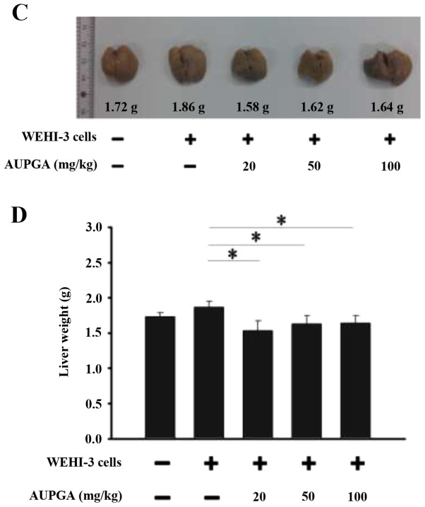
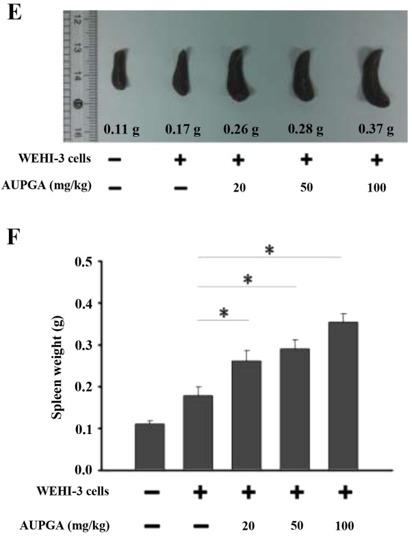
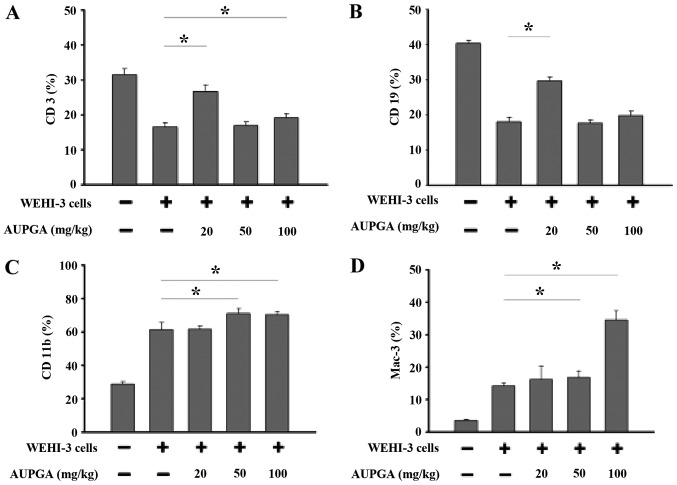
Rice is a staple food in numerous countries around the world. Anthocyanins found in black rice have been reported to reduce the risk of certain diseases, but the effects of crude extract of anthocyanins from Asia University-selected purple glutinous indica rice (AUPGA) on immune responses have not yet been demonstrated. The current study aimed to investigate whether AUPGA treatment could affect immune responses in murine leukemia cells in vivo. Murine acute myelomonocytic leukemia WEHI-3 cells were intraperitoneally injected into normal BALB/c mice to generate leukemia mice. A total of 50 mice were randomly divided into five groups (n=10 in each group) and were fed a diet supplemented with AUPGA at 0, 20, 50 or 100 mg/kg for three weeks. All mice were weighed and the blood, liver and spleen were collected for further experiments. The results indicated that AUPGA did not significantly affect animal body weight, but significantly increased spleen weight (P<0.05) and decreased liver weight (P<0.05) when compared with the control group. AUPGA significantly increased the T cell (CD3) population at treatments of 20 and 100 mg/kg (P<0.05). However, it only significantly increased the B cell (CD19) population at a treatment of 20 mg/kg (P<0.05). Furthermore, AUPGA at 50 and 100 mg/kg significantly increased the monocyte (CD11b) population and the level of macrophages (Mac-3; P<0.05 for both). AUPGA at 50 and 100 mg/kg significantly promoted macrophage phagocytosis in peripheral blood mononuclear cells (P<0.05), and all doses of AUPGA treatment significantly promoted macrophage phagocytotic activity in the peritoneum (P<0.05). AUPGA treatment significantly decreased natural killer cell activity from splenocytes (P<0.05). Finally, AUPGA treatment at 20 mg/kg treatment significantly promoted T cell proliferation (P<0.05), and treatment at 50 and 100 mg/kg significantly decreased B cell proliferation compared with the control group (P<0.05).
Keywords: WEHI-3 cells; crude extract of anthocyanins from black rice; macrophage; natural killer cell; phagocytosis.
Figures


Similar articles
-
Crude extract of Polygonum cuspidatum promotes immune responses in leukemic mice through enhancing phagocytosis of macrophage and natural killer cell activities in vivo.In Vivo. 2015 Mar-Apr;29(2):255-61. In Vivo. 2015. PMID: 25792654
-
Epigallocatechin gallate (EGCG), influences a murine WEHI-3 leukemia model in vivo through enhancing phagocytosis of macrophages and populations of T- and B-cells.In Vivo. 2013 Sep-Oct;27(5):627-34. In Vivo. 2013. PMID: 23988898
-
Safrole suppresses murine myelomonocytic leukemia WEHI-3 cells in vivo, and stimulates macrophage phagocytosis and natural killer cell cytotoxicity in leukemic mice.Environ Toxicol. 2013 Nov;28(11):601-8. doi: 10.1002/tox.20756. Epub 2011 Aug 24. Environ Toxicol. 2013. PMID: 24150866
-
Final report on the safety assessment of capsicum annuum extract, capsicum annuum fruit extract, capsicum annuum resin, capsicum annuum fruit powder, capsicum frutescens fruit, capsicum frutescens fruit extract, capsicum frutescens resin, and capsaicin.Int J Toxicol. 2007;26 Suppl 1:3-106. doi: 10.1080/10915810601163939. Int J Toxicol. 2007. PMID: 17365137 Review.
-
Amended final report on the safety assessment of Oryza Sativa (rice) Bran Oil, Oryza Sativa (rice) Germ Oil, Rice Bran Acid,Oryza Sativa (rice) Bran Wax, Hydrogenated Rice Bran Wax, Oryza Sativa (rice)Bran Extract, Oryza Sativa (rice) Extract, Oryza Sativa (rice) Germ Powder, Oryza Sativa (rice) Starch, Oryza Sativa (rice) Bran, Hydrolyzed Rice Bran Extract, Hydrolyzed Rice Bran Protein, Hydrolyzed Rice Extract, and Hydrolyzed Rice Protein.Int J Toxicol. 2006;25 Suppl 2:91-120. doi: 10.1080/10915810600964626. Int J Toxicol. 2006. PMID: 17090480 Review.
Cited by
-
Laminarin Promotes Immune Responses and Reduces Lactate Dehydrogenase But Increases Glutamic Pyruvic Transaminase in Normal Mice In Vivo.In Vivo. 2018 May-Jun;32(3):523-529. doi: 10.21873/invivo.11270. In Vivo. 2018. PMID: 29695555 Free PMC article.
-
Anthocyanins, delphinidin-3-O-glucoside and cyanidin-3-O-glucoside, inhibit immune checkpoints in human colorectal cancer cells in vitro and in silico.Sci Rep. 2019 Aug 9;9(1):11560. doi: 10.1038/s41598-019-47903-0. Sci Rep. 2019. PMID: 31399602 Free PMC article.
-
Gastroprotective Activities of Ethanol Extract of Black Rice Bran (Oryza sativa L.) in Rats.Molecules. 2021 Jun 22;26(13):3812. doi: 10.3390/molecules26133812. Molecules. 2021. PMID: 34206628 Free PMC article.
-
Laminarin Promotes Immune Responses and Normalizes Glutamic Oxaloacetic Transaminase and Glutamic Pyruvic Transaminase Levels in Leukemic Mice In Vivo.In Vivo. 2018 Jul-Aug;32(4):783-790. doi: 10.21873/invivo.11308. In Vivo. 2018. PMID: 29936459 Free PMC article.
-
Extract Derived From Black Rice Functions as a Photothermal Agent for Suppressing Tumor Growth and Metastasis.Front Bioeng Biotechnol. 2020 Aug 6;8:904. doi: 10.3389/fbioe.2020.00904. eCollection 2020. Front Bioeng Biotechnol. 2020. PMID: 32850748 Free PMC article.
References
-
- Juliusson G, Antunovic P, Derolf A, Lehmann S, Möllgård L, Stockelberg D, Tidefelt U, Wahlin A, Höglund M. Age and acute myeloid leukemia: Real world data on decision to treat and outcomes from the Swedish Acute Leukemia Registry. Blood. 2009;113:4179–4187. doi: 10.1182/blood-2008-07-172007. - DOI - PubMed
-
- Gundermann S, Klinker E, Kimmel B, Flierl U, Wilhelm M, Einsele H, Kunzmann V. A comprehensive analysis of primary acute myeloid leukemia identifies biomarkers predicting susceptibility to human allogeneic Vγ9Vδ2 T T cells. J Immunother. 2014;37:321–330. doi: 10.1097/CJI.0000000000000043. - DOI - PubMed
LinkOut - more resources
Full Text Sources
Other Literature Sources
Research Materials