

Cucurbitacin B inhibits cell proliferation and induces apoptosis in human osteosarcoma cells via modulation of the JAK2/STAT3 and MAPK pathways
- PMID: 28673003
- PMCID: PMC5488743
- DOI: 10.3892/etm.2017.4547
Cucurbitacin B inhibits cell proliferation and induces apoptosis in human osteosarcoma cells via modulation of the JAK2/STAT3 and MAPK pathways
Abstract
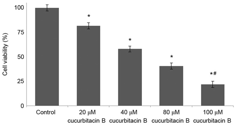
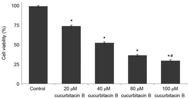
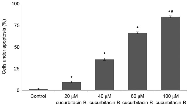
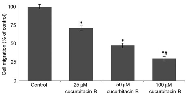
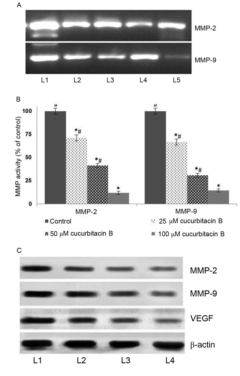
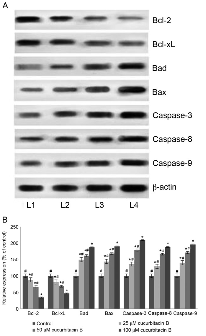
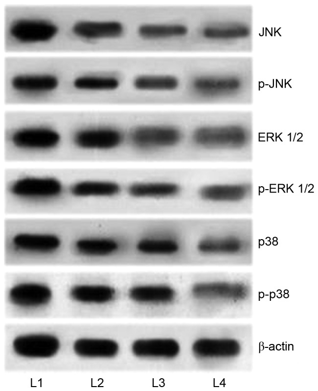
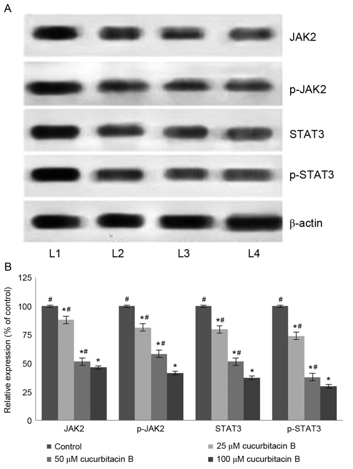
Osteosarcoma (OS) is the most commonly diagnosed tumor of the bones in children and young adults. Even with conventional therapies the 5-year survival rate is ~65% in patients with OS. Considering the side effects and aggressiveness of malignant bone tumors, research is focussing on multi-targeted strategies in treatment. Cucurbitacin B, a triterpenoid compound has been demonstrated to induce apoptosis in various cancer cell types. The Janus kinase 2/signal transducer and activator of transcription 3 (JAK2/STAT3) signalling cascades and mitogen activated protein kinases (MAPK) signalling cascades are critical regulators of tumorigenesis. The present study assessed the influence of cucurbitacin B on the viability and expression of MAPKs and proteins of the JAK2/STAT3 cascades in human OS cells (U-2 OS). Cucurbitacin B (20-100 µM) significantly reduced cell viability (P<0.05) and induced apoptosis, as assessed by MTT and Annexin V/propidium iodide staining, along with inhibiting cell migration. Gelatin zymography revealed supressed activities of matrix metalloproteinase (MMP-)2 and 9. Furthermore, cucurbitacin B effectively upregulated the apoptotic pathway and caused the effective inhibition of MAPK signalling and JAK2/STAT3 cascades. Multifold suppression of vascular endothelial growth factor by cucurbitacin B was also observed, indicating inhibition of angiogenesis. Thus, by downregulating major pathways-MAPK and JAK2/STAT3 and MMPs, cucurbitacin B has potent anti-proliferative and anti-metastatic effects that require further investigation with regards to cancer treatment.
Keywords: Janus kinase/signal transducer and activator of transcription pathway; apoptosis; cucurbitacin; mitogen activated protein kinases; osteosarcoma.
Figures
Similar articles
-
4-parvifuran inhibits metastatic and invasive actions through the JAK2/STAT3 pathway in osteosarcoma cells.Arch Pharm Res. 2017 May;40(5):601-609. doi: 10.1007/s12272-017-0911-4. Epub 2017 Apr 6. Arch Pharm Res. 2017. PMID: 28386742
-
Cucurbitacin B inhibits growth and induces apoptosis through the JAK2/STAT3 and MAPK pathways in SH‑SY5Y human neuroblastoma cells.Mol Med Rep. 2014 Jul;10(1):89-94. doi: 10.3892/mmr.2014.2175. Epub 2014 Apr 24. Mol Med Rep. 2014. PMID: 24789581
-
STAT3 inhibitor, cucurbitacin I, is a novel therapeutic agent for osteosarcoma.Int J Oncol. 2016 Dec;49(6):2275-2284. doi: 10.3892/ijo.2016.3757. Epub 2016 Nov 3. Int J Oncol. 2016. PMID: 27840900 Free PMC article.
-
Cucurbitacin IIa: A review of phytochemistry and pharmacology.Phytother Res. 2021 Aug;35(8):4155-4170. doi: 10.1002/ptr.7077. Epub 2021 Mar 16. Phytother Res. 2021. PMID: 33724593 Review.
-
Multifaceted Therapeutic Impacts of Cucurbitacin B: Recent Evidences From Preclinical Studies.Phytother Res. 2025 May;39(5):1966-1995. doi: 10.1002/ptr.8454. Epub 2025 Feb 18. Phytother Res. 2025. PMID: 39963741 Review.
Cited by
-
Apoptotic Pathway as the Therapeutic Target for Anticancer Traditional Chinese Medicines.Front Pharmacol. 2019 Jul 12;10:758. doi: 10.3389/fphar.2019.00758. eCollection 2019. Front Pharmacol. 2019. PMID: 31354479 Free PMC article. Review.
-
Recent Advances in the Application of Cucurbitacins as Anticancer Agents.Metabolites. 2023 Oct 14;13(10):1081. doi: 10.3390/metabo13101081. Metabolites. 2023. PMID: 37887406 Free PMC article. Review.
-
Sorafenib and CuB exert synergistic antitumor effects against hepatocellular carcinoma cells via inhibition of STAT3 phosphorylation.FEBS Open Bio. 2021 Jan;11(1):133-145. doi: 10.1002/2211-5463.13035. Epub 2020 Nov 27. FEBS Open Bio. 2021. PMID: 33176070 Free PMC article.
-
Liposome Delivery of Natural STAT3 Inhibitors for the Treatment of Cancer.Pharm Front. 2019;1:e190007. doi: 10.20900/pf20190007. Epub 2019 Nov 28. Pharm Front. 2019. PMID: 31886474 Free PMC article.
-
Fruit Extract of Sechium chinantlense (Lira & F. Chiang) Induces Apoptosis in the Human Cervical Cancer HeLa Cell Line.Nutrients. 2023 Jan 28;15(3):667. doi: 10.3390/nu15030667. Nutrients. 2023. PMID: 36771372 Free PMC article.
References
-
- Thayanithy V, Park C, Sarver AL, Kartha RV, Korpela DM, Graef AJ, Steer CJ, Modiano JF, Subramanian S. Combinatorial treatment of DNA and chromatin-modifying drugs cause cell death in human and canine osteosarcoma cell lines. PLoS One. 2012;7:e43720. doi: 10.1371/journal.pone.0043720. - DOI - PMC - PubMed
LinkOut - more resources
Full Text Sources
Other Literature Sources
Miscellaneous