Review
. 2017 Aug 1;38(4):302-324.
doi: 10.1210/er.2017-00025.

A Reappraisal of Testosterone's Binding in Circulation: Physiological and Clinical Implications

Affiliations
Review

A Reappraisal of Testosterone's Binding in Circulation: Physiological and Clinical Implications

Anna L Goldman et al. Endocr Rev. .

Abstract

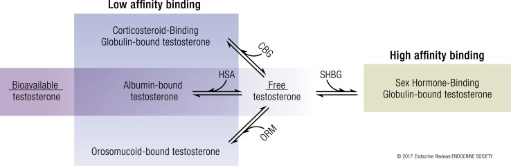
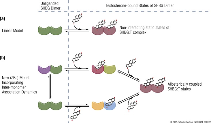
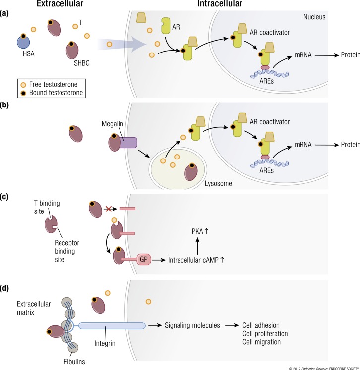
In the circulation, testosterone and other sex hormones are bound to binding proteins, which play an important role in regulating their transport, distribution, metabolism, and biological activity. According to the free hormone hypothesis, which has been debated extensively, only the unbound or free fraction is biologically active in target tissues. Consequently, accurate determination of the partitioning of testosterone between bound and free fractions is central to our understanding of how its delivery to the target tissues and biological activity are regulated and consequently to the diagnosis and treatment of androgen disorders in men and women. Here, we present a historical perspective on the evolution of our understanding of the binding of testosterone to circulating binding proteins. On the basis of an appraisal of the literature as well as experimental data, we show that the assumptions of stoichiometry, binding dynamics, and the affinity of the prevailing models of testosterone binding to sex hormone-binding globulin and human serum albumin are not supported by published experimental data and are most likely inaccurate. This review offers some guiding principles for the application of free testosterone measurements in the diagnosis and treatment of patients with androgen disorders. The growing number of testosterone prescriptions and widely recognized problems with the direct measurement as well as the computation of free testosterone concentrations render this critical review timely and clinically relevant.


Figures

Figure 1.
Figure 1.
Partitioning of testosterone in the systemic circulation. Circulating testosterone is bound tightly to SHBG (green = high affinity binding) and weakly to albumin, orosomucoid (ORM), and CBG (blue = low affinity binding) (11). Only 1% to 4% of circulating testosterone is unbound or free. The combination of free and albumin-bound testosterone is also referred to as the “bioavailable testosterone” fraction.
Figure 2.
Figure 2.
Multiple domains of HSA are involved in the binding and transport of biomolecules. (a) Crystal structure of HSA (PDB:1AO6) depicting multiple domains (I to III) of HSA that participate in the binding and transport of hormones, nutrients, other biomolecules, and drugs. Testosterone is thought to bind to a binding site in domain II of HSA (41, 62, 63). (b) A linear model of testosterone binding to HSA based on the assumption of 1:1 stoichiometry. Although structural and functional studies of HSA have shown several hydrophobic pockets for ligands, the prevailing models assume only 1:1 stoichiometry of testosterone binding to HSA.
Figure 3.
Figure 3.
Schematic representation of experimental models of testosterone binding to SHBG. (a) Linear model of testosterone (T) binding to SHBG as conceptualized by Vermeulen et al. (3), Södergard et al. (4), and Mazer (5). (b) New model (ZBJ, schematic adaptation) proposed by Zakharov et al. (34) incorporating the dynamics of allosteric regulation in testosterone binding to SHBG. The different shapes represent conformationally distinct states of SHBG in the dynamic repartitioning of free testosterone into bound forms. Recent evidence derived from new biophysical techniques indicates that the binding of testosterone to SHBG is a dynamic, multistep process. The binding of one molecule of testosterone to the first binding site on an SHBG dimer leads to conformational rearrangement and allostery between the two binding sites, such that the second testosterone molecule binds to the second binding site with a different binding affinity; there is readjustment of equilibria between these interconverting microstates. This multistep, allosteric model provides validated estimates of free testosterone, which have close correspondence with values measured using equilibrium dialysis.
Figure 4.
Figure 4.
Multiple hypothetical mechanisms for the cellular uptake of testosterone and downstream signaling. (a) The model depicting the “free” hormone hypothesis. In this model, testosterone (T) that is not bound to SHBG or HSA or other binding proteins diffuses across the plasma membrane and binds to the androgen receptor (AR). The liganded AR recruits coregulators and chaperone proteins, translocates to the nucleus, and binds to androgen response elements (AREs) on androgen-responsive target genes, which activates the transcription of target genes. (b) The megalin-dependent mode of testosterone entry. According to this model, SHBG-bound testosterone is internalized into the cell through an endocytic process mediated by the membrane protein megalin. Once internalized, SHBG-bound testosterone is released at the low pH within the lysosome. (c) The SHBG receptor-testosterone system. The SHBG dimer has multiple binding sites—two sites (simplified as one in this model) bind testosterone, and one site binds to a membrane receptor. It may be that only unbound SHBG is able to bind to the receptor, then the SHBG-receptor-testosterone complex is coupled to the activation of a G protein (GP), the accumulation of intracellular cyclic adenosine monophosphate (cAMP), and activation of protein kinase A (PKA). PKA may modulate AR function by activating AR through phosphorylation (not depicted) (92). (d) Steroid ligand−dependent interactions between SHBG and at least two matrix-associated proteins in the fibulin family (fibulin-1D and fibulin-2) contribute to the extravascular sequestration of SHBG in some tissues, such as the breast, prostate, and endometrial stroma. According to this model, ligand-dependent interactions between SHBG and fibulins modulate their binding to various signaling molecules, such as integrins, to modify signaling pathways that regulate cell adhesion, proliferation, and migration. mRNA, messenger RNA.

Comment in

Similar articles

Cited by

References

    1. Hammond GL. Plasma steroid-binding proteins: primary gatekeepers of steroid hormone action. J Endocrinol. 2016;230(1):R13–R25. - PMC - PubMed
    1. Manni A, Pardridge WM, Cefalu W, Nisula BC, Bardin CW, Santner SJ, Santen RJ. Bioavailability of albumin-bound testosterone. J Clin Endocrinol Metab. 1985;61(4):705–710. - PubMed
    1. Vermeulen A, Verdonck L, Kaufman JM. A critical evaluation of simple methods for the estimation of free testosterone in serum. J Clin Endocrinol Metab. 1999;84(10):3666–3672. - PubMed
    1. Södergard R, Bäckström T, Shanbhag V, Carstensen H. Calculation of free and bound fractions of testosterone and estradiol-17 beta to human plasma proteins at body temperature. J Steroid Biochem. 1982;16(6):801–810. - PubMed
    1. Mazer NA. A novel spreadsheet method for calculating the free serum concentrations of testosterone, dihydrotestosterone, estradiol, estrone and cortisol: with illustrative examples from male and female populations. Steroids. 2009;74(6):512–519. - PubMed

Publication types

MeSH terms