

Association of descending thoracic aortic plaque with brain atrophy and white matter hyperintensities: The Framingham Heart Study
- PMID: 28673480
- PMCID: PMC5617776
- DOI: 10.1016/j.atherosclerosis.2017.06.919
Association of descending thoracic aortic plaque with brain atrophy and white matter hyperintensities: The Framingham Heart Study
Abstract
Background and aims: Aortic atherosclerosis is an aggregate marker of vascular risk factor exposure and has been associated with intracranial atherosclerosis and stroke. We hypothesized that atherosclerosis of the descending aorta (DAo) could be a risk marker for brain aging and injury.
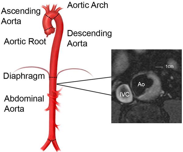
Methods: We evaluated 1527 participants (mean age 59.9 years, 53.5% women) in the Framingham Offspring cohort who underwent both aortic and brain MRI. Participants were free of clinical stroke, dementia, or other neurological illness at the time of axial MRI of the thoracic and abdominal DAo and subsequent brain MRI. We related the prevalence and burden of aortic plaque to total cerebral brain volume (TCBV) and white matter hyperintensity volume (WMHV). An additional analysis compared incidence of stroke or TIA in participants with and without DAo plaques.
Results: Presence of thoracic DAo plaque (8%) was associated with decreased TCBV in sex-pooled analysis (-0.77, SE 0.25, p = 0.002, equivalent to 4.5 years of aging) and with increased WMHV only in men (0.26, SE 0.12, p = 0.032, equivalent to 6.5 years aging). We observed similar associations of DAo plaque burden with TCBV and WMHV. There were 43 strokes and 11 TIAs in prospective follow-up (median 7 years). Presence of DAo plaque was not associated with subsequent stroke or TIA.
Conclusions: In this cross-sectional community-based study, we found DAo plaque is associated with accelerated brain aging. These data underscore the potential implications of incidentally identified subclinical aortic atherosclerosis and question whether targeted intervention in these high risk individuals can modulate cognitive decline.
Keywords: Aorta; Atherosclerosis; Brain; Cerebrovascular disorders; Magnetic resonance imaging; Neuroimaging; White matter.
Copyright © 2017 Elsevier B.V. All rights reserved.
Conflict of interest statement
The authors declared they do not have anything to disclose regarding conflict of interest with respect to this manuscript.
Figures
Comment in
-
Are structural brain changes driven by aortic atherosclerosis?Atherosclerosis. 2017 Oct;265:248-249. doi: 10.1016/j.atherosclerosis.2017.08.004. Epub 2017 Aug 19. Atherosclerosis. 2017. PMID: 28870632 No abstract available.
References
-
- Bos D, Vernooij MW, Elias-Smale SE, Verhaaren BF, Vrooman HA, Hofman A, et al. Atherosclerotic calcification relates to cognitive function and to brain changes on magnetic resonance imaging. Alzheimers Dement. 2012;8(5 Suppl):S104–11. - PubMed
-
- Atherosclerotic disease of the aortic arch as a risk factor for recurrent ischemic stroke. The French Study of Aortic Plaques in Stroke Group. N Engl J Med. 1996;334(19):1216–21. - PubMed
-
- Harloff A, Simon J, Brendecke S, Assefa D, Helbing T, Frydrychowicz A, et al. Complex plaques in the proximal descending aorta: an underestimated embolic source of stroke. Stroke. 2010;41(6):1145–50. - PubMed
Publication types
MeSH terms
Grants and funding
LinkOut - more resources
Full Text Sources
Other Literature Sources
