. 2017 Jul 18;114(29):E5796-E5804.
doi: 10.1073/pnas.1703998114. Epub 2017 Jul 3.

Quadrupling efficiency in production of genetically modified pigs through improved oocyte maturation

Affiliations

Quadrupling efficiency in production of genetically modified pigs through improved oocyte maturation

Ye Yuan et al. Proc Natl Acad Sci U S A. .

Abstract

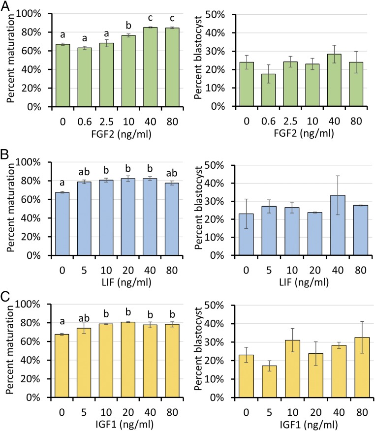
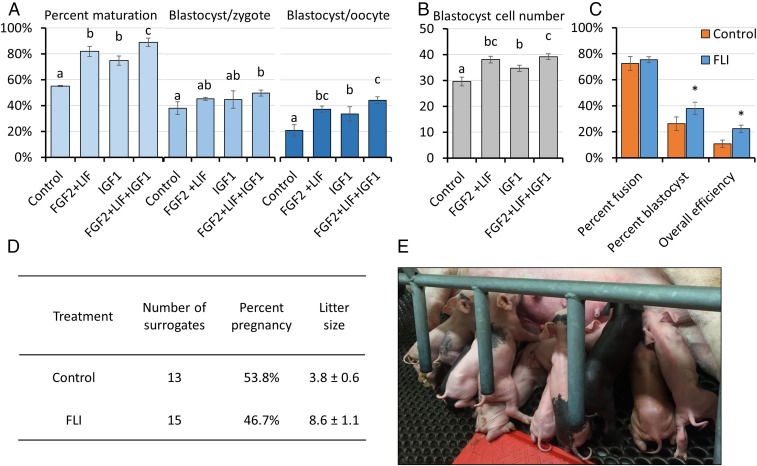
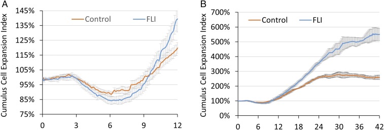
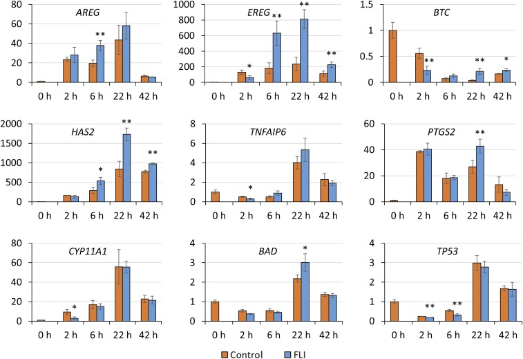
Assisted reproductive technologies in all mammals are critically dependent on the quality of the oocytes used to produce embryos. For reasons not fully clear, oocytes matured in vitro tend to be much less competent to become fertilized, advance to the blastocyst stage, and give rise to live young than their in vivo-produced counterparts, particularly if they are derived from immature females. Here we show that a chemically defined maturation medium supplemented with three cytokines (FGF2, LIF, and IGF1) in combination, so-called "FLI medium," improves nuclear maturation of oocytes in cumulus-oocyte complexes derived from immature pig ovaries and provides a twofold increase in the efficiency of blastocyst production after in vitro fertilization. Transfer of such blastocysts to recipient females doubles mean litter size to about nine piglets per litter. Maturation of oocytes in FLI medium, therefore, effectively provides a fourfold increase in piglets born per oocyte collected. As they progress in culture, the FLI-matured cumulus-oocyte complexes display distinctly different kinetics of MAPK activation in the cumulus cells, much increased cumulus cell expansion, and an accelerated severance of cytoplasmic projections between the cumulus cells outside the zona pellucida and the oocyte within. These events likely underpin the improvement in oocyte quality achieved by using the FLI medium.

Keywords: MAPK signaling; cumulus cell; embryo development; genetic modification; in vitro fertilization.


Conflict of interest statement

Conflict of interest statement: Y.Y., L.D.S., R.S.P., and R.M.R. have a pending patent application “Medium Supplement to Increase the Efficiency of Oocyte maturation and Embryo Culture in vitro.”

Figures

Fig. 1.
Fig. 1.
Effects of various concentrations of FGF2 (A), LIF (B), and IGF1 (C) in porcine oocyte maturation medium on nuclear maturation and subsequent developmental competence. Data are reported as means ± SEM. Different superscripts (a and b) denote a significant difference from the control, P < 0.05. The experiments were replicated four times with a total of 3,554 oocytes.
Fig. 2.
Fig. 2.
Supplementation of standard IVM medium with FLI improves porcine oocyte meiotic maturation and developmental competence. (A and B) Effects of FLI on nuclear maturation and subsequent blastocyst development following IVF. The experiments were replicated four times with a total of 796 oocytes. Different letters denote significant differences, P < 0.05. (C) Supplementation with FLI improves blastocyst development following SCNT. The experiments were replicated five times with a total of 488 oocytes. An asterisk (*) denotes a significant difference between control and treatment, P < 0.05. Percent fusion was calculated as fused embryos/enucleated oocytes; percent blastocyst was calculated as blastocysts/fused embryos; overall efficiency was calculated as blastocysts/enucleated oocytes. Embryo transfers were not performed in these experiments. (D) The in vivo developmental competence of the blastocysts produced from the FLI treated oocytes in terms of litter size after embryo transfer. Approximately 50 blastocysts that had been injected with CRISPR/Cas9 constructs at the zygote stage were transferred to surrogates on day 5 or 6. (E) Typical litter of 13 healthy piglets obtained after embryo transfer of blastocyst stage embryos derived from FLI-matured oocytes.
Fig. 3.
Fig. 3.
FLI regulation of MAPK1/3 activation in cumulus cells during IVM. (A) Representative images of Western blots showing the effects of FLI on MAPK1/3 activity in cumulus cells during IVM. (B) The relative expression of pMAPK/MAPK was quantified, and the ratios of the two forms compared between control and FLI treatments at the same time points. Each cumulus cell sample was pooled from 50 COCs. This experiment was replicated four times. Asterisks (*) denote a significant difference between control and treatment, P < 0.05. (C) Effects of FLI and each individual cytokine on MAPK1/3 activation in cumulus cells at 42 h after IVM as determined by Western blotting. (D) Effects of adding kinase inhibitors in FLI medium on oocyte meiotic maturation. The experiments were replicated four times with 1,208 oocytes. Different letters denote significant differences, P < 0.05.
Fig. 4.
Fig. 4.
Effects of FLI on cumulus cell expansion. (A) Cumulus cell expansion for the first 12 h of IVM. (B) Cumulus cell expansion occurring over the entire 42-h culture period of IVM. Images were taken every 7.5 min and recorded by the CytoSMART system. Size of COCs was estimated by dividing the total coverage of COCs provided by the system with the number of COCs within the images. Cumulus cell expansion index represents the relative size of COCs relative the median size of COCs at 0 h. The experiments were replicated six times for Fig. 3A, and three times for Fig. 3B.
Fig. 5.
Fig. 5.
Effects of FLI on TZP integrity during IVM. (A) Quantification of TZPs during IVM. Number of intact TZPs was compared within each time point. These experiments were replicated three times, with a total of 142 COCs. Asterisks (*) denote a significant difference between control and treatment, P < 0.05. (B) Maximum-intensity projections of the equatorial cross-section of the oocytes stained for F-actin (rhodamine phalloidin) at different time points during IVM. (Scale bar, 20 μm.)
Fig. 6.
Fig. 6.
Effects of FLI on mRNA abundance of EGF-like factors (AREG, EREG, BTC), cumulus cell expansion factors (HAS2, TNFAIP6, PTGS2), and stress related genes (CYP11A1, BAD, TP53) during IVM. The relative abundance of mRNA was examined by quantitative PCR and compared between control and FLI cumulus cells at the same time point. The mRNA level for each gene was arbitrarily set to 1 for controls at 0 h. **P < 0.05, significant differences in mRNA abundance between control and treatment; *P < 0.1, a trend to be different. The experiments were replicated four times for the 0-, 2-, and 6-h time points, and three times for the 22- and 42-h time points.

Similar articles

Cited by

References

    1. Yuan Y, Krisher RL. In vitro maturation (IVM) of porcine oocytes. Methods Mol Biol. 2012;825:183–198. - PubMed
    1. Lonergan P, Fair T. Maturation of oocytes in vitro. Annu Rev Anim Biosci. 2016;4:255–268. - PubMed
    1. Grupen CG. The evolution of porcine embryo in vitro production. Theriogenology. 2014;81:24–37. - PubMed
    1. Seyhan A, et al. Severe early ovarian hyperstimulation syndrome following GnRH agonist trigger with the addition of 1500 IU hCG. Hum Reprod. 2013;28:2522–2528. - PubMed
    1. Humaidan P, et al. Ovarian hyperstimulation syndrome: Review and new classification criteria for reporting in clinical trials. Hum Reprod. 2016;31:1997–2004. - PubMed

Publication types

MeSH terms

Substances