. 2017 Jul 18;114(29):7588-7593.
doi: 10.1073/pnas.1617589114. Epub 2017 Jul 3.

Brain regions and functional interactions supporting early word recognition in the face of input variability

Affiliations

Brain regions and functional interactions supporting early word recognition in the face of input variability

Silvia Benavides-Varela et al. Proc Natl Acad Sci U S A. .

Abstract

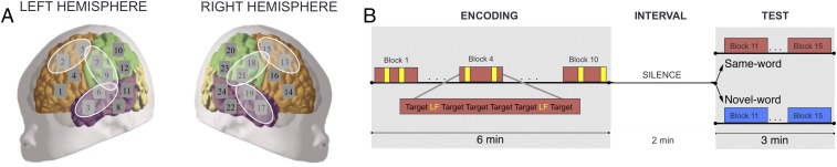
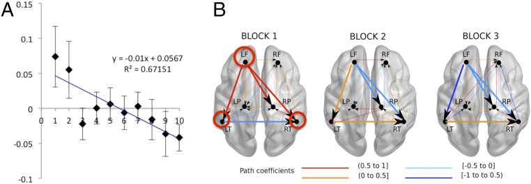
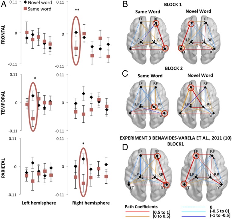
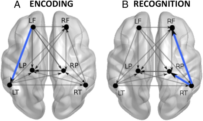
Perception and cognition in infants have been traditionally investigated using habituation paradigms, assuming that babies' memories in laboratory contexts are best constructed after numerous repetitions of the very same stimulus in the absence of interference. A crucial, yet open, question regards how babies deal with stimuli experienced in a fashion similar to everyday learning situations-namely, in the presence of interfering stimuli. To address this question, we used functional near-infrared spectroscopy to test 40 healthy newborns on their ability to encode words presented in concomitance with other words. The results evidenced a habituation-like hemodynamic response during encoding in the left-frontal region, which was associated with a progressive decrement of the functional connections between this region and the left-temporal, right-temporal, and right-parietal regions. In a recognition test phase, a characteristic neural signature of recognition recruited first the right-frontal region and subsequently the right-parietal ones. Connections originating from the right-temporal regions to these areas emerged when newborns listened to the familiar word in the test phase. These findings suggest a neural specialization at birth characterized by the lateralization of memory functions: the interplay between temporal and left-frontal regions during encoding and between temporo-parietal and right-frontal regions during recognition of speech sounds. Most critically, the results show that newborns are capable of retaining the sound of specific words despite hearing other stimuli during encoding. Thus, habituation designs that include various items may be as effective for studying early memory as repeated presentation of a single word.

Keywords: fNIRS effective connectivity; habituation; language; memory; newborns.


Conflict of interest statement

The authors declare no conflict of interest.

Figures

Fig. 1.
Fig. 1.
(A) Location of channels (gray squares) and ROIs (white ellipses) on a schematic neonate brain; see also Fig. S1 depicting a projection of the probes to a label map of a MRI neonate template and the SI Materials and Methods for a detailed description of the cortical regions underlying the channels. (B) Experimental design. Each rectangle represents a block consisting of a series words separated by short pauses of 0.5 or 1.5 s. During the encoding phase, all neonates heard 10 blocks composed of 6 repetitions of the target word and 2 repetitions of a potentially distracting word (low-frequency word, LF). In the test, half of the neonates heard the target word and the other half heard a completely novel word. A 2-min silent interval separated the encoding and test phases.
Fig. S1.
Fig. S1.
Photograph of a neonate with the probe set on the head and a projection from the scalp surface to the intensity model and label map of the neonate MRI templates (templates from ref. are publicly available at http://bric.unc.edu/ideagroup/free-softwares/). Red diamonds correspond to emitters/sources and blue diamonds to detectors. The yellow numbered squares, between adjacent emitter–detector pairs, correspond to the channels.
Fig. 2.
Fig. 2.
(A) Time course of the relative OxyHb changes in the LF area averaged across subjects. The plot depicts mean concentration changes in the encoding phase. The x axis shows block numbers. The y axis shows the changes in concentration of OxyHb in mmol∙mm. Error bars indicate standard error of the mean. (B) Effective connections observed among the bilateral frontal (LF, RF), temporal (LT, RT), and parietal (LP, RP) areas during the habituation response in the first three blocks of the encoding phase. The evaluation of different models with AMOS showed good data fit in all of the blocks. Block 1, χ2 (df = 2) =1.377, P = 0.502; block 2, χ2 (df = 3) = 4.03, P = 0.258; block 3, χ2 (df = 3) = 1.195, P = 0.754. Wider lines highlight the progressive weakening of the functional connections of the LF region and the strengthening of the connections between the temporal regions. Red circles highlight the regions with noteworthy connections. Fig. S2 depicts the hypothetical starting model.
Fig. S2.
Fig. S2.
Hypothetical starting model including the six ROIs and directional connections between them.
Fig. 3.
Fig. 3.
(A) Relative OxyHb changes measured in six ROIs during the 5 blocks comprising the test phase. The y axes show concentrations of OxyHb in mmol·mm. Red ellipses indicate the blocks in which significant differences between groups were found. Fig. S3 shows the hemodynamic curves of the same-word and novel-word groups in the RF area during the first block of the test phase. In all graphs, error bars indicate standard error of the mean (permutation test, *P < 0.01; **P < 0.001). (B–D) Functional connections observed during the first (B) and second block (C) of the recognition test in the current study and the first block (D) of the recognition test in Benavides-Varela et al., 2011 (10), experiment 3. The models appropriately fit the data: block 1 same-word group, χ2 (df = 4) = 5.011, P = 0.286; novel-word group, χ2 (df = 2) = 2.692; P = 0.260; block 2 same-word group, χ2 (df = 4) = 5.694; P = 0.223; novel-word group, χ2 (df = 1) = 1.697; P = 0.193; previous study: block 1 same-word group, χ2 (df = 1) = 1.109, P = 0.292; novel-word group, χ2 (df = 1) = 0.074, P = 0.786. Dotted lines depict connections that were absent in a given block-group model; solid lines highlight the corresponding connections between each pair of areas in the other group. Red circles highlight the regions with noteworthy connections. Fig. S2 depicts the hypothetical starting model.
Fig. S3.
Fig. S3.
Hemodynamic curves of the same-word and novel-word groups in the RF area during the first block of the test phase. The x axis shows time in seconds from the start of stimulation, and the gray rectangle indicates the stimulation period. Asterisks indicate Bonferroni-corrected P < 0.01.
Fig. 4.
Fig. 4.
Verbal memory network at birth and its interactions, mainly characterized by (A) the interplay between temporal and LF regions during encoding and (B) the interaction between temporal, parietal, and RF regions during recognition of words. Blue lines depict the most significant connections characterizing the two memory stages.

References

    1. Oakes LM. Using habituation of looking time to assess mental processes in infancy. J Cogn Dev. 2010;11:255–268. - PMC - PubMed
    1. Nordt M, Hoehl S, Weigelt S. The use of repetition suppression paradigms in developmental cognitive neuroscience. Cortex. 2016;80:61–75. - PubMed
    1. Turk-Browne NB, Scholl BJ, Chun MM. Babies and brains: Habituation in infant cognition and functional neuroimaging. Front Hum Neurosci. 2008;2:16. - PMC - PubMed
    1. Jardri R, et al. Fetal cortical activation to sound at 33 weeks of gestation: A functional MRI study. Neuroimage. 2008;42:10–18. - PubMed
    1. Draganova R, et al. Sound frequency change detection in fetuses and newborns, a magnetoencephalographic study. Neuroimage. 2005;28:354–361. - PubMed

Publication types

LinkOut - more resources