Review
. 2017 Jul 4;136(1):98-109.
doi: 10.1161/CIRCULATIONAHA.116.027039.

Is Cardioprotection Dead?

Affiliations
Review

Is Cardioprotection Dead?

David J Lefer et al. Circulation. .

Abstract

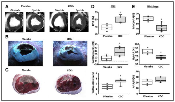
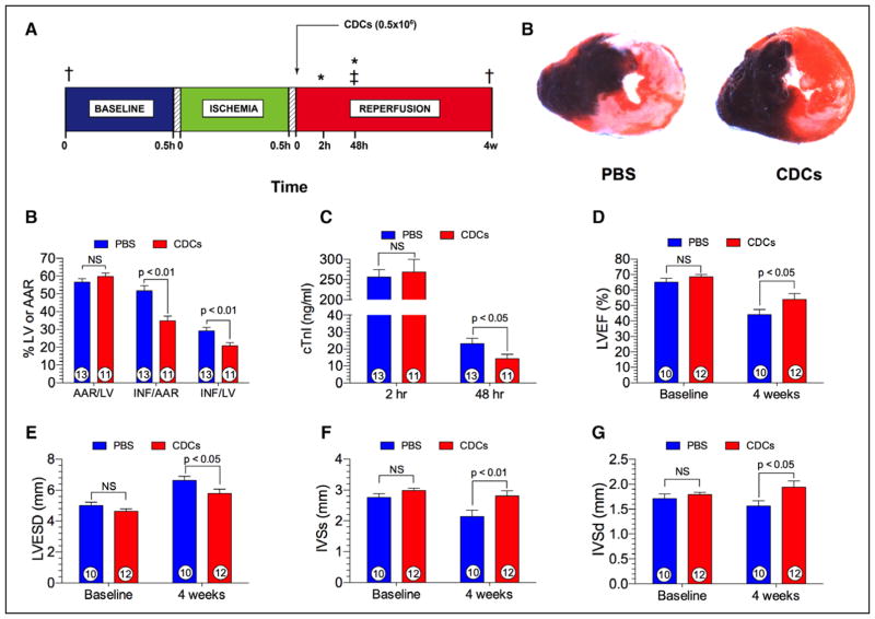
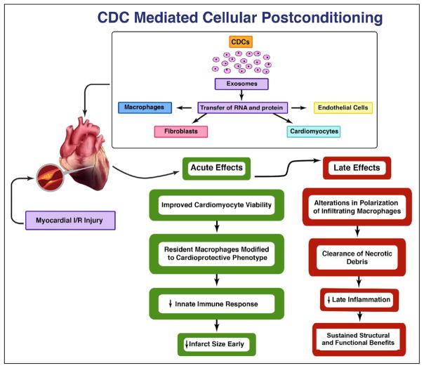
For >4 decades, the holy grail in the treatment of acute myocardial infarction has been the mitigation of lethal injury. Despite promising initial results and decades of investigation by the cardiology research community, the only treatment with proven efficacy is early reperfusion of the occluded coronary artery. The remarkable record of failure has led us and others to wonder if cardioprotection is dead. The path to translation, like the ascent to Everest, is certainly littered with corpses. We do, however, highlight a therapeutic principle that provides a glimmer of hope: cellular postconditioning. Administration of cardiosphere-derived cells after reperfusion limits infarct size measured acutely, while providing long-term structural and functional benefits. The recognition that cell therapy may be cardioprotective, and not just regenerative, merits further exploration before we abandon the pursuit entirely.

Keywords: cardiosphere derived cells; cell- and tissue-based therapy; heart failure; myocardial Infarction; ventricular function, left.


Figures

Figure 1
Figure 1. Validation of cellular postconditioning in pigs
A, MR short-axis images from a placebo and CDC-treated pig. Transverse cardiac slices stained with Thioflavin T and Gentian Violet (B), and triphenyl tetrazolium chloride (TTC) (C) in the same representative sample. B, The area of MVO appears nonfluorescent under UV light, whereas the area-at-risk (AAR) is unstained with Gentian Violet. C, Viable myocardium appears red and scar appears white/yellow. LVEF (D, top), infarct wall thickening (D, middle), and infarct wall motion (D, lower) are improved following CDC treatment. MVO/AAR (E, top), and infarct size (IS)/AAR (E, middle) are decreased following CDC treatment, whereas AAR is not different between groups (E, lower). Graphs depict mean±SEM. Statistical significance was determined by using the Student t test. *P<0.05. CDC indicates cardiosphere-derived cell; LVEF, left ventricular ejection fraction; MVO, microvascular occlusion; and SEM, standard error of the mean.
Figure 2
Figure 2. Cellular postconditioning in spontaneously hypertensive rats
A, Experimental protocol involving male spontaneously hypertensive rats (SHRs) subjected to 30 minutes of left coronary artery ischemia followed by either 48 hours (h) or 4 weeks (w) of reperfusion. Myocardial area-at-risk and infarct size were determined at 48 h postreperfusion. Plasma levels of cardiac troponin I (cTnI) was measured at 2 h and 48 h of reperfusion. At 20 minutes of reperfusion, rat CDCs (0.5×10⁶) or phosphate-buffered saline (PBS) were injected directly into the left ventricular lumen following aortic cross-clamping. B, Representative photomicrographs of SHRs receiving either PBS or CDCs at 20 minutes of reperfusion. Myocardial infarct size is significantly attenuated in the CDC-treated heart. C, Myocardial area-at-risk (AAR) as a percentage of the left ventricle (LV), infarct size (INF) per AAR, and INF as a percent of the LV in rats receiving either PBS or CDCs. Myocardial infarct size per area-at-risk or LV was significantly (P<0.01) reduced in the CDC group. D, Plasma cardiac troponin I (cTnI) levels at 2 and 48 h following reperfusion. cTnI levels are significantly (P<0.05) reduced at 48 h postreperfusion. E, Left ventricular ejection fraction (LVEF) at baseline and at 4 weeks following reperfusion. LVEF is similar at baseline and significantly (P<0.05) greater in animals receiving CDCs. F, Left ventricular end-systolic dimension (LVESD) at baseline and 4 weeks of reperfusion in the PBS and CDC groups. LVESD is significantly (P<0.05) reduced in the CDC group in comparison with PBS. G, Interventricular septal dimension at end-systole (IVSs) at baseline and 4 weeks following reperfusion. IVSs was significantly (P<0.01) greater in hearts treated with CDCs than with PBS. H, Interventricular septal dimension at end-diastole (IVSd) at baseline and 4 weeks postreperfusion. Similar to IVSs, IVSd was significantly (P<0.01) greater in the CDC group than in the PBS group. Numbers inside the bars represent the number of animals in each group. Statistical significance was determined by using the Student t test. CDC indicates cardiosphere-derived cell; and 2,3,5-TTC, 2,3,5-triphenyltetrazolium chloride. *Plasma samples for cTnI. ‡Myocardial infarct size analysis. †2-D Echocardiography, Visual Sonics Vevo 2100.
Figure 3
Figure 3. Mechanisms of CDC-mediated cellular postconditioning
Cardiosphere-derived cells (CDCs) release exosomes resulting in the transfer of RNA and proteins to macrophages, fibroblasts, endothelial cells, and cardiomyocytes, in turn, leading to both acute and late cardioprotective actions. In the acute phase of reperfusion injury, CDCs improve cardiomyocyte viability and reduce myocardial infarct size by the conversion of resident macrophages to a cardioprotective phenotype and dampening the innate immune response. During the later phases of myocardial re-perfusion injury, CDC therapy results in sustained infarct size reduction by alterations in polarization of infiltrating macrophages, accelerated clearance of necrotic debris, and significant attenuation of the late inflammatory response in the myocardium.

References

    1. Gross L, Blum L, Silverman G. Experimental attempts to increase the blood supply to the dog’s heart by means of coronary sinus occlusion. J Exp Med. 1937;65:91–108. - PMC - PubMed
    1. Keeley EC, Boura JA, Grines CL. Primary angioplasty versus intravenous thrombolytic therapy for acute myocardial infarction: a quantitative review of 23 randomised trials. Lancet. 2003;361:13–20. doi: 10.1016/S0140-6736(03)12113-7. - DOI - PubMed
    1. Pfeffer MA, Braunwald E. Ventricular remodeling after myocardial infarction. Experimental observations and clinical implications. Circulation. 1990;81:1161–1172. - PubMed
    1. St John Sutton M, Pfeffer MA, Plappert T, Rouleau JL, Moyé LA, Dagenais GR, Lamas GA, Klein M, Sussex B, Goldman S. Quantitative two-dimensional echocardiographic measurements are major predictors of adverse cardiovascular events after acute myocardial infarction. The protective effects of captopril. Circulation. 1994;89:68–75. - PubMed
    1. Reimer KA, Jennings RB. Failure of the xanthine oxidase inhibitor allopurinol to limit infarct size after ischemia and reperfusion in dogs. Circulation. 1985;71:1069–1075. - PubMed

MeSH terms