

Diagnosis and Treatment of Traumatic Cerebrovascular Injury: Pitfalls in the Management of Neurotrauma
- PMID: 28674346
- PMCID: PMC5566700
- DOI: 10.2176/nmc.oa.2017-0056
Diagnosis and Treatment of Traumatic Cerebrovascular Injury: Pitfalls in the Management of Neurotrauma
Abstract
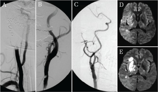
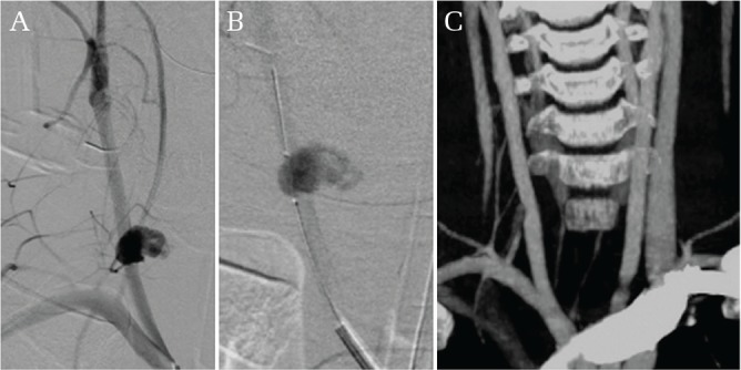
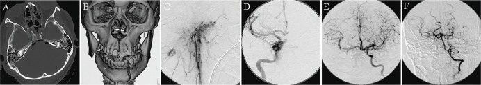
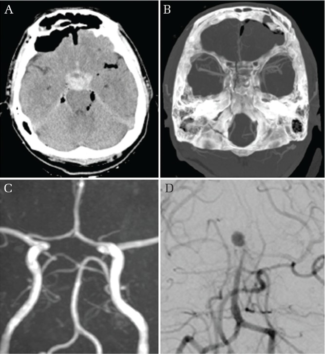
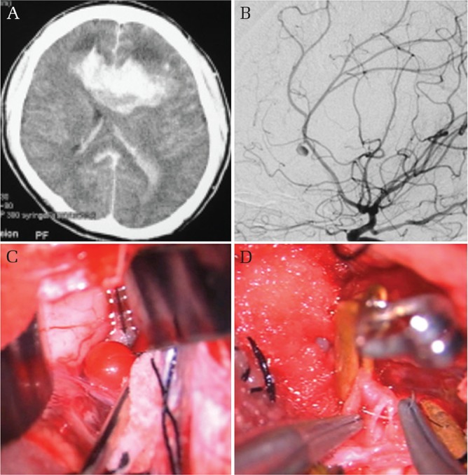
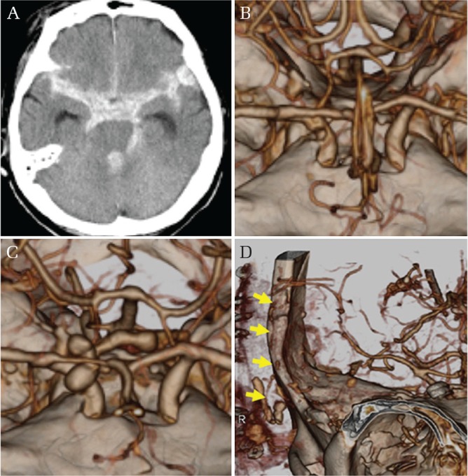
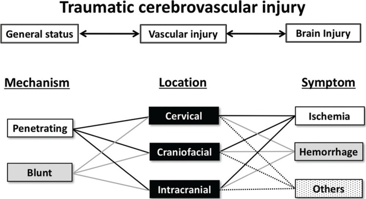
Traumatic cerebrovascular injury (TCVI) is an uncommon clinical entity in traumatic brain injury (TBI), yet it may cause devastating brain injury with high morbidity and mortality. Early recognition and prioritized strategic treatment are of paramount importance. A total of 1966 TBI patients admitted between 1999 and 2015 in our tertiary critical care center were reviewed. Screening of TCVI was based on the Guidelines for the Management of Severe Head Injury in Japan. TCVI was confirmed in 33 (1.7%) patients; 29 blunt and 4 penetrating injuries. The primary location of the injury included 16 cervical, 6 craniofacial, and 11 intracranial lesions. On arrival, 15 patients presented with hemorrhage, 5 of these arrived in shock status with massive hemorrhage. Ten presented with ischemic symptoms. Sixteen patients underwent surgical or endovascular intervention, 13 of whom required immediate treatment upon arrival. Surgical procedures included clipping or trapping for traumatic aneurysms, superficial temporal artery - middle cerebral artery bypass, carotid endarterectomy, and direct suture of the injured vessels. Endovascular intervention was undertaken in 7 patients; embolization with Gelfoam (Pharmacia and Upjohn Company, Kalamazoo, MI, USA) or coil for 6 hemorrhagic lesions and stent placement for 1 lesion causing ischemia. Patients' outcome assessed by the Glasgow Outcome Scale at 3 months were good recovery in 8, moderate disability in 3, severe disability in 9, persistent vegetative state in 1, and death in 12, respectively. In order to rescue potentially salvageable TCVI patients, neurosurgeons in charge should be aware of TCVI and master basic skills of cerebrovascular surgical and endovascular procedures to utilize in an emergency setting.
Keywords: emergency medicine; microneurosurgery; neurotrauma; traumatic brain injury; traumatic cerebrovascular injury.
Conflict of interest statement
The author hereby declares no conflict of interest regarding this article.
Figures
Similar articles
-
Blunt cerebrovascular injuries in severe traumatic brain injury: incidence, risk factors, and evolution.J Neurosurg. 2017 Jul;127(1):16-22. doi: 10.3171/2016.4.JNS152600. Epub 2016 Jul 29. J Neurosurg. 2017. PMID: 27471889
-
[Traumatic Cerebrovascular Injury].No Shinkei Geka. 2021 Sep;49(5):1041-1055. doi: 10.11477/mf.1436204488. No Shinkei Geka. 2021. PMID: 34615764 Japanese.
-
Extracranial traumatic aneurysms due to blunt cerebrovascular injury.J Neurosurg. 2014 Jun;120(6):1437-45. doi: 10.3171/2014.3.JNS131959. Epub 2014 Apr 4. J Neurosurg. 2014. PMID: 24702325
-
The endovascular treatment strategies of cerebrovascular injuries in traumatic brain injury.Chin J Traumatol. 2025 Mar;28(2):81-90. doi: 10.1016/j.cjtee.2025.01.001. Epub 2025 Jan 22. Chin J Traumatol. 2025. PMID: 39934036 Free PMC article. Review.
-
Clinical Characteristics and Outcome in Elderly Patients with Traumatic Brain Injury: For Establishment of Management Strategy.Neurol Med Chir (Tokyo). 2017 Aug 15;57(8):418-425. doi: 10.2176/nmc.st.2017-0058. Epub 2017 Jul 5. Neurol Med Chir (Tokyo). 2017. PMID: 28679968 Free PMC article. Review.
Cited by
-
Blunt Traumatic Cervical Vascular Injury Without any Modified Denver Criteria.Clin Pract Cases Emerg Med. 2018 Jun 26;2(3):200-202. doi: 10.5811/cpcem.2018.4.37719. eCollection 2018 Aug. Clin Pract Cases Emerg Med. 2018. PMID: 30083632 Free PMC article.
-
Bilateral Carotid and Vertebral Artery Dissection from Blunt Trauma.Case Rep Emerg Med. 2018 Mar 12;2018:1919034. doi: 10.1155/2018/1919034. eCollection 2018. Case Rep Emerg Med. 2018. PMID: 29721341 Free PMC article.
-
Cerebral vasospasm and hypoperfusion after traumatic brain injury: Combined CT angiography and CT perfusion imaging study.Surg Neurol Int. 2021 Jul 19;12:361. doi: 10.25259/SNI_859_2020. eCollection 2021. Surg Neurol Int. 2021. PMID: 34345501 Free PMC article.
-
Self-inflicted very-low-velocity penetrating head injury: A CARE-compliant case report.Medicine (Baltimore). 2024 May 3;103(18):e37896. doi: 10.1097/MD.0000000000037896. Medicine (Baltimore). 2024. PMID: 38701288 Free PMC article.
-
Navigating the Role of Surgery in Optimizing Patient Outcomes in Traumatic Brain Injuries (TBIs): A Comprehensive Review.Cureus. 2024 Oct 10;16(10):e71234. doi: 10.7759/cureus.71234. eCollection 2024 Oct. Cureus. 2024. PMID: 39525257 Free PMC article. Review.
References
-
- Fusco MR, Harrigan MR: Cerebrovascular dissections: a review. Part II: blunt cerebrovascular injury. Neurosurgery 68: 517–530; discussion 530, 2011 - PubMed
-
- Bromberg WJ, Collier BC, Diebel LN, et al. : Blunt cerebrovascular injury practice management guidelines: the Eastern Association for the Surgery of Trauma. J Trauma 68:471–477, 2010 - PubMed
-
- Biffl WL, Moore EE, Offner PJ, Brega KE, Franciose RJ, Burch JM: Blunt carotid arterial injuries: implications of a new grading scale. J Trauma 47: 845–853, 1999 - PubMed
Publication types
MeSH terms
LinkOut - more resources
Full Text Sources
Other Literature Sources