. 2017 Jul 3;7(1):4546.
doi: 10.1038/s41598-017-04874-4.

HDAC6: A Novel Histone Deacetylase Implicated in Pulmonary Arterial Hypertension

Affiliations

HDAC6: A Novel Histone Deacetylase Implicated in Pulmonary Arterial Hypertension

Olivier Boucherat et al. Sci Rep. .

Abstract

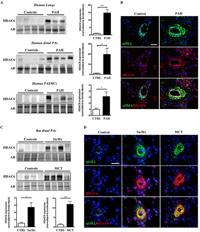
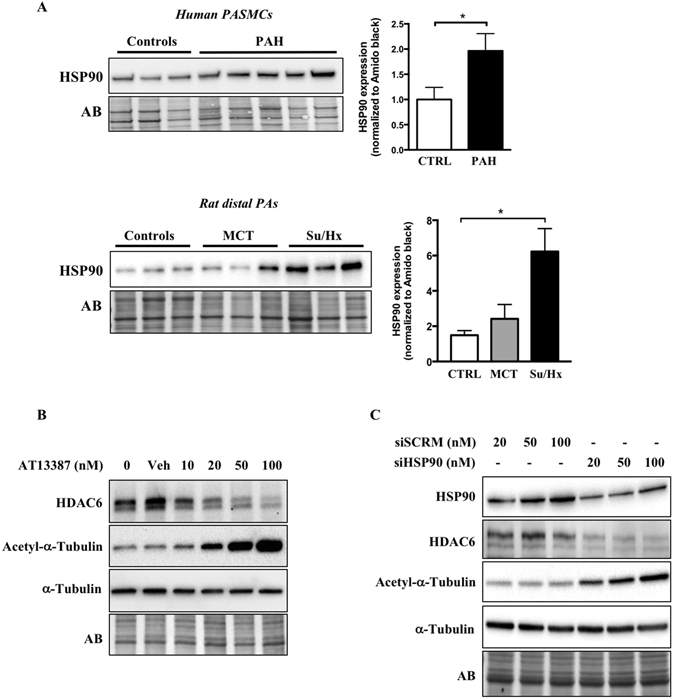
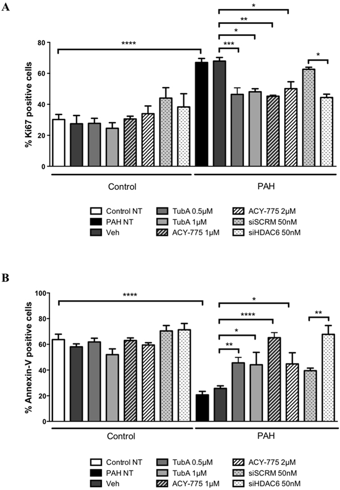
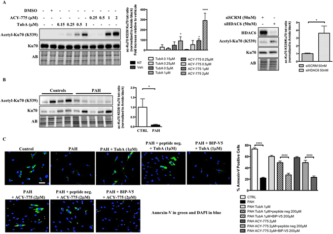
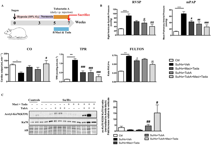
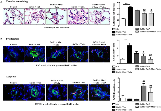
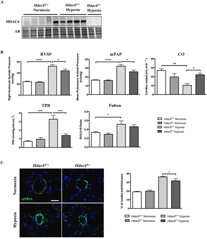
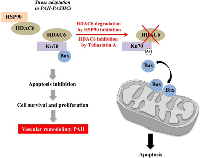
Pulmonary arterial hypertension (PAH) is a vascular remodeling disease with limited therapeutic options. Although exposed to stressful conditions, pulmonary artery (PA) smooth muscle cells (PASMCs) exhibit a "cancer-like" pro-proliferative and anti-apoptotic phenotype. HDAC6 is a cytoplasmic histone deacetylase regulating multiple pro-survival mechanisms and overexpressed in response to stress in cancer cells. Due to the similarities between cancer and PAH, we hypothesized that HDAC6 expression is increased in PAH-PASMCs to face stress allowing them to survive and proliferate, thus contributing to vascular remodeling in PAH. We found that HDAC6 is significantly up-regulated in lungs, distal PAs, and isolated PASMCs from PAH patients and animal models. Inhibition of HDAC6 reduced PAH-PASMC proliferation and resistance to apoptosis in vitro sparing control cells. Mechanistically, we demonstrated that HDAC6 maintains Ku70 in a hypoacetylated state, blocking the translocation of Bax to mitochondria and preventing apoptosis. In vivo, pharmacological inhibition of HDAC6 improved established PAH in two experimental models and can be safely given in combination with currently approved PAH therapies. Moreover, Hdac6 deficient mice were partially protected against chronic hypoxia-induced pulmonary hypertension. Our study shows for the first time that HDAC6 is implicated in PAH development and represents a new promising target to improve PAH.


Conflict of interest statement

The authors declare that they have no competing interests.

Figures

Figure 1
Figure 1
HDAC6 is overexpressed in lungs and distal pulmonary arteries (PAs) from PAH patients and animal models. (A) Representative Western blots and corresponding densitometric analyses of HDAC6 expression in lung tissues, distal PAs and isolated PA smooth muscle cells (PASMCs) from control (n = 4–8) and PAH patients (n = 4–12). (B) Double immunofluorescence staining for αSMA (green) and HDAC6 (red) and DAPI nuclear staining in lungs from control donors (n = 5) and PAH patients (n = 5), confirming the overexpression of HDAC6 in remodeled PAs. (C) Representative Western blots and corresponding densitometric analyses of HDAC6 expression in distal PAs of non-treated rats (n = 5 for each model) as well as rats exposed to Sugen-hypoxia (Su/Hx, n = 6) or monocrotaline (MCT, n = 5). (D) Double immunofluorescence staining for αSMA (green) and HDAC6 (red) and DAPI nuclear staining showing increased expression of HDAC6 in remodeled distal PAs after Su/Hx exposure or MCT injury compared to non-treated rats (n = 5 per group). Protein expression was normalized by Amido black (AB). Scale bar = 25 μm *P < 0.05 and ***P < 0.001.
Figure 2
Figure 2
HSP90 is increased in PAH-PASMCs and stabilizes HDAC6 expression. (A) HSP90 expression was assessed by Western blot in pulmonary artery smooth muscle cells (PASMCs) from control (n = 3) and PAH patients (n = 5) and distal PAs from control (n = 3), MCT (n = 3) and Su/Hx (n = 3) rats. Protein band densitometry is reported in the corresponding graph. (B) Expression of HDAC6, acetylated-α-Tubulin, and α-Tubulin was assessed in PAH-PASMCs by Western blot after treatment or not with a pharmacological HSP90 inhibitor (AT13387, 10–100 nM for 48 hours) or vehicle (dimethyl sulfoxide). (C) Expression of HSP90, HDAC6, acetylated-α-Tubulin, and α-Tubulin was assessed in PAH-PASMCs by Western blot after treatments with a siHSP90 or siSCRM (20–100 nM for 48 hours). Blots are representative of two independent experiments. *P < 0.05.
Figure 3
Figure 3
Inhibition of HDAC6 diminishes PAH-PASMC proliferation and resistance to apoptosis. (A) Proliferation (Ki67) was measured after treatments of PAH-PASMCs with two pharmacological HDAC6 inhibitors (Tubastatin A and ACY-775) or vehicle (DMSO), as well as siHDAC6 or siSCRM (50 nM) for 48 hours. (B) Apoptosis was similarly assessed following HDAC6 inhibition. *P < 0.05, **P < 0.01, ***P < 0.001 and ****P < 0.001. Experiments were performed in triplicate in 3 control and 4 PAH-PASMC cell lines.
Figure 4
Figure 4
HDAC6 inhibition in PAH-PASMCs causes Bax-induced cell death by increasing acetylation of cytosolic Ku70. (A) Representative Western blots of acetyl(K539)-Ku70 and total Ku70 in PAH-PASMCs after treatments with two HDAC6 inhibitors Tubastatin A and ACY-775 or siHDAC6 for 48 hours. Data were expressed as the ratio of acetyl(K539)-Ku70/total Ku70. (B) Expression of acetylated(K539)-Ku70 and Ku70 was assessed in distal PAs from control (n = 4) and PAH patients (n = 5) by Western blot. Data were expressed as the ratio of acetyl(K539)-Ku70/total Ku70. (C) Apoptosis was measured using Annexin V staining (green) in control (n = 3 cell lines) and PAH-PASMCs (n = 3 cell lines) pre-treated or not with Bax inhibitory peptides (BIP-V5, 200 μM) or control peptides before being exposed to indicated HDAC6 inhibitors for 48 hours. Representative immunofluorescence images of apoptotic Annexin-V-positive cells (green) stained for DAPI (blue) and corresponding quantification are shown. Scale bar = 50 μm. Protein expression was normalized by Amido black (AB). ***P < 0.001 and ****P < 0.001.
Figure 5
Figure 5
Tubastatin A (TubA) improves pulmonary arterial hypertension in the Sugen/Hypoxia (Su/Hx) rat model and provides a therapeutic effect comparable to the combination of standard PAH therapies. (A) Schematic of the experimental design. (B) RVSP, mPAP, CO, TPR and Fulton index were measured in control, Su/Hx + Veh (dimethyl sulfoxide), Su/Hx + Macitentan (30 mg/kg/d) + Tadalafil (10 mg/kg/d), Su/Hx + TubA (25 mg/kg/d) and Su/Hx + TubA + Macitentan + Tadalafil; n = 5 to 7 rats/group. (C) Expression of acetylated(K539)-Ku70 and Ku70 was assessed by Western blot in distal PAs from control and Su/Hx exposed rats treated or not with Tubastatin A (TubA, 25 mg/kg/d), combination of Macitentan (Maci, 30 mg/kg/day) and Tadalafil (Tada, 10 mg/kg/day) or combination of Tubastastatin A, Macitentan and Tadalafil for 2 weeks. TubA-treated Su/Hx rats display increased expression of acetylated(K539)-Ku70 confirming successful inhibitory effect of TubA on HDAC6 activity in distal PAs. Protein expression was normalized by Amido black and protein band densitometry was reported in the corresponding graph. *P < 0.05, ***P < 0.001 and ****P < 0.001 (vs control) and #P < 0.05, ##P < 0.01, and ###P < 0.001 (vs Su/Hx + Veh).
Figure 6
Figure 6
HDAC6 inhibition using Tubastatin A decreases vascular remodeling in Su/Hx-induced rats. (A) Representative images of distal pulmonary vessels and corresponding quantification of vascular remodeling as determined by the measure of the medial wall thickness using Hematoxylin and Eosin (H&E) stain and αSMA labeling. (B) Proliferation (Ki67) and apoptosis (TUNEL) were studied in lungs of control, Su/Hx-PAH + Veh, Su/Hx + Macitentan + Tadalafil, Su/Hx + TubA and Su/Hx + TubA + Macitentan + Tadalafil rats. Representative images of distal pulmonary vessels labeled with Ki67 (top) and TUNEL (bottom) in red. Vascular smooth muscle cells were labeled using alpha smooth muscle actin staining (green). Graphs represent the percentage of PASMCs positive for of Ki67- or TUNELin distal pulmonary vessels. Arrowheads mark positive cells. Scale bar = 20 μm; n = 5 to 7 rats/group (mean of 20 vessels/rat). **P < 0.01 (vs control) and #P < 0.05, ##P < 0.01, and ###P < 0.001 (vs Su/Hx + Veh).
Figure 7
Figure 7
Hdac6 loss of function in mice confers protection against chronic hypoxia-induced pulmonary hypertension. (A) HDAC6 expression was measured by Western blot in lungs from wild-type (Hdac6 Y/+) and Hdac6 mutant (Hdac6 Y/−) mice exposed or not to hypoxia for 3 weeks. (B) RVSP, mPAP, CO, TPR and Fulton index were measured in wild-type and Hdac6 mutant mice exposed to normoxia or hypoxia (10% O2) for 3 weeks. (C) Representative images of distal pulmonary vessels and corresponding quantification of vascular remodeling as determined by the measure of the medial wall thickness using αSMA labeling. (mean of 10 vessels/mice). Scale bar = 20 μm. Protein expression was normalized by Amido black (AB). n = 5 to 10 mice/group. *P < 0.05, **P < 0.01, ***P < 0.001 and ****P < 0.0001.
Figure 8
Figure 8
Proposed model depicting the molecular mechanisms by which HDAC6 promotes vascular remodeling in PAH. To deal with stressful conditions, HSP90 is upregulated in PAH-PASMCs and stabilizes HDAC6. HDAC6 maintains cytosolic Ku70 in a hypoacetylated state preventing Bax translocation to mitochondria and subsequent apoptosis. This results in increased cell survival contributing to vascular remodeling and PAH progression. Conversely, HSP90 or HDAC6 inhibition increases Ku70 acetylation and Bax release promoting mitochondrial membrane depolarization and programmed cell death.

References

    1. Leopold, J. A. & Maron, B. A. Molecular Mechanisms of Pulmonary Vascular Remodeling in Pulmonary Arterial Hypertension. Int J Mol Sci17, doi:10.3390/ijms17050761 (2016). - PMC - PubMed
    1. Lajoie AC, et al. Combination therapy versus monotherapy for pulmonary arterial hypertension: a meta-analysis. The Lancet. Respiratory medicine. 2016;4:291–305. doi: 10.1016/S2213-2600(16)00027-8. - DOI - PubMed
    1. Bonnet, S. et al. Translating Research into Improved Patient Care in Pulmonary Arterial Hypertension. Am J Respir Crit Care Med, doi:10.1164/rccm.201607-1515PP (2016). - PMC - PubMed
    1. Pullamsetti, S. S., Savai, R., Seeger, W. & Goncharova, E. A. From Cancer Biology to New PAH Therapeutics: Targeting Cell Growth and Proliferation Signaling Hubs. American Journal of Respiratory and Critical Care Medicine, doi:10.1164/rccm.201606-1226PP (2016). - PMC - PubMed
    1. Meloche J, et al. Bromodomain-Containing Protein 4: The Epigenetic Origin of Pulmonary Arterial Hypertension. Circ Res. 2015;117:525–535. doi: 10.1161/CIRCRESAHA.115.307004. - DOI - PubMed

Publication types

MeSH terms