Mobile nucleic acid amplification testing (mobiNAAT) for Chlamydia trachomatis screening in hospital emergency department settings
- PMID: 28674410
- PMCID: PMC5495747
- DOI: 10.1038/s41598-017-04781-8
Mobile nucleic acid amplification testing (mobiNAAT) for Chlamydia trachomatis screening in hospital emergency department settings
Abstract
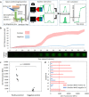
Management of curable sexually-transmitted infections (STI) such as Chlamydia can be revolutionized by highly sensitive nucleic acid testing that is deployable at the point-of-care (POC). Here we report the development of a mobile nucleic acid amplification testing (mobiNAAT) platform utilizing a mobile phone and droplet magnetofluidics to deliver NAAT in a portable and accessible format. By using magnetic particles as a mobile substrate for nucleic acid capture and transport, fluid handling is reduced to particle translocation on a simple magnetofluidic cartridge assembled with reagents for nucleic acid purification and amplification. A mobile phone user interface operating in tandem with a portable Bluetooth-enabled cartridge-processing unit facilitates process integration. We tested 30 potentially Chlamydia trachomatis (CT)-infected patients in a hospital emergency department and confirmed that mobiNAAT showed 100% concordance with laboratory-based NAAT. Concurrent evaluation by a nontechnical study coordinator who received brief training via an embedded mobile app module demonstrated ease of use and reproducibility of the platform. This work demonstrates the potential of mobile nucleic acid testing in bridging the diagnostic gap between centralized laboratories and hospital emergency departments.
Conflict of interest statement
The authors declare that they have no competing interests.
Figures


Similar articles
-
Extragenital self-collection testing for gonorrhea and chlamydia: A feasibility study for expanding STI screening in the Veterans Health Administration.Am J Clin Pathol. 2025 Mar 8;163(3):406-410. doi: 10.1093/ajcp/aqae135. Am J Clin Pathol. 2025. PMID: 39394953
-
Self-Collected versus Clinician-Collected Sampling for Chlamydia and Gonorrhea Screening: A Systemic Review and Meta-Analysis.PLoS One. 2015 Jul 13;10(7):e0132776. doi: 10.1371/journal.pone.0132776. eCollection 2015. PLoS One. 2015. PMID: 26168051 Free PMC article.
-
Accuracy of self-collection versus cervical sampling for the molecular diagnosis of Chlamydia trachomatis and Neisseria gonorrhoeae in women attending gynecological services.Rev Bras Ginecol Obstet. 2025 Jul 15;47:e-rbgo31. doi: 10.61622/rbgo/2025rbgo31. eCollection 2025. Rev Bras Ginecol Obstet. 2025. PMID: 40673035 Free PMC article.
-
Ophthalmia Neonatorum.2025 Jul 7. In: StatPearls [Internet]. Treasure Island (FL): StatPearls Publishing; 2025 Jan–. 2025 Jul 7. In: StatPearls [Internet]. Treasure Island (FL): StatPearls Publishing; 2025 Jan–. PMID: 31855399 Free Books & Documents.
-
The accuracy and efficacy of screening tests for Chlamydia trachomatis: a systematic review.J Med Microbiol. 2002 Dec;51(12):1021-1031. doi: 10.1099/0022-1317-51-12-1021. J Med Microbiol. 2002. PMID: 12466399
Cited by
-
A Portable Droplet Magnetofluidic Device for Point-of-Care Detection of Multidrug-Resistant Candida auris.Front Bioeng Biotechnol. 2022 Mar 24;10:826694. doi: 10.3389/fbioe.2022.826694. eCollection 2022. Front Bioeng Biotechnol. 2022. PMID: 35425764 Free PMC article.
-
Magnetic particles for integrated nucleic acid purification, amplification and detection without pipetting.Trends Analyt Chem. 2020 Jun;127:115912. doi: 10.1016/j.trac.2020.115912. Epub 2020 May 6. Trends Analyt Chem. 2020. PMID: 32382202 Free PMC article. Review.
-
Smartphone-Enabled Personalized Diagnostics: Current Status and Future Prospects.Diagnostics (Basel). 2021 Jun 9;11(6):1067. doi: 10.3390/diagnostics11061067. Diagnostics (Basel). 2021. PMID: 34207908 Free PMC article. Review.
-
Sample-to-Answer Droplet Magnetofluidic Platform for Point-of-Care Hepatitis C Viral Load Quantitation.Sci Rep. 2018 Jun 28;8(1):9793. doi: 10.1038/s41598-018-28124-3. Sci Rep. 2018. PMID: 29955160 Free PMC article.
-
Development and Evaluation of a Point-of-Care Test in a Low-Resource Setting with High Rates of Chlamydia trachomatis Urogenital Infections in Fiji.J Clin Microbiol. 2021 Jun 18;59(7):e0018221. doi: 10.1128/JCM.00182-21. Epub 2021 Jun 18. J Clin Microbiol. 2021. PMID: 33910964 Free PMC article.
References
-
- K. Owusu-Edusei Jr. et al. The estimated direct medical cost of selected sexually transmitted infections in the United States, 2008. Sex Transm Dis. 40(13) (2013). - PubMed
-
- Centers for Disease Control and Prevention. Sexually transmitted diseases treatment guidelines. 2010 MMWR Morb Mortal Wkly Rep. 2010; No.59(RR-12):1–110. Erratum in: MMWR Recomm Rep. 2011; 60 (1):18. - PubMed
-
- American Congress of Obstetricians and Gynecologists. Chlamydia Screening Rates Too Low, Reinfection Rates Too High. Available at http://www.acog.org/About_ACOG/News_Room/News_Releases/2012/Chlamydia_Sc... Accessed April 20, 2016.
Publication types
MeSH terms
Grants and funding
LinkOut - more resources
Full Text Sources
Other Literature Sources
Medical
Miscellaneous

