

Serial femtosecond crystallography structure of cytochrome c oxidase at room temperature
- PMID: 28674417
- PMCID: PMC5495810
- DOI: 10.1038/s41598-017-04817-z
Serial femtosecond crystallography structure of cytochrome c oxidase at room temperature
Abstract
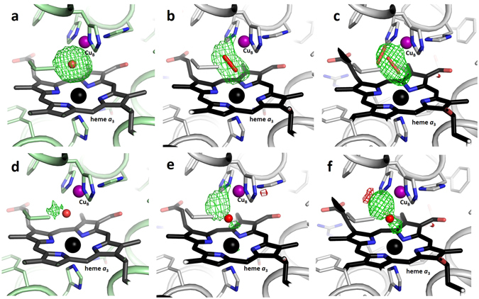
Cytochrome c oxidase catalyses the reduction of molecular oxygen to water while the energy released in this process is used to pump protons across a biological membrane. Although an extremely well-studied biological system, the molecular mechanism of proton pumping by cytochrome c oxidase is still not understood. Here we report a method to produce large quantities of highly diffracting microcrystals of ba 3-type cytochrome c oxidase from Thermus thermophilus suitable for serial femtosecond crystallography. The room-temperature structure of cytochrome c oxidase is solved to 2.3 Å resolution from data collected at an X-ray Free Electron Laser. We find overall agreement with earlier X-ray structures solved from diffraction data collected at cryogenic temperature. Previous structures solved from synchrotron radiation data, however, have shown conflicting results regarding the identity of the active-site ligand. Our room-temperature structure, which is free from the effects of radiation damage, reveals that a single-oxygen species in the form of a water molecule or hydroxide ion is bound in the active site. Structural differences between the ba 3-type and aa 3-type cytochrome c oxidases around the proton-loading site are also described.
Conflict of interest statement
The authors declare that they have no competing interests.
Figures
Similar articles
-
Structure and mechanism of the aberrant ba(3)-cytochrome c oxidase from thermus thermophilus.EMBO J. 2000 Apr 17;19(8):1766-76. doi: 10.1093/emboj/19.8.1766. EMBO J. 2000. PMID: 10775261 Free PMC article.
-
High resolution structure of the ba3 cytochrome c oxidase from Thermus thermophilus in a lipidic environment.PLoS One. 2011;6(7):e22348. doi: 10.1371/journal.pone.0022348. Epub 2011 Jul 21. PLoS One. 2011. PMID: 21814577 Free PMC article.
-
Timing of electron and proton transfer in the ba(3) cytochrome c oxidase from Thermus thermophilus.Biochemistry. 2012 Jun 5;51(22):4507-17. doi: 10.1021/bi300132t. Epub 2012 May 24. Biochemistry. 2012. PMID: 22624600
-
Proton transfer in ba(3) cytochrome c oxidase from Thermus thermophilus.Biochim Biophys Acta. 2012 Apr;1817(4):650-7. doi: 10.1016/j.bbabio.2011.11.015. Epub 2011 Dec 7. Biochim Biophys Acta. 2012. PMID: 22172736 Review.
-
Respiratory conservation of energy with dioxygen: cytochrome C oxidase.Met Ions Life Sci. 2015;15:89-130. doi: 10.1007/978-3-319-12415-5_4. Met Ions Life Sci. 2015. PMID: 25707467 Review.
Cited by
-
Segmented flow generator for serial crystallography at the European X-ray free electron laser.Nat Commun. 2020 Sep 9;11(1):4511. doi: 10.1038/s41467-020-18156-7. Nat Commun. 2020. PMID: 32908128 Free PMC article.
-
Well-based crystallization of lipidic cubic phase microcrystals for serial X-ray crystallography experiments.Acta Crystallogr D Struct Biol. 2019 Oct 1;75(Pt 10):937-946. doi: 10.1107/S2059798319012695. Epub 2019 Oct 1. Acta Crystallogr D Struct Biol. 2019. PMID: 31588925 Free PMC article.
-
Structural insights into functional properties of the oxidized form of cytochrome c oxidase.Nat Commun. 2023 Sep 16;14(1):5752. doi: 10.1038/s41467-023-41533-x. Nat Commun. 2023. PMID: 37717031 Free PMC article.
-
Proton Pumping and Non-Pumping Terminal Respiratory Oxidases: Active Sites Intermediates of These Molecular Machines and Their Derivatives.Int J Mol Sci. 2021 Oct 7;22(19):10852. doi: 10.3390/ijms221910852. Int J Mol Sci. 2021. PMID: 34639193 Free PMC article. Review.
-
Cryo-EM structures of intermediates suggest an alternative catalytic reaction cycle for cytochrome c oxidase.Nat Commun. 2021 Nov 25;12(1):6903. doi: 10.1038/s41467-021-27174-y. Nat Commun. 2021. PMID: 34824221 Free PMC article.
References
-
- Ganesan K, Gennis RB. Blocking the K-pathway still allows rapid one-electron reduction of the binuclear center during the anaerobic reduction of the aa3-type cytochrome c oxidase from Rhodobacter sphaeroides. Biochimica et biophysica acta. 2010;1797:619–624. doi: 10.1016/j.bbabio.2010.03.012. - DOI - PMC - PubMed
Publication types
MeSH terms
Substances
LinkOut - more resources
Full Text Sources
Other Literature Sources
