Review
. 2017 Jun 19:8:274.
doi: 10.3389/fneur.2017.00274. eCollection 2017.

Aspects on the Physiological and Biochemical Foundations of Neurocritical Care

Affiliations
Review

Aspects on the Physiological and Biochemical Foundations of Neurocritical Care

Carl-Henrik Nordström et al. Front Neurol. .

Abstract

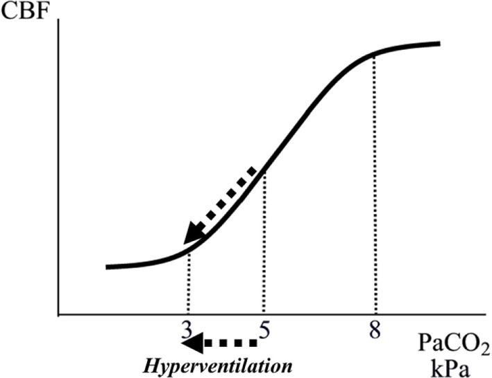
Neurocritical care (NCC) is a branch of intensive care medicine characterized by specific physiological and biochemical monitoring techniques necessary for identifying cerebral adverse events and for evaluating specific therapies. Information is primarily obtained from physiological variables related to intracranial pressure (ICP) and cerebral blood flow (CBF) and from physiological and biochemical variables related to cerebral energy metabolism. Non-surgical therapies developed for treating increased ICP are based on knowledge regarding transport of water across the intact and injured blood-brain barrier (BBB) and the regulation of CBF. Brain volume is strictly controlled as the BBB permeability to crystalloids is very low restricting net transport of water across the capillary wall. Cerebral pressure autoregulation prevents changes in intracranial blood volume and intracapillary hydrostatic pressure at variations in arterial blood pressure. Information regarding cerebral oxidative metabolism is obtained from measurements of brain tissue oxygen tension (PbtO2) and biochemical data obtained from intracerebral microdialysis. As interstitial lactate/pyruvate (LP) ratio instantaneously reflects shifts in intracellular cytoplasmatic redox state, it is an important indicator of compromised cerebral oxidative metabolism. The combined information obtained from PbtO2, LP ratio, and the pattern of biochemical variables reveals whether impaired oxidative metabolism is due to insufficient perfusion (ischemia) or mitochondrial dysfunction. Intracerebral microdialysis and PbtO2 give information from a very small volume of tissue. Accordingly, clinical interpretation of the data must be based on information of the probe location in relation to focal brain damage. Attempts to evaluate global cerebral energy state from microdialysis of intraventricular fluid and from the LP ratio of the draining venous blood have recently been presented. To be of clinical relevance, the information from all monitoring techniques should be presented bedside online. Accordingly, in the future, the chemical variables obtained from microdialysis will probably be analyzed by biochemical sensors.

Keywords: cerebral blood flow; cerebral energy metabolism; intracranial pressure; microdialysis; neurocritical care.


Figures

Figure 1
Figure 1
Schematic illustration of water exchange across cerebral capillaries in three hypothetical situations: (A) the normal brain with intact blood–brain barrier (BBB); (B) the injured brain with a BBB permeable for crystalloids but not colloids; (C) the injured brain with a ruptured BBB permeable for crystalloids as well as colloids. Gray area represents crystalloids in the capillary; black circles represent large (colloidal) molecules; filled gray circles represent blood cells.
Figure 2
Figure 2
Schematic illustration of the brain and its surroundings being enclosed in a rigid shell (CSF, cerebrospinal fluid). The vessels responsible for precapillary vascular resistance (1) as well as the intracerebral venous compartment (2) are indicated in the figure.
Figure 3
Figure 3
Schematic illustration of the relation between arterial tension of CO2 (PaCO2) and cerebral blood flow (CBF). The interrupted arrows indicate the decrease in CBF during a decrease in PaCO2 due to hyperventilation.
Figure 4
Figure 4
Illustration of the relation between cerebral metabolic rate and cerebral blood flow (CBF). The figure gives a schematic summary of data obtained from experimental studies during induced epileptic seizures (41, 42) and immobilization stress (43, 44), and after administration of amphetamine (45) and phenobarbitone (46).
Figure 5
Figure 5
Schematic illustration of cerebral pressure autoregulation of blood flow: the relation between mean arterial blood pressure (MAP)/cerebral perfusion pressure (CPP) and cerebral blood flow (CBF)/intracapillary hydrostatic pressure. Line A indicates intact autoregulation; line C indicates absence of autoregulation; line B indicates the description of absence of autoregulation often given in neurosurgical literature. The point A/B illustrates that in neurosurgical literature intact autoregulation is often interpreted as a mechanism preventing a decrease in CBF at a decrease in MAP/CPP.
Figure 6
Figure 6
(A–F) Percentage changes in global cerebral blood flow (CBF), cerebral arterio-venous difference in oxygen (AVDO), intracranial pressure (ICP), and cerebro-vascular resistance (CVR) in patients with severe brain trauma. After the initial test with increased controlled ventilation (hyperventilation), the patients were assigned either to the group “Preserved CO2-reactivity” (A) or “Impaired CO2-reactivity” (B). After restoration of normoventilation, the patients were given either a bolus of thiopentone (C,D) (5–11 mg/kg intravenously) or a bolus of dihydroergotamine (E,F) (DHE; 4 mg/kg intravenously). *p < 0.05; **p < 0.01; ***p < 0.001 for significance of difference from control value. Data from Ref. (48, 156).
Figure 7
Figure 7
Schematic illustration of the theoretical principles behind the “Vasoconstrictor Cascade” according to Rosner et al. (172).
Figure 8
Figure 8
Schematic diagram of cerebral intermediary metabolism, with a focus on the glycolytic chain and its relation to glycerol and glycerophospholipids and to the citric acid cycle (Krebs cycle). F-1,6-DP: fructose-1,6-diposphate; DHAP, dihydroxyacetone-phosphate; GA-3P, glyceraldehyde-3-phosphate; G-3-P, glycerol-3-phosphate; FFA, free fatty acids; α-KG, α-ketoglutarate. Underlined metabolites are measured at the bedside with enzymatic techniques. References levels of the various metabolites for normal human brain obtained from Ref. (182).
Figure 9
Figure 9
Changes in intracerebral biochemistry during transient global cerebral ischemia. Levels of glucose, lactate, and pyruvate (mean ± SD). Data from Ref. (190).
Figure 10
Figure 10
Changes in intracerebral biochemistry during transient global cerebral ischemia. Levels of lactate/pyruvate (LP) ratio, glutamate, and glycerol (mean ± SD). Data from Ref. (190).
Figure 11
Figure 11
Bar graphs demonstrating mean (±SD) lactate/pyruvate (LP) ratio in the better (white bar) and worse (gray bar) microdialysis catheter positions in relation to four ranges of cerebral perfusion pressure (CPP) in patients with severe traumatic brain lesions. Interrupted lines indicate the range (mean ± SD) in healthy brains in humans during wakefulness. Data from Ref. (182, 194), respectively.
Figure 12
Figure 12
Schematic illustration of cerebral tissue oxygenation (PbtO2) and changes in the levels of lactate (La), pyruvate (Py), and the lactate/pyruvate (LP) ratio in experimental cerebral ischemia (A) and mitochondrial dysfunction (B). Data from Ref. (200).

References

    1. Berthelsen PG, Cronquist M. The first intensive care unit in the world: Copenhagen 1953. Acta Anaesthesiol Scand (2003) 47:1190–5. 10.1046/j.1399-6576.2003.00256.x - DOI - PubMed
    1. Weil MH, Tang W. From intensive care to critical care medicine: a historical perspective. Am J Respir Crit Care Med (2011) 183:1451–3. 10.1164/rccm.201008-1341OE - DOI - PubMed
    1. Nordström CH, Nielsen TH, Jacobsen A. Techniques and strategies in neurocritical care originating from southern Scandinavia. J Rehabil Med (2013) 45:710–7. 10.2340/16501977-1157 - DOI - PubMed
    1. Lundberg N. Continuous recording and control of ventricular pressure in neurosurgical practice. Acta Psychiatr Scand Suppl (1960) 36:1–93. - PubMed
    1. Hawthorne C, Piper I. Monitoring of intracranial pressure in patients with traumatic brain injury. Front Neurol (2014) 5:121. 10.3389/fneur.2014.00121 - DOI - PMC - PubMed