. 2017 Jun 19:8:699.
doi: 10.3389/fimmu.2017.00699. eCollection 2017.

Human CD56dimCD16dim Cells As an Individualized Natural Killer Cell Subset

Affiliations

Human CD56dimCD16dim Cells As an Individualized Natural Killer Cell Subset

Mathieu Amand et al. Front Immunol. .

Abstract

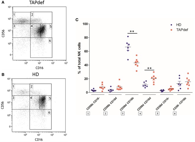
Human natural killer (NK) cells can be subdivided in several subpopulations on the basis of the relative expression of the adhesion molecule CD56 and the activating receptor CD16. Whereas blood CD56brightCD16dim/- NK cells are classically viewed as immature precursors and cytokine producers, the larger CD56dimCD16bright subset is considered as the most cytotoxic one. In peripheral blood of healthy donors, we noticed the existence of a population of CD56dimCD16dim NK cells that was frequently higher in number than the CD56bright subsets and even expanded in occasional control donors but also in transporter associated with antigen processing-deficient patients, two familial hemophagocytic lymphohistiocytosis type II patients, and several common variable immunodeficiency patients. This population was detected but globally reduced in a longitudinal cohort of 18 HIV-1-infected individuals. Phenotypically, the new subset contained a high percentage of relatively immature cells, as reflected by a significantly stronger representation of NKG2A+ and CD57- cells compared to their CD56dimCD16bright counterparts. The phenotype of the CD56dimCD16dim population was differentially affected by HIV-1 infection as compared to the other NK cell subsets and only partly restored to normal by antiretroviral therapy. From the functional point of view, sorted CD56dimCD16dim cells degranulated more than CD56dimCD16bright cells but less than CD56dimCD16- NK cells. The population was also identified in various organs of immunodeficient mice with a human immune system ("humanized" mice) reconstituted from human cord blood stem cells. In conclusion, the CD56dimCD16dim NK cell subpopulation displays distinct phenotypic and functional features. It remains to be clarified if these cells are the immediate precursors of the CD56dimCD16bright subset or placed somewhere else in the NK cell differentiation and maturation pathway.

Keywords: CD56dim natural killer cells; human; humanized mouse model; natural killer cells; subsets.


Figures

Figure 1
Figure 1
Flow cytometry dot plot of CD56 versus CD16 after gating on alive, single, CD3CD14CD19 blood cells from fresh peripheral blood mononuclear cells (PBMC) of a representative healthy donor (A). Percentages of the different natural killer (NK) cell subsets relative to the total NK cell population (100%) from fresh PBMC of a series of healthy donors (n = 5) (B).
Figure 2
Figure 2
Flow cytometry dot plot of CD56 versus CD16 after gating on alive, single, CD3CD14CD19 blood cells from frozen peripheral blood mononuclear cells (PBMC) of representative healthy donor (A), HIV-1-infected viremic patient (B), and HIV-1-infected aviremic patient under combined antiretroviral therapy (cART) (C). Percentages relative to the total natural killer (NK) cell population (100%) (D,E) of different NK cells subsets from frozen PBMC of a series of healthy donors (n = 12) and longitudinal paired samples of HIV-1-infected patients when viremic or aviremic under cART (n = 18) (*p < 0.05; **p < 0.01; ***p < 0.001).
Figure 3
Figure 3
Percentages relative to the total natural killer (NK) cell population (100%) of different blood NK cells subsets expressing the markers KIR2DL2/DL3/DS2, KIR2DL1/DS1, KIR3DL1, NKG2A, and CD57 from frozen peripheral blood mononuclear cells of a cohort of healthy donors (n = 12) (*p < 0.05; **p < 0.01; ***p < 0.001).
Figure 4
Figure 4
Percentages relative to the total natural killer (NK) cell population (100%) of different blood NK cells subsets expressing the markers SIGLEC-7, NKp30, KIR2DL1/DS1, CD62L, CD27, and NKG2A from frozen peripheral blood mononuclear cells of a series of healthy donors (n = 12) and longitudinal paired samples of HIV-1-infected patients when viremic or aviremic under combined antiretroviral therapy (n = 18) (*p < 0.05; **p < 0.01; ***p < 0.001).
Figure 5
Figure 5
Percentages relative to the total natural killer (NK) cell population (100%) of different blood NK cells subsets expressing CD107a from frozen peripheral blood mononuclear cells (PBMC) of a series of healthy donors (HD) (n = 12) and longitudinal paired samples of HIV-1-infected patients when viremic or aviremic under combined antiretroviral therapy (cART) (n = 18) at basal levels (A) or following stimulation with K562 cells (B). Percentages relative to the total NK cell population (100%) of different blood NK cells subsets producing IFNγ from frozen PBMC of a series of HD (n = 12) and longitudinal paired samples of HIV-1-infected patients when viremic or aviremic under cART (n = 18) at basal levels (C) or following stimulation with K562 cells (D) (*p < 0.05; **p < 0.01; ***p < 0.001).
Figure 6
Figure 6
(A) Flow cytometry dot plot CD56 versus CD16 after gating on alive, single, CD3CD14CD19 blood cells from fresh peripheral blood mononuclear cells (PBMC) of a representative healthy donor prior to cell sorting (A). The gates 1, 2, and 3 represent the sorted CD56dimCD16, CD56dimCD16dim, and CD56dimCD16bright subsets, respectively. Each subset was stimulated with K562 cells, and their degranulation and IFNγ production were assessed by flow cytometry. (B,C) Percentages of blood natural killer cells from sorted CD56dimCD16bright, CD56dimCD16dim, and CD56dimCD16 subtypes expressing CD107a (B) and IFNγ (C) from fresh PBMC of a series of healthy donors (n = 4) at basal levels or following stimulation with K562 cells (*p < 0.05).
Figure 7
Figure 7
Flow cytometry dot plot of CD56 versus CD16 after gating on alive, single, CD3CD14CD19 blood cells from a representative TAP-deficient patient (A) and a representative healthy donor (B). Percentages of the different natural killer (NK) cell subsets relative to the total NK cell population (100%) in a cohort of TAP-deficient patients (n = 7) compared with healthy donors (n = 6) (C) (*p < 0.05; **p < 0.01).
Figure 8
Figure 8
(A) Peripheral blood cells from a representative humanized NSG mouse were stained with antibodies as described in Material and Methods. The gate was set on alive single cells positive for human CD45 to exclude contaminating mouse cells. The CD3 versus CD14/CD19 dot plot allows the identification and discrimination of human T cells, B cells, and monocytes. The remaining triple-negative cells were analyzed in a CD56 versus CD16 dot plot to identify the natural killer (NK) cells. CD56dimCD16dim NK cells are clearly present. (B) Percentages of CD56dim (a, b) and CD56bright (c, d) NK cell subsets in various organs of humanized NSG (NSG; a, c; n = 3) and NSG/human leukocyte antigen (HLA)-A2 (HLA-A2; b, d; n = 2) mice. BM: bone marrow; LN: lymph nodes.

References

    1. Yoon SR, Kim TD, Choi I. Understanding of molecular mechanisms in natural killer cell therapy. Exp Mol Med (2015) 47:e141. 10.1038/emm.2014.114 - DOI - PMC - PubMed
    1. O’Sullivan TE, Sun JC, Lanier LL. Natural killer cell memory. Immunity (2015) 43(4):634–45. 10.1016/j.immuni.2015.09.013 - DOI - PMC - PubMed
    1. Cerwenka A, Lanier LL. Natural killer cell memory in infection, inflammation and cancer. Nat Rev Immunol (2016) 16(2):112–23. 10.1038/nri.2015.9 - DOI - PubMed
    1. Adams NM, O’Sullivan TE, Geary CD, Karo JM, Amezquita RA, Joshi NS, et al. NK cell responses redefine immunological memory. J Immunol (2016) 197(8):2963–70. 10.4049/jimmunol.1600973 - DOI - PMC - PubMed
    1. Morandi F, Horenstein AL, Chillemi A, Quarona V, Chiesa S, Imperatori A, et al. CD56brightCD16-NK cells produce adenosine through a CD38-mediated pathway and act as regulatory cells inhibiting autologous CD4+ T cell proliferation. J Immunol (2015) 195(3):965–72. 10.4049/jimmunol.1500591 - DOI - PubMed