. 2018 Jul;68(4):503-520.
doi: 10.1007/s12576-017-0557-5. Epub 2017 Jul 3.

Investigating β-adrenergic-induced cardiac hypertrophy through computational approach: classical and non-classical pathways

Affiliations

Investigating β-adrenergic-induced cardiac hypertrophy through computational approach: classical and non-classical pathways

Ali Khalilimeybodi et al. J Physiol Sci. 2018 Jul.

Abstract

The chronic stimulation of β-adrenergic receptors plays a crucial role in cardiac hypertrophy and its progression to heart failure. In β-adrenergic signaling, in addition to the well-established classical pathway, Gs/AC/cAMP/PKA, activation of non-classical pathways such as Gi/PI3K/Akt/GSK3β and Gi/Ras/Raf/MEK/ERK contribute in cardiac hypertrophy. The signaling network of β-adrenergic-induced hypertrophy is very complex and not fully understood. So, we use a computational approach to investigate the dynamic response and contribution of β-adrenergic mediators in cardiac hypertrophy. The proposed computational model provides insights into the effects of β-adrenergic classical and non-classical pathways on the activity of hypertrophic transcription factors CREB and GATA4. The results illustrate that the model captures the dynamics of the main signaling mediators and reproduces the experimental observations well. The results also show that despite the low portion of β2 receptors out of total cardiac β-adrenergic receptors, their contribution in the activation of hypertrophic mediators and regulation of β-adrenergic-induced hypertrophy is noticeable and variations in β1/β2 receptors ratio greatly affect the ISO-induced hypertrophic response. The model results illustrate that GSK3β deactivation after β-adrenergic receptor stimulation has a major influence on CREB and GATA4 activation and consequent cardiac hypertrophy. Also, it is found through sensitivity analysis that PKB (Akt) activation has both pro-hypertrophic and anti-hypertrophic effects in β-adrenergic signaling.

Keywords: CREB transcription factor; GATA4 transcription factor; Gi/PI3K/Akt/GSK3β pathway; Gi/Ras/Raf/MEK/ERK pathway; Non-classical pathways; β-Adrenergic signaling.


Figures
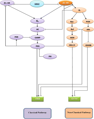
Fig. 1
Fig. 1
The model of β2-AR signaling pathways involved in the cardiac hypertrophy. The left is classical and the right is non-classical β-adrenergic pathway. Activation and inhibition of the components are indicated by arrows and T-bars, respectively
Fig. 2
Fig. 2
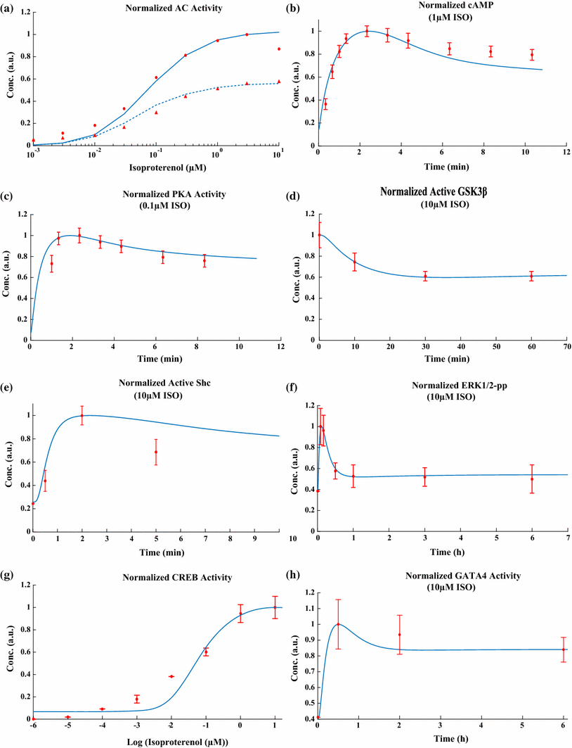
Estimation of kinetic parameters for β-adrenergic classical and non-classical signaling pathways. a The sensitivity of normalized AC activity over basal to isoproterenol for maximal AC activity (simulation solid line and experimental filled circle) and AC activity after 30 min of ISO stimulation (simulation dashed line and experimental filled triangle). The experimental data were obtained from Freedman et al.’s study [21] for β1-AR stimulation and its desensitization through GRK2 mechanism. b The time course of normalized cAMP level after indicated ISO stimulation. The experimental data were obtained from De Arcangelis et al.’s study [18]. c The time course of normalized PKA activity after indicated ISO stimulation. The experimental data were obtained from De Arcangelis et al.’s study [18]. d The time course of normalized active GSK3β after indicated ISO stimulation. The experimental data were obtained from Morisco et al.’s study [41]. e The time course of normalized active Shc after indicated ISO stimulation. The experimental data were obtained from Zou et al.’s study [69]. f The time course of normalized ERK1/2 pp concentration after indicated ISO stimulation. The experimental data were obtained from Shin et al.’s study [56]. g The sensitivity of normalized steady-state activity of CREB to isoproterenol. The experimental data were obtained from Yang et al.’s study [66]. h The time course of normalized GATA4 activity after indicated ISO stimulation. The experimental data were obtained from Morisco et al.’s study [42]. For bh, simulation results are indicated by solid lines and experimental data are indicated by filled circles. The experimental data are mean ± SE of at least three independent experiments
Fig. 3
Fig. 3
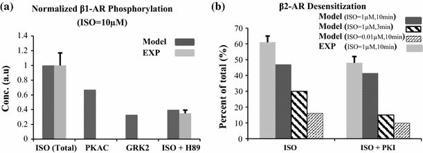
The β-ARs desensitization by isoproterenol. a Phosphorylation of β1-AR by ISO through GRK2 and PKA. The experimental data were obtained from Freedman et al.’s study [21] for HEK293 cells with and without PKA inhibitor (H89) after 3-min ISO stimulation. b Desensitization of β2-AR by ISO after different ISO concentration and stimulation times. The experimental data were obtained from Lohse et al.’s study [36] for A431 cells with and without PKI (1 µM). The experimental data are mean ± SE of at least three independent experiments
Fig. 4
Fig. 4
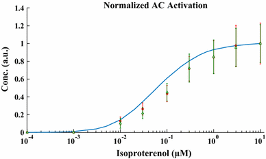
Normalized AC activity above basal level after ISO stimulation at different concentrations. The simulation result is indicated by a solid line and experimental data for mouse hearts [59] and ventricular myocytes [33] are indicated by filled circles and hollow triangles, respectively. The experimental data represent mean ± SE from five individual experiments
Fig. 5
Fig. 5
Effects of β-ARs stimulation by isoproterenol on the cellular cAMP level. a The cAMP accumulation in cardiomyocytes after ISO stimulation. The experimental data were obtained from O’Connell et al.’s study [44] for mouse cardiac myocytes and are mean ± SE of three independent experiments. b Effects of β-AR subtype specific-antagonists and PDEs inhibitor, IBMX, (100 µM) on the cellular cAMP level. The experimental data were obtained from Rochais et al.’s study [46] for rat ventricular myocytes after 3 min ISO stimulation and represent mean ± SE of at least eight separate experiments
Fig. 6
Fig. 6
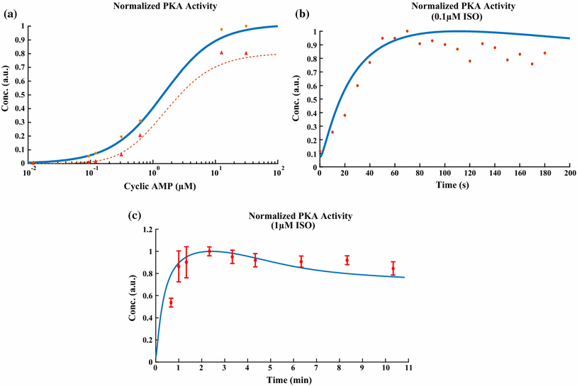
The PKA activity after β-ARs stimulation by ISO (a) normalized PKA activity as a function of cAMP level with (simulation dashed line and experimental hollow triangle) and without PKI (1 µM) (simulation solid line and experimental filled circle). The experimental data were obtained from Beavo et al.’s study [5]. b The time course of normalized PKA activity after indicated ISO stimulation. The experimental data were obtained from Amanfu and Saucerman’s study [2] for cardiac myocytes. In a and b, no error bar was provided for experimental data in references. c The time course of normalized PKA activity after indicated ISO stimulation. The experimental data were obtained from De Arcangelis et al.’s study [18] for cardiac myocytes and are mean ± SE from three separate experiments
Fig. 7
Fig. 7
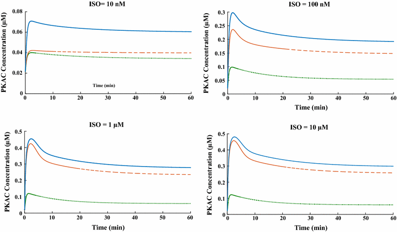
The simulated PKA activity after different indicated ISO stimulation. Simulated time courses of PKAc concentration for control condition (normal cardiomyocytes), selective β1-AR and β2-AR stimulation are shown by solid, dashed, and dotted lines, respectively
Fig. 8
Fig. 8
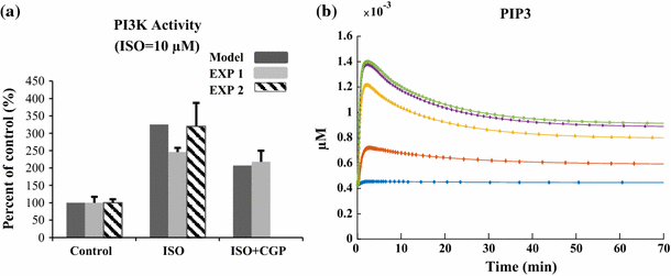
The effect of β-AR stimulation by ISO on PI3 K activity. a The β-AR-induced increase in PI3 K activity after 2-min stimulation by ISO with and without CGP antagonist. The experimental data were obtained from Yano et al.’s study [67] for H9c2 cardiomyocytes and represent mean ± SE from three separate experiments. b The simulated time course of PIP3 concentration after stimulation with different isoproterenol concentrations (0.001 µM filled circle, 0.01 µM filled diamond, 0.1 µM filled triangle, 1 µM star and 10 µM hollow square)
Fig. 9
Fig. 9
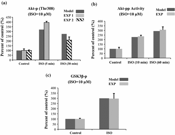
Effects of β-AR stimulation on Akt and GSK3β activity. a ISO-induced Akt phosphorylation at Thr308. The increase in Akt phosphorylation after stimulation by ISO for 2 min EXP1 and 30 min EXP2. The experimental data were obtained from Yano et al.’s study [67] for H9c2 cardiomyocytes (EXP1) and from Zhang et al.’s study [68] for mouse cardiac cells (EXP 2). The experimental data represent mean ± SE from three (EXP1) and six (EXP2) individual experiments. b ISO-induced Akt-pp kinase activity. The experimental data were obtained from Morisco et al. study [41] for rat cardiac myocytes and represent mean ± SE from four separate experiments. c GSK3β phosphorylation (deactivation) after ISO stimulation through PI3 K/Akt pathway. The experimental data were obtained from Zhang et al.’s study [68] for mouse cardiac cells at 30 min after ISO stimulation. Data are mean ± SE from six individual experiments
Fig. 10
Fig. 10
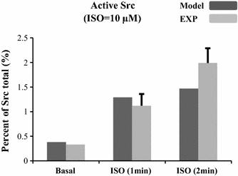
Activation of Src after indicated ISO stimulation. The experimental data were obtained from Luttrell et al.’s study [38] for HEK 293 cells and are mean ± SE of three independent experiments
Fig. 11
Fig. 11
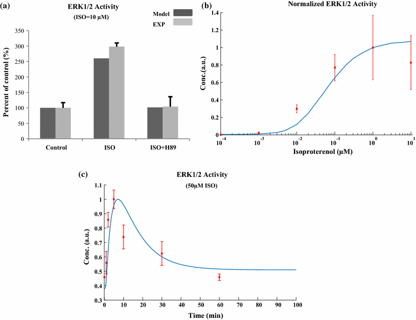
Effects of β-AR stimulation on ERK1/2 activity. a The experimental data for ERK1/2 pp after 8 min of ISO stimulation with and without PKA inhibition (H89) were obtained from Zou et al. study [69] for rat cardiac myocytes. Data represent mean ± SE of three individual experiments. b Normalized ERK1/2 activity as a function of isoproterenol concentration. The simulation result is indicated by a solid line and the experimental data for ERK1/2 pp after 5 min of ISO stimulation were obtained from Shenoy et al.’s study [55] for HEK-293 cells. Data represent mean ± SE of three individual experiments. c The time course of ERK1/2 pp after indicated ISO stimulation. The simulation result is indicated by a solid line and the experimental data were obtained from Bogoyevitch et al.’s study [7] for rat cardiac myocytes. Data represent mean ± SE of at least three independent experiments
Fig. 12
Fig. 12
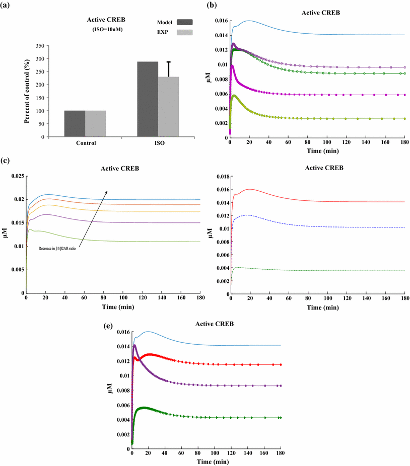
Effects of β-AR stimulation on CREB activity. a ISO-induced CREB activity. The experimental data were obtained from Shin et al.’s study [56] for rat cardiac myocytes after 5 min of indicated ISO stimulation. Data represent mean ± SE of at least three independent experiments. b Effects of β-AR subtypes on CREB activity. The time course of ISO-induced CREB activity in physiological (solid line), β1-AR inhibition by 50% (hollow circle) and 100% (filled circle), and β2-AR inhibition by 50% (hollow square) and 100% (filled square). c Effects of β1/ β2-AR ratio on CREB activity The ratios are 9:1, 8:2, 7:3, 6:4, and 5:5. d Simulated time course of CREB activity after ISO stimulation (0.01 µM dash-dotted line, 0.1 µM dashed line, and 1 µM solid line). e Effects of β-adrenergic mediators on CREB activity. The time course of ISO-induced CREB activity in physiological (solid line) and artificial states including blocking direct effect of PKA (triangle), and Aktpp (circle), and blocking ISO-induced variation in GSK3β (square)
Fig. 13
Fig. 13
Effects of β-AR stimulation on GATA4 activity. a Effects of β-AR subtypes on GATA4 activity. The time course of ISO-induced GATA4 activity in the following states physiological (solid line), β1-AR inhibition by 50% (hollow circle) and 100% (filled circle), and β2-AR inhibition by 50% (hollow square). b Effects of β1/ β2-AR ratio on GATA4 activity. The ratios are 9:1, 8:2, 7:3, 6:4, and 5:5. c Effects of β-adrenergic mediators on GATA4 activity. The time course of ISO-induced GATA activity in the following states physiological (solid line), and artificial [blocking ISO-induced variation in GSK3β (square)]. d Simulated time course of GATA4 transcription factor after ISO stimulation for different concentrations (0.01 µM dash-dotted line, 0.1 µM dashed line, and 1 µM solid line)
Fig. 14
Fig. 14
Contribution of model parameters in mediating hypertrophic response. Each column represents the sensitivity of the outputs (+1 maximum activation and −1 maximum inhibition) to the perturbation in indicated parameter. The associated parameter of each number is shown in Table S10 (supplementary materials)

Similar articles

Cited by

References

    1. Aass H, Skomedal T, Osnes J-B. Increase of cyclic AMP in subcellular fractions of rat heart muscle after β-adrenergic stimulation: prenalterol and isoprenaline caused different distribution of bound cyclic AMP. J Mol Cell Cardiol. 1988;20:847–860. doi: 10.1016/S0022-2828(88)80009-9. - DOI - PubMed
    1. Amanfu RK, Saucerman JJ. Modeling the effects of β1-adrenergic receptor blockers and polymorphisms on cardiac myocyte Ca2+ handling. Mol Pharmacol. 2014;86:222–230. doi: 10.1124/mol.113.090951. - DOI - PMC - PubMed
    1. Anguelova M, Karlsson J, Jirstrand M. Minimal output sets for identifiability. Math Biosci. 2012;239:139–153. doi: 10.1016/j.mbs.2012.04.005. - DOI - PubMed
    1. Barki-Harrington L, Perrino C, Rockman HA. Network integration of the adrenergic system in cardiac hypertrophy. Cardiovasc Res. 2004;63:391–402. doi: 10.1016/j.cardiores.2004.03.011. - DOI - PubMed
    1. Beavo JA, Bechtel P, Krebs E. Activation of protein kinase by physiological concentrations of cyclic AMP. Proc Natl Acad Sci. 1974;71:3580–3583. doi: 10.1073/pnas.71.9.3580. - DOI - PMC - PubMed

MeSH terms

Substances