. 2018 Jan;247(1):212-221.
doi: 10.1002/dvdy.24542. Epub 2017 Aug 2.

Separate and coincident expression of Hes1 and Hes5 in the developing mouse eye

Affiliations

Separate and coincident expression of Hes1 and Hes5 in the developing mouse eye

Amy N Riesenberg et al. Dev Dyn. 2018 Jan.

Abstract

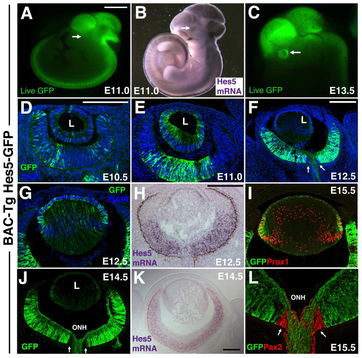
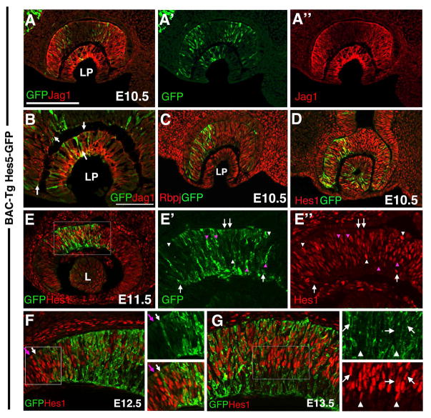
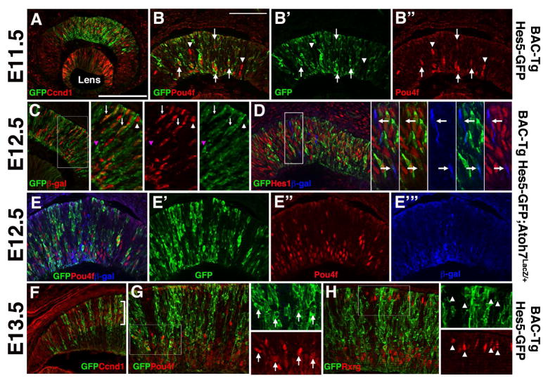
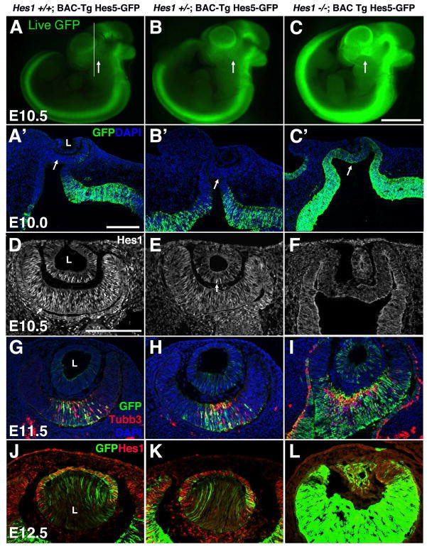
Background: Notch signaling is broadly required during embryogenesis, frequently activating the transcription of two basic helix-loop-helix transcription factors, Hes1 and Hes5. But, it remains unresolved when and where Hes1 and Hes5 act alone or together during development. Here, we analyzed a Hes5-green fluorescent protein (GFP) bacterial artificial chromosome (BAC) transgenic mouse, as a proxy for endogenous Hes5. We directly compared transgenic GFP expression with Hes1, and particular markers of embryonic lens and retina development.

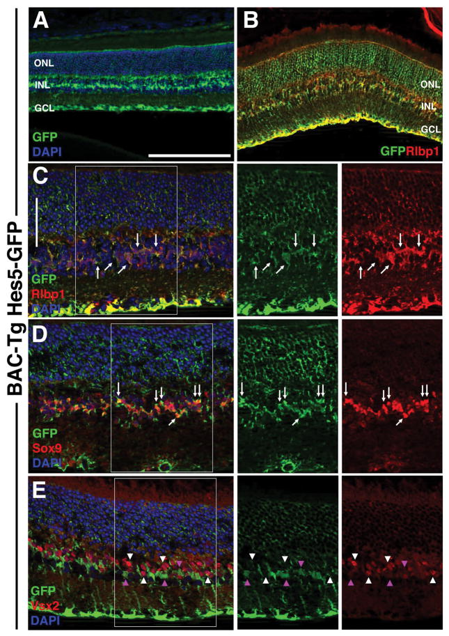
Results: Hes5-GFP is dynamic within subsets of retinal and lens progenitor cells, and differentiating retinal ganglion neurons, in contrast to Hes1 found in all progenitor cells. In the adult retina, only Müller glia express Hes5-GFP. Finally, Hes5-GFP is up-regulated in Hes1 germline mutants, consistent with previous demonstration that Hes1 suppresses Hes5 transcription.

Conclusions: Hes5-GFP BAC transgenic mice are useful for identifying Hes5-expressing cells. Although Hes5-GFP and Hes1 are coexpressed in particular developmental contexts, we also noted cohorts of lens or retinal cells expressing just one factor. The dynamic Hes5-GFP expression pattern, coupled with its derepressed expression in Hes1 mutants, suggests that this transgene contains the relevant cis-regulatory elements that regulate endogenous Hes5 in the mouse lens and retina. Developmental Dynamics 247:212-221, 2018. © 2017 Wiley Periodicals, Inc.

Keywords: Hes1; Hes5; Notch signaling; lens development; retina, neurogenesis.


Figures

Figure 1
Figure 1. Hes5-GFP BAC-Tg reporter expression relative to Hes5 mRNA
A,B) Whole embryo Hes5-GFP fluorescence, compared to Hes5 mRNA in situ expression. Arrows point to forming eye. C) At E13.5 GFP fluorescence in the developing eye is obvious (arrow). In all other panels, GFP expression was visualized by anti-GFP labeling of fixed cryosections, collected in the horizontal plane of sectioning. D–F) Dynamic changes in Hes5-GFP during early eye formation. F–H) Comparison of Hes5-GFP expression at E12.5 in the developing lens and retina. Arrows in F point to the optic nerve head, which lacks anti-GFP labeling. I) E15.5 Hes5-GFP expression in lens AEL and transition zone(bracket), compared to Prox1 nuclear expression (red) within transition zone and lens fiber cell nuclei. J,K) Comparison of Hes5-GFP and Hes5 mRNA expression at E14.5. Hes5-GFP is present in RPCs (like the endogenous gene), but also within RGCs, including their axons extending into the optic nerve head (ONH). J,L) Pax2+ optic nerve head (arrows) and optic stalk cells do not express GFP. Dorsal is up in A–C; Anterior is up, and nasal left in panels D–L. L = lens in D–F,J; ONH = optic nerve head in J,L. Scale bar in A=1mm (for A–C); in D (for D,E,G,L) or H=20 microns; in F for F,I,J) or K=10 microns.
Figure 2
Figure 2. Hes5-GFP expression in the early eye relative to other Notch signaling pathway components
A,A′A″, B) Hes5-GFP and Jag1 protein overlap in subsets of distal optic cup and lens pit cells. Arrows in B point to cells with Jag1 expression (red) at the cell membrane, surrounding cytoplasmic GFP. Nasal is to the left here, and in C,D. C) All Hes5-GFP cells co-express Rbpj. D) At this same age, every Hes5-GFP lens, optic cup and stalk cell also coexpresses Hes1 protein. E, E′,E″) Starting at E11.5, some GFP + only (pink arrowhead) and some Hes1+ only (white arrowheads) cells can be identified, although the majority of Hes5-GFP+ cells coexpress Hes1 (white arrows). E′ and E″ are 8X zoomed magnifications of boxed area in E. F) Comparison of Hes5-GFP and Hes1 expression at the E12.5 ciliary margin. 4X zoom of boxed area is shown in adjacent panels. Hes1 is present in RPCs and throughout the forming ciliary margin (left boxed area), but, Hes5-GFP+ is confined to the retina, like Hes5 mRNA (Fig 1H). White arrow points to distal-most extent of the Hes5-GFP domain, and the pink arrow denotes adjacent distal row of Hes1+ cells. G) Comparison of Hes5-GFP and Hes1 protein in E13.5 central retina. Arrows denote coexpressing cells, whereas white arrowheads point to Hes1+ only progenitors. Nascent RGCs in the inner retina express Hes5-GFP+. All images are in the horizontal plane of section. Scleral/apical is up in all panels; LP = lens pit; L= lens vesicle; Mag bar in A (for A,B,C,E) = 20 microns; in D (for D,E′,E″,F,I) = 50 microns.
Figure 3
Figure 3. Hes5-GFP expression correlates with RGC but not cone neurogenesis
A) Most Hes5-GFP cells in the E11.5 central optic cup coexpress the mitotic marker Ccnd1. B,B′B″) But a subset of nascent Pou4f+ RGCs also express Hes5-GFP (arrows). Arrowheads denote Pou4f+ nuclei that are Hes5-GFP-negative. C) In Hes5-GFP/+;Atoh7LacZ/+ eyes, anti-GFP and anti-βgal colabeling highlight three types of RPCs: double positive (white arrows), βgal-only (white arrowhead) GFP-only (pink arrowhead). D) Anti-GFP, anti-Hes1 and anti-βgal triple labeling further distinguishes a subset of Atoh7LacZ/+ cells that do not express Hes1 or Hes5-GFP proteins (arrows in zoomed insets of boxed area). E,E′E″, E‴) Subpopulations of E12.5 RPCs express different combinations of GFP, Pou4f (red) and β-gal (blue). F) At E13.5, a proportion of Hes5-GFP cells reside in the outer, neuroblast layer of the retina (bracket), as marked by Ccnd1 expression. G) Nearly all Pou4f+ RGCs in the E13.5 central retina coexpress Hes5-GFP. Arrows in 4X zoomed inset point to double positive cells. H) The Rxrg transcription factor is expressed by early RGCs (inner retina) and cone photoreceptors (outer retina) (Roberts et al., 2005). Although inner Rxrg+ RGCs coexpress Hes5-GFP, Rxrg+ cones were devoid of Hes5-GFP. Arrowheads in 4X zoomed insets point to Rxrg+ cones. Scleral/apical is up in all panels; mag bars in A (for A,C,F) = 20 microns; in B (for B, D, E, G, H) = 50 microns.
Figure 4
Figure 4. Specific Hes5-GFP expression in adult Müller glia
A) Anti-GFP labeling of P21 transgene hemizygous cryosection showing Hes5-GFP localization within particular INL cells and their cellular processes spanning the retina (from outer limiting membrane to nerve fiber layer). B) GFP coexpression with Rlbp1/Cralbp (arrows) in Müller cell bodies. C) Arrows point to coexpressing Müller cell bodies at higher magnification. D) Hes5-GFP was also co-expressed with Sox9 (arrows), another Müller cell nuclear marker. The circular structure within the GCL of boxed area is a blood vessel. E) No GFP+ INL cells (pink arrowheads) coexpress Vsx2, a bipolar marker (white arrowheads). Boxed areas in C,D,E are shown at same magnification at right, with marker channels separated. Scleral is up in all panels; bar in A,B = 20 microns; C–E = 50 microns, n ≥ 3 adult eyes analyzed per marker. ONL = outer nuclear layer, INL = inner nuclear layer, GCL = ganglion cell layer.
Figure 5
Figure 5. Hes5-GFP BAC Tg expression is regulated by Hes1
In each experiment (row), although not quantitative, labeling conditions and imaging time/parameters were held constant among genotypes. A–C) Live GFP expression among littermates. Line in A indicates sectioning plane for A′–C′ and arrows point to forming eye. Hes5-GFP expression is upregulated in Hes1 mutants, consistent with Hes5 mRNA expression (ref). A′–C′) Anti-GFP labeling of fixed ocular sections also highlights changes in Hes5-GFP expression in Hes1 mutants. Arrows point to forming optic cup. D–F) Loss of Hes1 protein expression from E10.5 Hes1 mutants. Arrows in D,E point to Hes1 nuclear protein expression. G–I) In the absence of Hes1 activity, neuronal differentiation (Tubb3+ cells in red) is also derepressed in the optic cup and RPE. J–L) Removal of Hes1 results in lens microphthalmia, although some Hes5-GFP expression persists (L). Bar in C (for A–C) = 1 mm; in A′ (for A′–C′) = 10 microns and in D (for D–L) = 20 microns; n ≥ 3 embryos per age and genotype analyzed.

References

    1. Baek JH, Hatakeyama J, Sakamoto S, Ohtsuka T, Kageyama R. Persistent and high levels of Hes1 expression regulate boundary formation in the developing central nervous system. Development. 2006;133:2467–2476. - PubMed
    1. Bao ZZ, Cepko CL. The expression and function of Notch pathway genes in the developing rat eye. J Neurosci. 1997;17:1425–1434. - PMC - PubMed
    1. Basak O, Taylor V. Identification of self-replicating multipotent progenitors in the embryonic nervous system by high Notch activity and Hes5 expression. Eur J Neurosci. 2007;25:1006–1022. - PubMed
    1. Bernardos RL, Lentz SI, Wolfe MS, Raymond PA. Notch-Delta signaling is required for spatial patterning and Muller glia differentiation in the zebrafish retina. Dev Biol. 2005;278:381–395. - PubMed
    1. Brown NL, Kanekar S, Vetter ML, Tucker PK, Gemza DL, Glaser T. Math5 encodes a murine basic helix-loop-helix transcription factor expressed during early stages of retinal neurogenesis. Development. 1998;125:4821–4833. - PubMed

MeSH terms

Substances