. 2017 Jul 5;18(1):213.
doi: 10.1186/s12882-017-0628-0.

White matter damage in maintenance hemodialysis patients: a diffusion tensor imaging study

Affiliations

White matter damage in maintenance hemodialysis patients: a diffusion tensor imaging study

David A Drew et al. BMC Nephrol. .

Abstract

Background: Patients treated with dialysis have high rates of brain infarcts, brain atrophy, and white matter disease. There are limited data regarding the presence of more subtle damage to brain white matter.

Methods: In the Cognition and Dialysis Study, we compared brain structure using diffusion tensor imaging in hemodialysis (HD) patients to individuals without known kidney disease, using tract based spatial statistics (TBSS) to compare Fractional Anisotropy (FA) and Mean Diffusivity (MD). Statistical comparison of each overlaid voxel was age controlled using a permutation based corrected p value of <0.05.

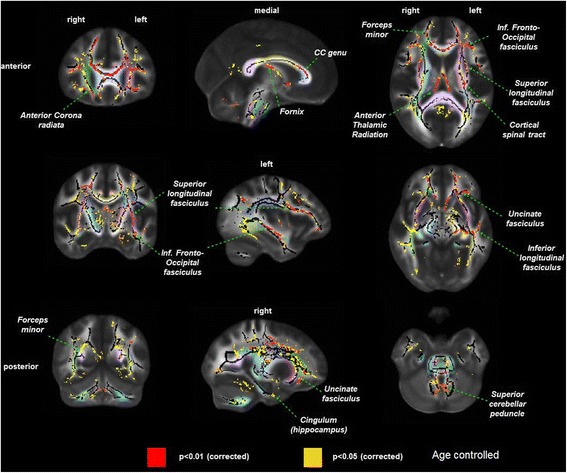
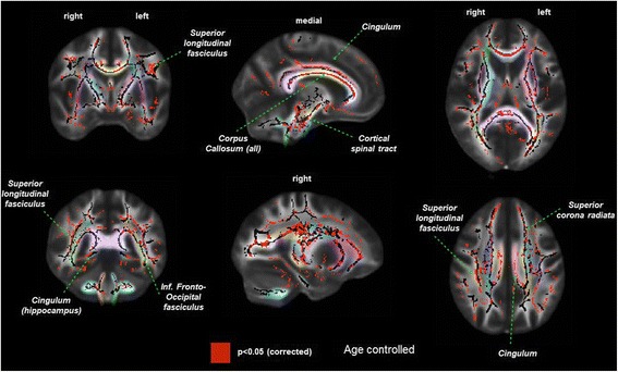
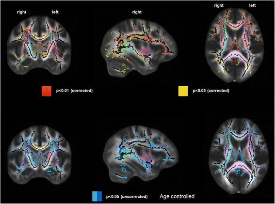
Results: Thirty-four HD patients and twenty six controls (52 vs 51 years for HD vs control) had adequate magnetic resonance imaging for analysis. The HD group had fewer women (38% vs 23%) and a higher prevalence of diabetes (29% vs 8%), heart failure (29% vs 0%) and clinical stroke (15% vs 0%). Hemodialysis patients had significantly lower FA across multiple white matter fiber tracts, with fronto-temporal connections, the genu of the corpus callosum and the fornix more significantly affected than posterior regions of the brain. Similarly, HD patients had significantly higher mean diffusivity in multiple anterior brain regions. Results remained similar when those with a prior history of stroke were excluded.

Conclusions: In HD patients, there is more white matter disease in the anterior than posterior parts of the brain compared to controls without kidney disease. This pattern of injury is most similar to that seen in aging, suggesting that developing chronic kidney disease and ultimately kidney failure may result in a phenotype consistent with accelerated aging.


Conflict of interest statement

Ethics approval and consent to participate

The Tufts MC Institutional Review Board approved the study, and all participants signed informed consent allowing for review of individual medical records as well as participation in the study.

Consent for publication

Not applicable.

Competing interests

The authors declare that they have no competing interests.

Publisher’s Note

Springer Nature remains neutral with regard to jurisdictional claims in published maps and institutional affiliations.

Figures

Fig. 1
Fig. 1
Difference in white matter damage assessed by fractional anisotropy in prevalent hemodialysis patients (n = 34) versus controls without kidney disease (n = 26), controlled for age. p values are corrected to account for multiple testing. Left column = coronal view, middle column = sagittal view, right column = axial view. Red = areas with most significant differences in FA (p < 0.01), yellow = remaining areas with differences in FA (p < 0.05). Pastel colors indicate distinct white matter fibers/bundles, which are labeled within the figure
Fig. 2
Fig. 2
Difference in white matter damage assessed by mean diffusivity in prevalent hemodialysis patients (n = 34) versus controls without kidney disease (n = 26). Left column = coronal view, middle column = sagittal view, right column = axial view. Red = areas with significant differences in FA (p < 0.05). Pastel colors indicate distinct white matter fibers/bundles, which are labeled within the figure
Fig. 3
Fig. 3
Difference in white matter damage assessed by fractional anisotropy in prevalent hemodialysis patients, separated by number of cardiovascular risk factors (zero or one vs more than one) compared to controls without kidney disease. Top row = Hemodialysis group with more than one vascular disease risk factor (n = 17) vs controls without kidney disease (n = 17). Bottom row = Hemodialysis group with zero or one vascular disease risk factor (n = 17) vs controls without kidney disease (n = 17). Left column = coronal view, middle column = sagittal view, right column = axial view. Red = areas with the most significant differences in FA, yellow = remaining areas with significant differences in FA. Pastel colors indicate distinct white matter fibers/bundles

References

    1. Khatri M, Wright CB, Nickolas TL, Yoshita M, Paik MC, Kranwinkel G, Sacco RL, DeCarli C. Chronic kidney disease is associated with white matter Hyperintensity volume. Stroke. 2007;38(12):3121–3126. doi: 10.1161/STROKEAHA.107.493593. - DOI - PMC - PubMed
    1. Fazekas G, Fazekas F, Schmidt R, Kapeller P, Offenbacher H, Krejs GJ. Brain MRI findings and cognitive impairment in patients undergoing chronic hemodialysis treatment. J Neurol Sci. 1995;134(1–2):83. doi: 10.1016/0022-510X(95)00226-7. - DOI - PubMed
    1. Nakatani T, Naganuma T, Uchida J, Masuda C, Wada S, Sugimura T, Sugimura K. Silent cerebral infarction in hemodialysis patients. Am J Nephrol. 2003;23(2):86. doi: 10.1159/000068034. - DOI - PubMed
    1. Watanabe A. Cerebral microbleeds and intracerebral hemorrhages in patients on maintenance hemodialysis. J Stroke Cerebrovasc Dis. 2007;16(1):30–33. doi: 10.1016/j.jstrokecerebrovasdis.2006.08.004. - DOI - PubMed
    1. Drew DA, Bhadelia R, Tighiouart H, Novak V, Scott TM, Lou KV, Shaffi K, Weiner DE, Sarnak MJ. Anatomic brain disease in hemodialysis patients: a cross-sectional study. Am J Kidney Dis. 2013;61(2):271–278. doi: 10.1053/j.ajkd.2012.08.035. - DOI - PMC - PubMed